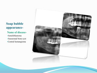
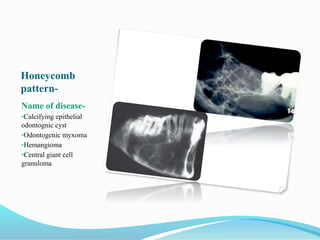
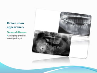
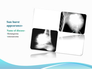
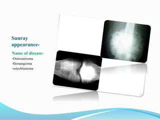
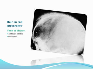
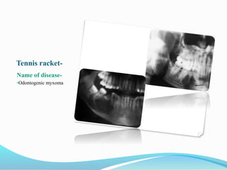
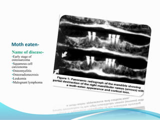
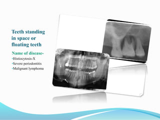
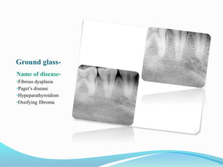
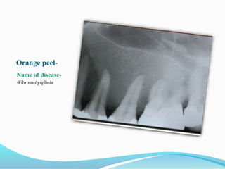
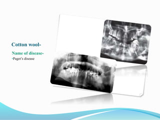
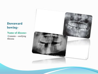
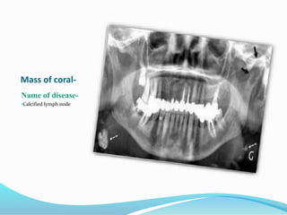
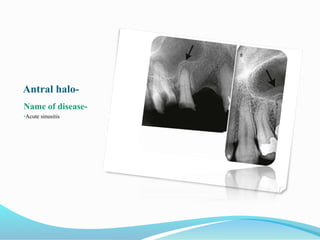
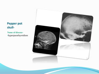
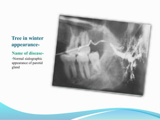
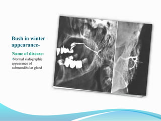
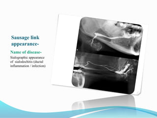
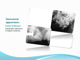
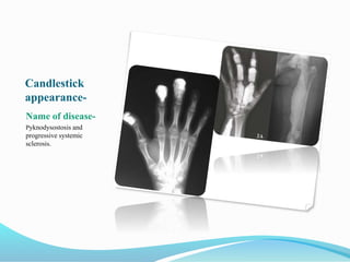
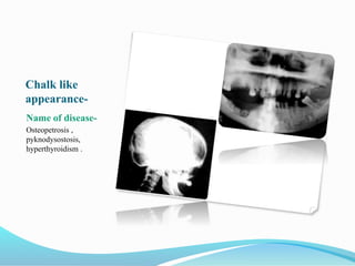
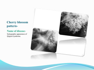
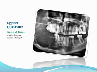
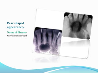
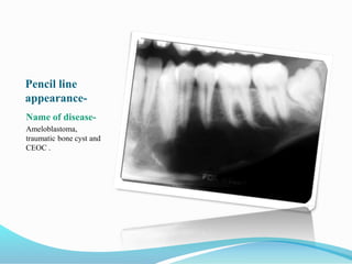
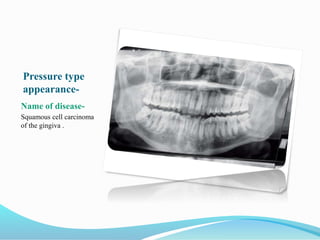
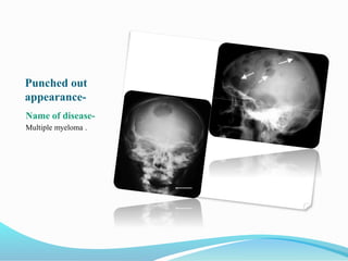
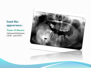
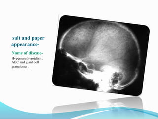
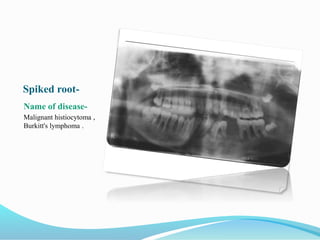
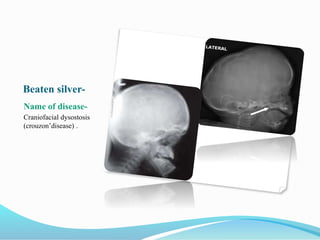
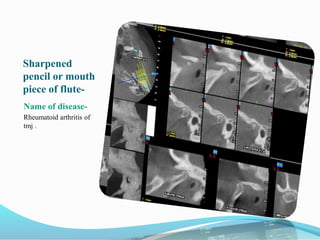
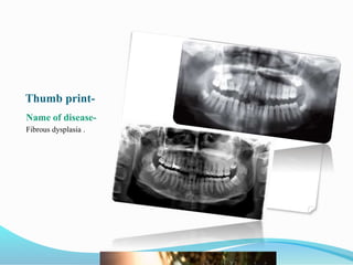
Typical radiological
feature Presented by-
Dr.Himanshu Gorawat
Dental surgeon
Soap bubble
appearance-
Name of disease-
•Ameloblastoma
•Aneursmal bone cyst
•Central hemangioma
Honeycomb
pattern-
Name of disease-
•Calcifying epithelial
odontognic cyst
•Odontogenic myxoma
•Hemangioma
•Central giant cell
granuloma
Driven snow
appearance-
Name of disease-
•Calcifying epithelial
odontogenic cyst
Sun burst
appearance-
Name of disease-
•Hemangioma
•osteosarcoma
Sunray
appearance-
Name of disease-
•Osteosarcoma
•Hemangioma
•osteoblastoma
Hair on end
appearance-
Name of disease-
•Sickle cell anemia
•thalassemia
Tennis racket-
Name of disease-
•Odontogenic myxoma
Moth eaten-
Name of disease-
•Early stage of
osteosarcoma
•Squamous cell
carcionoma
•Osteomyelitis
•Osteoradionecrosis
•Leukemia
•Malognant lymphoma
Teeth standing
in space or
floating teeth
Name of disease-
•Histiocytosis-X
•Severe periodontitis
•Malignant lymphoma
Ground glass-
Name of disease-
•Fibrous dysplasia
•Paget’s disease
•Hypeparathyroidism
•Ossifying fibroma
Orange peel-
Name of disease-
•Fibrous dysplasia
Cotton wool-
Name of disease-
•Paget’s disease
Downward
bowing-
Name of disease-
•Cemento – ossifying
fibroma
Mass of coral-
Name of disease-
•Calcified lymph node
Antral halo-
Name of disease-
•Acute sinusitis
Hanging drop
appearance in
maxillary sinus-
Name of disease-
•Blow out orbital fracture
Ely’s cyst-
Name of disease-
•osteoarthritis
Pepper pot
skull-
Name of disease-
•hyperparathyroidism
Step ladder
pattern-
Name of disease-
•Sickle cell anemia
Tree in winter
appearance-
Name of disease-
•Normal sialographic
appearance of parotid
gland
Bush in winter
appearance-
Name of disease-
•Normal sialographic
appearance of
submandibular gland
Sausage link
appearance-
Name of disease-
Sialographic appearance
of sialodochitis (ductal
inflammation / infection)
Snowstorm
appearance-
Name of disease-
Sialographic appearance
of Sjӧgren’syndrome.
Sialectasis-
Name of disease-
Sialographic appearance
of sialadenitis.
Ball in hand
appearance-
Name of disease-
Sialographic appearance
of intrinsic benign tumor.
Codman’s
triangle-
Name of disease-
Osteogenic sarcoma.
Balloon like
appearance-
Name of disease-
Follicular cyst.
Candlestick
appearance-
Name of disease-
Pyknodysostosis and
progressive systemic
sclerosis.
Chalk like
appearance-
Name of disease-
Osteopetrosis ,
pyknodysostosis,
hyperthyroidism .
Cherry blossom
pattern-
Name of disease-
Sialographic appearance of
Sjӧgren’syndrome.
Eggshell
appearance-
Name of disease-
Ameloblastoma ,
multilocular cyst .
Filling defect-
Name of disease-
Salivary gland tumor .
Mottled
appearance-
Name of disease-
Fibrous dysplasia ,
ossifying fibroma.
Onion peel (skin)
appearance-
Name of disease-
Chronic osteomyelitis ,
eosinophilic granuloma ,
Ewing's sarcoma .
Permeated
type-
Name of disease-
Carcinoma of gingiva ,
squampous cell
carcinoma of maxilla.
Pear shaped
appearance-
Name of disease-
Globulomaxillary cyst.
Pencil line
appearance-
Name of disease-
Ameloblastoma,
traumatic bone cyst and
CEOC .
Pressure type
appearance-
Name of disease-
Squamous cell carcinoma
of the gingiva .
Punched out
appearance-
Name of disease-
Multiple myeloma .
Sand like
appearance-
Name of disease-
Adenoameloblastoma ,
CEOC and CEOT .
salt and paper
appearance-
Name of disease-
Hyperparathyroidism ,
ABC and giant cell
granuloma .
Scalloping pattern
(margins)-
Name of disease-
Dentigerous cyst ,
traumatic bone cyst ,
ABC and giant cell
tumor .
Spiked root-
Name of disease-
Malignant histiocytoma ,
Burkitt's lymphoma .
Beaten silver-
Name of disease-
Craniofacial dysostosis
(crouzon’disease) .
Sharpened
pencil or mouth
piece of flute-
Name of disease-
Rheumatoid arthritis of
tmj .
Thumb print-
Name of disease-
Fibrous dysplasia .
Typical radiological feature

Typical radiological feature