1) The document reports a case study of a 48-year-old male polytrauma patient who was admitted to the ICU after a serious traffic accident with multiple injuries including cardiac and pulmonary contusions.
2) Due to rapidly worsening cardiogenic shock and refractory hypoxemia, the patient was placed on venoarterial extracorporeal membrane oxygenation (ECMO) as a rescue procedure.
3) ECMO successfully supported the patient's heart and lungs until respiratory and cardiac recovery occurred 4 days later, however the patient ultimately died on day 7 from an extensive brain infarction caused by the trauma.
PowerPoint presentation on ECMO (Extracorporeal Membrane Oxygenation). Part 2 focuses on Monitoring ECMO patients
Ventilatory strategies, Sedation and pain control, Weaning, Complications and recent advances in ECMO. For better understanding please have a look at ECMO part 1 before going through part 2.
It is a rare but potentially catastrophic event that is associated with high mortality. The reported incidence of ICA varies considerably across studies.
ECMO and its emerging role in trauma ICU 15th ECCC Dubai April 2019mansoor masjedi
Although there are some special considerations & important obstacles , extra-corporeal life support is increasingly used in multiple trauma patients admitted in ICU , with acceptable results.
Anesth considerations of pediatric patient with cardiac shunt for non cardiac...Bhavna Gupta
The large and growing population of patients who are living with CHD requires anaesthesia for non-cardiac surgeries and other procedures.
Knowledge of the pathophysiology of the common CHD lesions, as well as careful preoperative assessment and preparation, and communication with the patient’s cardiologist and surgeon, are essential to provide optimal care in the best setting for these patients.
Early experience of low flow extracorporeal carbon dioxide removal in managem...alungtech
Dr. Ravi Tiruvoipati presented the initial Australian experience with low-flow extracorporeal carbon dioxide removal (Hemolung RAS) at the 2015 Australian and New Zealand Intensive Care Society (ANZICS) meeting.
A Case Report of Hypothermia Rescued by Veno-Arterial Extracorporeal Membrane...semualkaira
Severe hypothermia is a life-threatening condition that often causes hemodynamic instability or cardiac arrest
and carries a high risk of mortality. The use of VA-ECMO in this
indication has greatly improved the prognosis of patients.
A Case Report of Hypothermia Rescued by Veno-Arterial Extracorporeal Membrane...semualkaira
Severe hypothermia is a life-threatening condition that often causes hemodynamic instability or cardiac arrest and carries a high risk of mortality. The use of VA-ECMO in this indication has greatly improved the prognosis of patients
A Case Report of Hypothermia Rescued by Veno-Arterial Extracorporeal Membrane...semualkaira
Severe hypothermia is a life-threatening condition that often causes hemodynamic instability or cardiac arrest
and carries a high risk of mortality. The use of VA-ECMO in this
indication has greatly improved the prognosis of patients
A study to assess the effectiveness of structured teaching program on knowledge regarding care of patients after cardiac surgery among staff nurses at Shree Narayana, Hospital, Raipur, chhattisgarh.
Worsening Tension Pneumocephalus from Late Post-traumatic Ventriculo-bronchia...asclepiuspdfs
The objective of the study was to report a case of tension pneumocephalus presenting as status epilepticus and outcome of treatment following emergency hyperbaric oxygen therapy. The data were collected from electronic medical record. The study was a case report. The data were extracted from medical record review and literature search. A 41-year-old male presented with status epilepticus and was found to have pneumocephalus within the cerebral venous sinuses. Before presentation he was complaining of intermittent hemoptysis attributed to a lung injury from a remote trauma due to a stab wound in the chest. At the time of his chest injury, he underwent multiple operations. His recovery was complicated by formation of left ventricular aneurysm and ventriculopleural fistula which was successfully repaired 5 years before presentation. Before determining the exact etiology of pneumocephalus, the patient was emergently treated with hyperbaric oxygen therapy (HBOT) to help with the management of intractable status epilepticus. During the HBOT therapy, the patient developed hemodynamic instability and the therapy was aborted. Repeat computed tomography (CT) scan showed worsening pneumocephalus with massive brain swelling and herniation. An echocardiogram showed bubbles crossing the left ventricle to the aorta. A CT thorax showed evidence of communication between the left ventricle and lung parenchyma at the site of the Gore-Tex confirming a ventriculo-bronchial fistula. Despite aggressive measures to control intracranial hypertension, the patient deteriorated and was declared brain dead. In cases of pneumocephalus where the exact cause is not well documented, an extensive investigation is recommended to ascertain the etiology before the institution of hyperbaric oxygen therapy.
Pulmonary Thromboembolism - etilogy, types, medical- Surgical and nursing man...VarunMahajani
Disruption of blood supply to lung alveoli due to blockage of one or more pulmonary blood vessels is called as Pulmonary thromboembolism. In this presentation we will discuss its causes, types and its management in depth.
ARTIFICIAL INTELLIGENCE IN HEALTHCARE.pdfAnujkumaranit
Artificial intelligence (AI) refers to the simulation of human intelligence processes by machines, especially computer systems. It encompasses tasks such as learning, reasoning, problem-solving, perception, and language understanding. AI technologies are revolutionizing various fields, from healthcare to finance, by enabling machines to perform tasks that typically require human intelligence.
Flu Vaccine Alert in Bangalore Karnatakaaddon Scans
As flu season approaches, health officials in Bangalore, Karnataka, are urging residents to get their flu vaccinations. The seasonal flu, while common, can lead to severe health complications, particularly for vulnerable populations such as young children, the elderly, and those with underlying health conditions.
Dr. Vidisha Kumari, a leading epidemiologist in Bangalore, emphasizes the importance of getting vaccinated. "The flu vaccine is our best defense against the influenza virus. It not only protects individuals but also helps prevent the spread of the virus in our communities," he says.
This year, the flu season is expected to coincide with a potential increase in other respiratory illnesses. The Karnataka Health Department has launched an awareness campaign highlighting the significance of flu vaccinations. They have set up multiple vaccination centers across Bangalore, making it convenient for residents to receive their shots.
To encourage widespread vaccination, the government is also collaborating with local schools, workplaces, and community centers to facilitate vaccination drives. Special attention is being given to ensuring that the vaccine is accessible to all, including marginalized communities who may have limited access to healthcare.
Residents are reminded that the flu vaccine is safe and effective. Common side effects are mild and may include soreness at the injection site, mild fever, or muscle aches. These side effects are generally short-lived and far less severe than the flu itself.
Healthcare providers are also stressing the importance of continuing COVID-19 precautions. Wearing masks, practicing good hand hygiene, and maintaining social distancing are still crucial, especially in crowded places.
Protect yourself and your loved ones by getting vaccinated. Together, we can help keep Bangalore healthy and safe this flu season. For more information on vaccination centers and schedules, residents can visit the Karnataka Health Department’s official website or follow their social media pages.
Stay informed, stay safe, and get your flu shot today!
263778731218 Abortion Clinic /Pills In Harare ,sisternakatoto
263778731218 Abortion Clinic /Pills In Harare ,ABORTION WOMEN’S CLINIC +27730423979 IN women clinic we believe that every woman should be able to make choices in her pregnancy. Our job is to provide compassionate care, safety,affordable and confidential services. That’s why we have won the trust from all generations of women all over the world. we use non surgical method(Abortion pills) to terminate…Dr.LISA +27730423979women Clinic is committed to providing the highest quality of obstetrical and gynecological care to women of all ages. Our dedicated staff aim to treat each patient and her health concerns with compassion and respect.Our dedicated group ABORTION WOMEN’S CLINIC +27730423979 IN women clinic we believe that every woman should be able to make choices in her pregnancy. Our job is to provide compassionate care, safety,affordable and confidential services. That’s why we have won the trust from all generations of women all over the world. we use non surgical method(Abortion pills) to terminate…Dr.LISA +27730423979women Clinic is committed to providing the highest quality of obstetrical and gynecological care to women of all ages. Our dedicated staff aim to treat each patient and her health concerns with compassion and respect.Our dedicated group of receptionists, nurses, and physicians have worked together as a teamof receptionists, nurses, and physicians have worked together as a team wwww.lisywomensclinic.co.za/
Ethanol (CH3CH2OH), or beverage alcohol, is a two-carbon alcohol
that is rapidly distributed in the body and brain. Ethanol alters many
neurochemical systems and has rewarding and addictive properties. It
is the oldest recreational drug and likely contributes to more morbidity,
mortality, and public health costs than all illicit drugs combined. The
5th edition of the Diagnostic and Statistical Manual of Mental Disorders
(DSM-5) integrates alcohol abuse and alcohol dependence into a single
disorder called alcohol use disorder (AUD), with mild, moderate,
and severe subclassifications (American Psychiatric Association, 2013).
In the DSM-5, all types of substance abuse and dependence have been
combined into a single substance use disorder (SUD) on a continuum
from mild to severe. A diagnosis of AUD requires that at least two of
the 11 DSM-5 behaviors be present within a 12-month period (mild
AUD: 2–3 criteria; moderate AUD: 4–5 criteria; severe AUD: 6–11 criteria).
The four main behavioral effects of AUD are impaired control over
drinking, negative social consequences, risky use, and altered physiological
effects (tolerance, withdrawal). This chapter presents an overview
of the prevalence and harmful consequences of AUD in the U.S.,
the systemic nature of the disease, neurocircuitry and stages of AUD,
comorbidities, fetal alcohol spectrum disorders, genetic risk factors, and
pharmacotherapies for AUD.
Title: Sense of Taste
Presenter: Dr. Faiza, Assistant Professor of Physiology
Qualifications:
MBBS (Best Graduate, AIMC Lahore)
FCPS Physiology
ICMT, CHPE, DHPE (STMU)
MPH (GC University, Faisalabad)
MBA (Virtual University of Pakistan)
Learning Objectives:
Describe the structure and function of taste buds.
Describe the relationship between the taste threshold and taste index of common substances.
Explain the chemical basis and signal transduction of taste perception for each type of primary taste sensation.
Recognize different abnormalities of taste perception and their causes.
Key Topics:
Significance of Taste Sensation:
Differentiation between pleasant and harmful food
Influence on behavior
Selection of food based on metabolic needs
Receptors of Taste:
Taste buds on the tongue
Influence of sense of smell, texture of food, and pain stimulation (e.g., by pepper)
Primary and Secondary Taste Sensations:
Primary taste sensations: Sweet, Sour, Salty, Bitter, Umami
Chemical basis and signal transduction mechanisms for each taste
Taste Threshold and Index:
Taste threshold values for Sweet (sucrose), Salty (NaCl), Sour (HCl), and Bitter (Quinine)
Taste index relationship: Inversely proportional to taste threshold
Taste Blindness:
Inability to taste certain substances, particularly thiourea compounds
Example: Phenylthiocarbamide
Structure and Function of Taste Buds:
Composition: Epithelial cells, Sustentacular/Supporting cells, Taste cells, Basal cells
Features: Taste pores, Taste hairs/microvilli, and Taste nerve fibers
Location of Taste Buds:
Found in papillae of the tongue (Fungiform, Circumvallate, Foliate)
Also present on the palate, tonsillar pillars, epiglottis, and proximal esophagus
Mechanism of Taste Stimulation:
Interaction of taste substances with receptors on microvilli
Signal transduction pathways for Umami, Sweet, Bitter, Sour, and Salty tastes
Taste Sensitivity and Adaptation:
Decrease in sensitivity with age
Rapid adaptation of taste sensation
Role of Saliva in Taste:
Dissolution of tastants to reach receptors
Washing away the stimulus
Taste Preferences and Aversions:
Mechanisms behind taste preference and aversion
Influence of receptors and neural pathways
Impact of Sensory Nerve Damage:
Degeneration of taste buds if the sensory nerve fiber is cut
Abnormalities of Taste Detection:
Conditions: Ageusia, Hypogeusia, Dysgeusia (parageusia)
Causes: Nerve damage, neurological disorders, infections, poor oral hygiene, adverse drug effects, deficiencies, aging, tobacco use, altered neurotransmitter levels
Neurotransmitters and Taste Threshold:
Effects of serotonin (5-HT) and norepinephrine (NE) on taste sensitivity
Supertasters:
25% of the population with heightened sensitivity to taste, especially bitterness
Increased number of fungiform papillae
NVBDCP.pptx Nation vector borne disease control programSapna Thakur
NVBDCP was launched in 2003-2004 . Vector-Borne Disease: Disease that results from an infection transmitted to humans and other animals by blood-feeding arthropods, such as mosquitoes, ticks, and fleas. Examples of vector-borne diseases include Dengue fever, West Nile Virus, Lyme disease, and malaria.
Title: Sense of Smell
Presenter: Dr. Faiza, Assistant Professor of Physiology
Qualifications:
MBBS (Best Graduate, AIMC Lahore)
FCPS Physiology
ICMT, CHPE, DHPE (STMU)
MPH (GC University, Faisalabad)
MBA (Virtual University of Pakistan)
Learning Objectives:
Describe the primary categories of smells and the concept of odor blindness.
Explain the structure and location of the olfactory membrane and mucosa, including the types and roles of cells involved in olfaction.
Describe the pathway and mechanisms of olfactory signal transmission from the olfactory receptors to the brain.
Illustrate the biochemical cascade triggered by odorant binding to olfactory receptors, including the role of G-proteins and second messengers in generating an action potential.
Identify different types of olfactory disorders such as anosmia, hyposmia, hyperosmia, and dysosmia, including their potential causes.
Key Topics:
Olfactory Genes:
3% of the human genome accounts for olfactory genes.
400 genes for odorant receptors.
Olfactory Membrane:
Located in the superior part of the nasal cavity.
Medially: Folds downward along the superior septum.
Laterally: Folds over the superior turbinate and upper surface of the middle turbinate.
Total surface area: 5-10 square centimeters.
Olfactory Mucosa:
Olfactory Cells: Bipolar nerve cells derived from the CNS (100 million), with 4-25 olfactory cilia per cell.
Sustentacular Cells: Produce mucus and maintain ionic and molecular environment.
Basal Cells: Replace worn-out olfactory cells with an average lifespan of 1-2 months.
Bowman’s Gland: Secretes mucus.
Stimulation of Olfactory Cells:
Odorant dissolves in mucus and attaches to receptors on olfactory cilia.
Involves a cascade effect through G-proteins and second messengers, leading to depolarization and action potential generation in the olfactory nerve.
Quality of a Good Odorant:
Small (3-20 Carbon atoms), volatile, water-soluble, and lipid-soluble.
Facilitated by odorant-binding proteins in mucus.
Membrane Potential and Action Potential:
Resting membrane potential: -55mV.
Action potential frequency in the olfactory nerve increases with odorant strength.
Adaptation Towards the Sense of Smell:
Rapid adaptation within the first second, with further slow adaptation.
Psychological adaptation greater than receptor adaptation, involving feedback inhibition from the central nervous system.
Primary Sensations of Smell:
Camphoraceous, Musky, Floral, Pepperminty, Ethereal, Pungent, Putrid.
Odor Detection Threshold:
Examples: Hydrogen sulfide (0.0005 ppm), Methyl-mercaptan (0.002 ppm).
Some toxic substances are odorless at lethal concentrations.
Characteristics of Smell:
Odor blindness for single substances due to lack of appropriate receptor protein.
Behavioral and emotional influences of smell.
Transmission of Olfactory Signals:
From olfactory cells to glomeruli in the olfactory bulb, involving lateral inhibition.
Primitive, less old, and new olfactory systems with different path
Ozempic: Preoperative Management of Patients on GLP-1 Receptor Agonists Saeid Safari
Preoperative Management of Patients on GLP-1 Receptor Agonists like Ozempic and Semiglutide
ASA GUIDELINE
NYSORA Guideline
2 Case Reports of Gastric Ultrasound
New Drug Discovery and Development .....NEHA GUPTA
The "New Drug Discovery and Development" process involves the identification, design, testing, and manufacturing of novel pharmaceutical compounds with the aim of introducing new and improved treatments for various medical conditions. This comprehensive endeavor encompasses various stages, including target identification, preclinical studies, clinical trials, regulatory approval, and post-market surveillance. It involves multidisciplinary collaboration among scientists, researchers, clinicians, regulatory experts, and pharmaceutical companies to bring innovative therapies to market and address unmet medical needs.
1. 374
Rev Bras Ter Intensiva. 2011; 23(3):374-379
Hemodynamic and respiratory support using
venoarterial extracorporeal membrane oxygenation
(ECMO) in a polytrauma patient
Uso de suporte hemodinâmico e respiratório por meio de
oxigenação extracorpórea por membrana (ECMO) venoarterial
em um paciente politraumatizado
INTRODUCTION
The use of extracorporeal membrane oxygenation (ECMO) as respiratory
support has been widely acknowledged as a rescue technique for refractory
hypoxemia in H1N1-infected patients.(1)
Recently, our institution adopted
the use of ECMO in the intensive care unit (ICU) for selected refractory
cardiopulmonary dysfunction cases.
In the venoarterial modality, venous blood is oxygenated and pumped
back into the arterial system, providing total/nearly total cardiorespiratory
support. This method is mostly used in patients who are difficult to wean
from cardiopulmonary bypass following acute myocardial infarction or in
cases of refractory cardiac arrest.(2)
Few investigators have reported the use of ECMO for simultaneous post-
traumatic cardiac and pulmonary dysfunctions.(3,4)
In this article, we report
the case of a 48-year-old male patient with cardiogenic shock and hypoxemia
due to cardiac and pulmonary contusions, who was successfully supported by
venoarterial ECMO until cardiorespiratory recovery.
CASE REPORT
A 48-year-old male patient, with no previous comorbidities, was brought
to the emergency service of Hospital das Clínicas after a serious traffic accident
(automobile versus motorcycle). The patient was a motorcycle rider who was
not wearing a helmet. At the accident site, he had an oxygen saturation of
Estevão Bassi1
, Luciano César
Pontes Azevedo1,2,3
, Eduardo Leite
Vieira Costa2,3,4
, Alexandre Toledo
Maciel1,2,3
, Edzangela Vasconcelos1,4
,
César Biselli Ferreira5
, Luiz Marcelo
Sá Malbouisson5
, Marcelo Park1,2,3
1. Intensive Care Unit, Discipline of
Emergency Medicine, Hospital das
Clínicas da Faculdade de Medicina da
Universidade de São Paulo – USP – São
Paulo (SP), Brazil.
2. Research and Education Institute,
Hospital Sírio-Libanês – São Paulo (SP),
Brazil.
3. Intensive Care Unit, Hospital Sírio-
Libanês – São Paulo (SP), Brazil.
4. Respiratory Intensive Care Unit,
Discipline of Pulmonology, Hospital das
Clínicas da Faculdade de Medicina
da Universidade de São Paulo – USP –
São Paulo (SP), Brazil.
5. Intensive Care Unit, Discipline of
General Surgery and Trauma, Hospital
das Clínicas da Faculdade de Medicina
da Universidade de São Paulo – USP –
São Paulo (SP), Brazil.
ABSTRACT
Therearefewreportsintheliterature
regarding the use of venoarterial
extracorporeal membrane oxygenation
(ECMO) for double-dysfunction
from both heart and lung contusions
in polytrauma patients. This article
reports a 48-year-old patient admitted
after a traffic accident. He rapidly
progressed to shock with low cardiac
output due to myocardial contusion
and refractory hypoxemia due to
pulmonary contusion, an unstable
chest wall and bilateral pneumothorax.
ECMO was an effective rescue
procedure in this dramatic situation
and was successfully discontinued on
the fourth day after the trauma. The
patient also developed an extensive
brain infarction and eventually died
on the seventh day after admission.
Keywords: Oxygenation; Shock,
cardiogenic; Acute lung injury;
Craniocerebral trauma; Case reports
Study conducted at the Intensive Care
Unit of the Emergency Medicine Service
of Hospital das Clínicas da Faculdade de
Medicina da Universidade de São Paulo –
USP – São Paulo (SP), Brazil.
Conflicts of interest: The ECMO
membranes were donated by Maquet
Cardiopulmonary.
Submitted on June 10, 2011
Accepted on August 18, 2011
Corresponding author:
Marcelo Park
Rua Enéas Carvalho de Aguiar, 255
Disciplina de Emergências – 5º andar
Zip Code: 05403-000 - São Paulo (SP),
Brazil.
Email: mpark@uol.com.br
CASE REPORT
2. Venoarterial ECMO in polytrauma patients 375
Rev Bras Ter Intensiva. 2011; 23(3):374-379
90% (while breathing room air), an unstable chest wall,
a heart rate of 130 beats per minute, an arterial blood
pressure of 80/40 mmHg, and a Glasgow coma scale
(GCS) rating of 12. During transport to the hospital,
orotracheal intubation, right hemithorax relief puncture
and volume expansion with 1,000 mL of saline solution
were performed.
Upon admission to the emergency room, the patient
was lying on a rigid bed with cervical collar in place. He
had a level 3 GCS and miotic pupils and was mechanically
ventilated. He had reduced breath sounds and severe
chest subcutaneous emphysema and was hypotensive.
Focused assessment with sonography for trauma (FAST)
was negative. Chest tubes were placed bilaterally, and
350 mL of blood had drained from the right side. The
patient was persistently hypotensive, despite volume
expansion with crystalloid solutions and administration
of vasopressor drugs.
Computed tomography (CT) imaging of the head,
chest, abdomen and pelvis showed a small left frontal
contusion (without an indication for surgical treatment)
with mild lateral ventricular asymmetry, suggesting
probable right brain edema; multiple costal fractures;
extensive pneumothorax and pneumopericardium;
bilateral pulmonary contusions; vertebral spinous
process fractures; fractures of the lumbar transverse
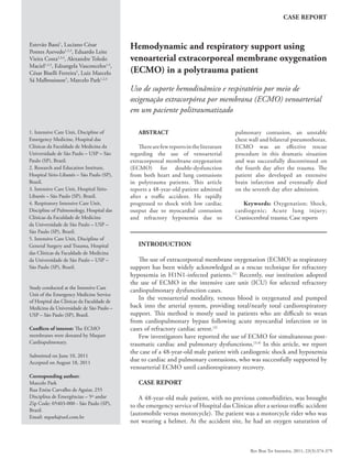
Figure 1 - A) Admission chest radiograph showing bilateral pneumothorax and extensive right lung consolidation, in addition
to several costal fractures. B) Initial chest computed tomography showing pneumothorax and extensive bilateral consolidation,
compatible with polytrauma and pulmonary contusion. C) Chest radiograph upon withdrawal of extracorporeal support
showing improvement in the pulmonary contusion and pneumothorax.
Figure 2 - A) Head computed tomography showing the initially mild asymmetric lateral ventricles, suggesting brain edema likely
from ischemia. B) Head computed tomography on the 5th
day from admission, already without extracorporeal support, showing
extensive left hemisphere infarction. C) Hemorrhagic transformation of the infarction after left frontoparietal craniectomy.
3. 376 Bassi E, Azevedo LCP, Costa ELV,
Maciel AT, Vasconcelos E, Ferreira CB et al.
Rev Bras Ter Intensiva. 2011; 23(3):374-379
processes; and a fracture of the left ilium extending to
the pubis and acetabulum (chest and head CT shown in
figures 1 and 2).
After admission to the ICU, difficulty with adequate
ventilation persisted due to the patient’s extensive
pulmonary contusions, even after effective bilateral lung
drainage. Additionally, the patient required increasing
doses of noradrenalin and dobutamine due to persistent
signs of low cardiac output (diaphoresis, coldness
and slow capillary filling). Bedside echocardiography
was performed, and the subcostal window showed
an extremely dilated (diastolic diameter 6 cm) and
hypokinetic left ventricle, with an estimated ejection
fraction of 0.08 (Teicholz). The esophageal Doppler
measured a cardiac index of 0.8 L/m2
.
About 18 hours after the trauma, despite the
administration of 4 mcg/kg/min noradrenalin and 20
mcg/kg/min dobutamine, the patient’s hemodynamics
progressively worsened, with a mean blood pressure of
50 mmHg, profuse sweating and delayed peripheral
perfusion. The patient was then placed under assisted
pressure controlled mechanical ventilation with an
inspired oxygen fraction (FiO2
) of 1.0, a positive end-
expiratory pressure (PEEP) of 10 cmH2
O, an inspiratory
pressure of 25 cmH2
O (15 cmH2
O driving pressure), an
inspiratory time of 0.75 seconds and a respiratory rate
of 30. Using these parameters, arterial blood gas showed
a PaO2
of 56 mmHg, an oxygen saturation of 84% and
3.1 mEq/L (28 mg/dL) lactate. Subsequent tests showed
progressive worsening of the physiological parameters,
with a central venous saturation of 57% (see Baseline
column in Table 1).
Given the imminent risk of death from cardiogenic
shock and refractory hypoxemia, our institution’s
ECMO team chose to start venoarterial ECMO support
as a rescue procedure. Using the Seldinger technique,
22 Fr draining cannulas were inserted into the right
common femoral vein. A return cannula was placed in
the right femoral artery with an 8F catheter for distal
perfusion of the right lower limb. A centrifuge magnetic
pump with a polymethylpentene oxygenation membrane
(Rotaflow/Jostra Quadrox, Maquet Cardiopulmonary
AG, Hirrlinger, Germany) was used. The blood flow was
initially at 4,500 mL/min with a 6,000 mL/min gas flow
(pure oxygen Sweeper).
The ECMO team of Hospital das Clínicas de São
Paulo and Hospital Sirio-Libanês consists of nurses,
physicians and physiotherapists. The entire shift team
Table 1 – Clinical progression including hemodynamic, respiratory, neurological (Sedation-Agitation Scale) and organ
dysfunction (Sequential Organ Failure Assessment score – SOFA) parameters
Baseline ECMO start 1st
ECMO day Last ECMO day After decannulation
PEEP (cmH2
O) 10 10 10 10 5
Ventilator FIO2
1 0.4 0.3 0.21 0.6
Respiratory rate (ipm) 30 10 10 10 14-30
Noradrenalin (mcg/kg/min) 4 0.5 0.19-1.38 0.06-0.03 0
Dobutamine (mcg/kg/min) 20 0 0 0 8
ECMO blood flow (L/min) na 4-4.5 4 4-4.5 na
ECMO gas flow (L/min) na 4-6 4 4 na
FIO2
ECMO na 1 0.4 0.4 na
Heart rate (bpm) 160 120 64-156 104-120 100-127
Mean blood pressure (mmHg) 50 70 64-71 65-80 53-91
pH 7.28 7.19 7.11 7.4 7.42
pO2
56 316 126 58 62
Arterial O2
saturation 84 99.8 97 90 92
pCO2
31 32 25 43 44
Bicarbonate 14 12 14 27 28
Base excess -11 -15 -19 2 3.1
Lactate 28 59 96 32 19
SAS 1-2 1-2 1-2 2 2
SOFA 17 17 16 14 14
ECMO – extracorporeal membrane oxygenation; PEEP – positive end-expiratory pressure; FiO2
– inspired oxygen fraction; SAS - Sedation-Agitation
Scale; SOFA - Sequential Organ Failure Assessment score.
Baseline column data represent the patient’s condition 18 hours after the trauma, immediately before the start of ECMO.
4. Venoarterial ECMO in polytrauma patients 377
Rev Bras Ter Intensiva. 2011; 23(3):374-379
rather than one specific person was responsible for
managing the device.
Progressivehemodynamicandrespiratoryimprovement
occurred about 8 hours after ECMO was started. This
allowed us to wean the patient from dobutamine and taper
the noradrenaline dose to 0.5 mcg/kg/min. a mean blood
pressure of 70 mmHg was maintained. The absence of a
pressure curve and a pulse pressure led us to infer that the
entire blood flow was mediated by the ECMO. Minimal
mechanical ventilation parameters were maintained, with
a PEEP of 10 cmH2
O, an inspiratory pressure of 20
cmH2
O and a FiO2
of 0.3 (controlled pressure mode).(5)
Blood gas analysis showed that the patient’s hypoxia had
been corrected; however, he continued to have metabolic
acidosis and significant hyperlactatemia. The ECMO
parameters were adjusted according to the perfusion and
oxygenation indices (Table 1).
During the ICU stay, this patient was given analgesia
with continuous fentanyl (0.25 – 0.5 mcg/kg/minute).
He continued to be obtunded (GCS 5T; Sedation
Agitation Scale (SAS) 1-2). On the second day following
admission, bedside cranial ultrasonography showed optic
sheath widening (6 mm) and midline shift; however, due
to the patient’s critical clinical status, no new CT scans
were possible during ECMO. Because the nature of the
intracranial event could not be precisely established,
we chose to maintain analgesia with fentanyl while
monitoring the consciousness level until imaging could
be performed. Pain was assessed based on behavioral and
physiological reactions.
Because of heavy bleeding from the chest tube (1.5 L
during the first day), which required multiple transfusions,
and the above described neurological conditions, an
anticoagulant was not given during the ECMO.
Despite the presence of acute renal failure (requiring
hemodialysis), low platelet counts and signs of extremity
ischemia (worse in the right leg where the arterial return
cannula was located), cardiac and pulmonary functions
progressively improved. On the 4th
day of support, a
pulse pressure curve was detected by invasive blood
pressure monitoring, and the echocardiogram-estimated
left ventricle ejection fraction was 0.3. The pulmonary
condition also improved, as assessed by chest x-ray.
Dobutamine inotropic support was restarted, and the
patient was successfully decannulated (Table 1).
The day after decannulation, the patient had
anisocoria and a decreased consciousness level (GCS
3T, SAS1). Repeat head CTs showed a left hemisphere
infarction (Figure 2). Left frontotemporal decompressive
craniectomy with duraplasty was performed. Postoperative
imaging showed significant post-decompression bleeding
(Figure 2) with neurological deterioration. The next day,
clinical examinations were compatible with brain death,
which could not be confirmed due to intraoperative use
of thionembutal. About 24 hours later, somatic death was
diagnosed.
DISCUSSION
The use of extracorporeal support for severe hypoxemia
in children is supported by relatively strong clinical
evidence.(2)
Little evidence has supported the use of this
technique in adult patients.(5)
Recently, however, interest
in the use of ECMO in adults has intensified, partly
due to technological advances (such as biocompatible
and durable membranes) and especially due to the large
number of refractory hypoxemia cases that occurred
during the H1N1 influenza epidemics.(1)
Extracorporeal support was a key component of the
successful Australian treatment regimen for H1N1-related
refractory hypoxemia.(1)
A recent randomized trial showed
a possible benefit from extracorporeal support in patients
with severe acute respiratory failure secondary to acute
lung injury/adult acute respiratory distress syndrome. In
this trial, ECMO was used to prevent lung injury caused
by mechanical ventilation using low ventilation volumes
and pressures. However, this study has been criticized
because ECMO was only used in 68 of the 90 randomized
patients, and there was a relatively high rate of death
among patients during transfer to ECMO-specialized
sites.(5)
The lack of well-designed clinical trials of ECMO in
adult patients prevents us from drawing clear conclusions
about the utility of the procedure in adult critical care
patients, as highlighted in a recent systematic review.(6)
Therefore, the current status of ECMO is that of a rescue
measure for failed traditional therapeutics. Because of
this, a multidisciplinary team was established for using
ECMO in selected refractory hypoxemia cases. With
similar indications to those proposed by CESAR,(5)
this team aims to offer an alternative for patients in
whom usual hypoxemia management measures (e.g.,
alveolar recruitment maneuvers, nitric oxide and high-
frequency ventilation) are ineffective and/or harmful
(e.g., barotrauma or high airway pressure needed for
maintaining acceptable ventilation).
Significant hypoxemia is a common complication
of pulmonary contusion, and the use of extracorporeal
oxygenation in some of these patients has been reported.(7)
However, the case reported here differs from prior
5. 378 Bassi E, Azevedo LCP, Costa ELV,
Maciel AT, Vasconcelos E, Ferreira CB et al.
Rev Bras Ter Intensiva. 2011; 23(3):374-379
reports in several important ways. This patient had
refractory hypoxemia from several injury mechanisms
(pulmonary contusion, bilateral extensive pneumothorax
and an unstable chest wall) and had a PaO2
/FiO2
ratio
of 56. Measures frequently used in this context would
be inappropriate or even harmful. Alveolar recruitment
maneuvers could worsen the bilateral air fistulae, and
prone positioning is contraindicated due to the extreme
hemodynamic instability.
Additionally, the patient had shock that was refractory
to volume expansion, vasopressors and inotropic drugs.
Echocardiography revealed clear cardiogenic shock, likely
due to myocardial contusion. Considering the imminent
risk of death and that there were no other therapeutic
options, we chose to use venoarterial ECMO with total
cardiorespiratory support to simultaneously support
the patient’s respiratory and hemodynamic functions.
Significant improvement was quickly achieved (Table 1),
allowing the progressive recovery of cardiac and pulmonary
functions, and we were able to discontinue extracorporeal
support after 4 days.
During this time, clinical (consciousness level) and
imagery signs (transcranial ultrasound and widened optical
sheath) were indicative of intracranial hypertension.
However, the patient’s complete dependency on
hemodynamic and respiratory support prevented him
from undergoing a head CT. Unfortunately, the patient
died from his intracranial injury, which could not be
assessed and treated in a timely fashion.
Few reports discuss full cardiopulmonary support
with venoarterial ECMO in trauma patients. Perchinsky
et al. reported 50% survival in a series of 6 patients using
this procedure as a rescue measure for severe polytrauma
patients deteriorating in spite of the conventional
therapy.(3)
More recently, Masiakos et al. reported the
successful management of a patient with pulmonary
and myocardial contusions in addition to right
ventricular papillary muscle rupture with significant
tricuspid regurgitation, who presented with significant
hypoxemia, hemodynamic instability and difficult-to-
manage ventricular arrhythmias.(4)
In our report, we successfully used extracorporeal
support as a rescue measure for cardiorespiratory
dysfunction that would otherwise have been rapidly fatal.
ECMO was effective as a bridging strategy, allowing
decannulation on the 4th
day after the trauma. The patient
died from head trauma, which was not related to nor
treated with extracorporeal support. Unlike the report by
Masiakos et al.,(4)
no additional ICU professionals (e.g.,
a perfusionist) were necessary during the extracorporeal
support; only our institutional ECMO team was involved.
In summary, this article reports a polytrauma patient
with refractory hypoxemia due to pulmonary contusion
and refractory cardiogenic shock due to cardiac contusion.
Venoarterial ECMO was successfully used as a bridging
strategy for cardiac and pulmonary recovery, and the
extracorporeal support was discontinued on the 4th
day
after trauma. This extracorporeal support method can be
lifesaving in selected patients. However, additional studies
are necessary to evaluate how this promising technology
may best be used clinically.
Participants of the Hospital das Clínicas de São
Paulo and Hospital Sírio-Libanês ECMO team:
Luciano Cesar Pontes Azevedo, Marcelo Park, André
Luiz de Oliveira Martins, Eduardo Leite Vieira Costa,
Guilherme Paula Pinto Schettino, Marcelo Brito Passos
Amato, Carlos Roberto Ribeiro Carvalho, Mauro Tucci,
Alexandre Toledo Maciel, Fernanda Maria Queiroz Silva,
Leandro Utino Taniguchi, Edzângela Vasconcelos, Raquel
de Nardi, Cláudio Machtans, Michele Nardi and Adriana
Sayuri Hirota.
RESUMO
Existem poucos relatos na literatura sobre o uso de oxige-
nação extracorpórea por membrana venoarterial por dupla dis-
função decorrente de contusão cardíaca e pulmonar no paciente
politraumatizado. Relatamos o caso de um paciente de 48 anos,
vítima de acidente de motocicleta e automóvel, que evoluiu ra-
pidamente com choque refratário com baixo débito cardíaco
por contusão miocárdica e hipoxemia refratária decorrente de
contusão pulmonar, tórax instável e pneumotórax bilateral. O
suporte extracorpóreo foi uma medida efetiva de resgate para
esse caso dramático, e o seu uso pôde ser interrompido com su-
cesso no 4º dia após o trauma. O paciente evoluiu com extenso
infarto cerebral, morrendo no 7º dia de internação.
Descritores: Oxigenação; Choque cardiogênico; Lesão
pulmonar aguda; Traumatismos craniocerebrais; Relatos de casos
6. Venoarterial ECMO in polytrauma patients 379
Rev Bras Ter Intensiva. 2011; 23(3):374-379
REFERENCES
1. Australia and New Zealand Extracorporeal Membrane
Oxygenation(ANZECMO)InfluenzaInvestigators, Davies
A, Jones D, Bailey M, Beca J, Bellomo R, Blackwell N, et
al. Extracorporeal Membrane Oxygenation for 2009
Influenza A(H1N1) Acute Respiratory Distress Syndrome.
JAMA. 2009;302(17):1888-95.
2. Sidebotham D, McGeorge A, McGuinness S, Edwards
M, WillcoxT, BecaJ.Extracorporealmembraneoxygenation
for treating severe cardiac and respiratory disease in adults:
Part 1--overview of extracorporeal membrane oxygenation.
J Cardiothorac Vasc Anesth. 2009;23(6):886-9.
3. Perchinsky MJ, Long WB, Hill JG, Parsons
JA, Bennett JB. Extracorporeal cardiopulmonary
life support with heparin-bonded circuitry in the
resuscitation of massively injured trauma patients. Am
J Surg. 1995;169(5):488-91.
4. Masiakos PT, Hirsch EF, Millham FH. Management
of severe combined pulmonary and myocardial
contusion with extracorporeal membrane oxygenation. J
Trauma. 2003;54(5):1012-5.
5. Peek GJ, Mugford M, Tiruvoipati R, Wilson A, Allen
E, Thalanany MM, Hibbert CL, Truesdale A, Clemens
F, Cooper N, Firmin RK, Elbourne D; CESAR trial
collaboration. Efficacy and economic assessment of
conventional ventilatory support versus extracorporeal
membrane oxygenation for severe adult respiratory failure
(CESAR): a multicentre randomised controlled trial.
Lancet. 2009;374(9698):1351-63. Erratum in Lancet.
2009;374(9698):1330.
6. Mitchell MD, Mikkelsen ME, Umscheid CA, Lee
I, Fuchs BD, Halpern SD. A systematic review to inform
institutional decisions about the use of extracorporeal
membrane oxygenation during the H1N1 influenza
pandemic. Crit Care Med. 2010;38(6):1398-404.
7. Keel M, Meier C. Chest injuries - what is new? Curr Opin
Crit Care. 2007;13(6):674-9. Review.