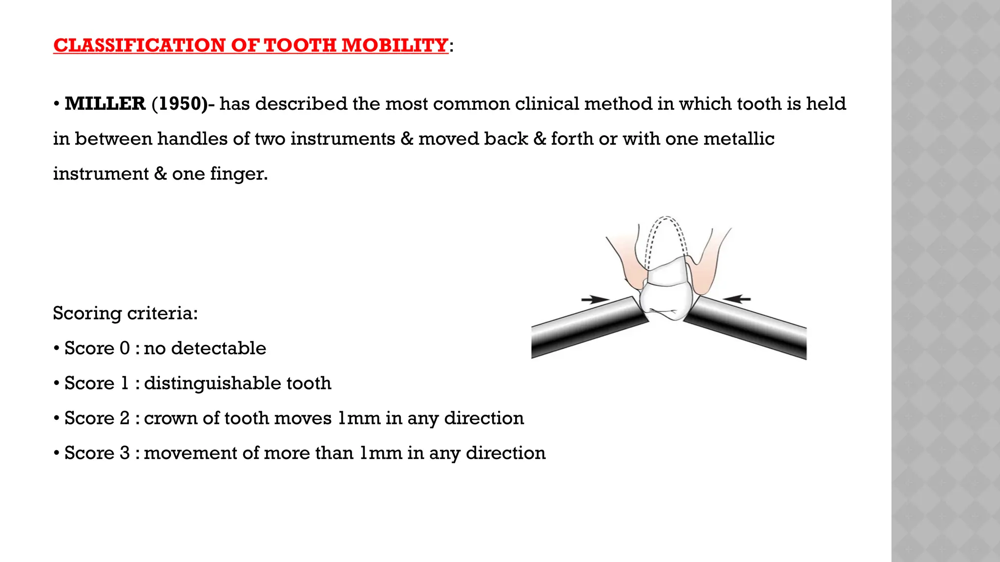
Good morning
Tooth mobility and
periodontal disease
 Introduction
 Causes of tooth mobility
 Types of mobility
 Tooth mobility curve
 Stages of tooth mobility
 Classifications of tooth mobility
 Methods of assesing tooth mobility
periodontometer
periotest
 Radiographic changes
 Reduction in tooth mobility
 Clinical studies
 Conclusion
 References
Contents:
Tooth mobility is defined as a visually perceptible movement
of the tooth away from its normal position when a light force
is applied.
( Gher 1996)
Tooth mobility
Introduction:
Physiologic mobility is movement up to 0.2 mm horizontally and
0.02 mm axially.
.
CAUSES
•Loss of tooth support
•Extension of inflammation from the gingival or from the periapex into the PDL.
•Periodontal surgery
•Tooth mobility increases in pregnancy
•Pathologies of the jaw
•Trauma from occlusion.
•In health, physiological or functional mobility of tooth exists & every tooth with
healthy periodontal support will have a physiologic range of mobility .
•Mobility is a measurement of horizontal & vertical tooth displacement in the
socket.
•One study has suggested that pockets around mobile teeth harbor higher
proportions of Campylobacter rectus and Peptostreptococcus micros and
possibly Porphyromonas gingivalis as compared with nonmobile teeth.
(CARRANZA 13TH
EDITION)
•MOBILITY CAN BE OF TWO TYPES:
1. PHYSIOLOGIC TOOTH MOBILITY
2. PATHOLOGIC TOOTH MOBILITY
PHYSIOLOGIC TOOTH MOBILITY
• It refers to moderate force exerted on the crown of tooth surrounded by a
healthy & intact periodontium & tooth will show tipping movement until a
closer contact has been established between root & marginal bony tissue
(MUHLEMAN,1951 KORBER,1971 LINDHE ,1989 )
• Normal tooth mobility varies between different types teeth:
Incisors - 10- 12 mm/ 100 mm
Canines - 5 - 9mm/100mm
Premolars - 8 - 10mm/100mm
Molars - 4 - 8mm/100mm
•A large number of assessments showed that this force/displacement relationship had a
typical pattern which could be illustrated by a double sloped curve, the so called tooth
mobility (TM) curve.
•The 2 parts of this curve were defined as "initial" (ITM) and "secondary" (STM) tooth
mobility components .
(Muhlemann 1954, 1960).
•In this 2nd phase, a comparatively larger force increment was needed to obtain a
certain additional tooth movement .
(Muhlemann 1954, 1960)
•"initial tooth mobility" (ITM), corresponded with a movement of the root within the
periodontal ligament space.
•The movement progressed until the complete elongation of the fibers was achieved. but
slower, root displacement (STM), obtained by the use of heavier (>100 g) forces, was
accompanied by the distortion of the marginal bone and compression of the soft tissues.
Factors affecting physiologic tooth mobility:
Daily variations:
• Tooth mobility will be greatest in the morning,which progressively decreases due to
slight extrusion of tooth & minimal during sleep.
• During walking hours mobility is reduced by chewing & swallowing forces which intrude
teeth into socket .
•Greater tooth mobility values were found in children and females than in adults and
males. (Mtlhlemann 1960)
Tooth contact during deglutition:
• functional forces received by teeth during deglutition resulted in tooth contact
which maintains the tooth in proper positions.
Effect of stress-inducing conditions:
• Habits like bruxism & clenching activities affect tooth mobility as well
• Females > males
• Pregnancy
It is increased in pregnancy & sometimes associated with menstrual cycle or
use of hormonal contraceptives . As a result of the hormonal influences on collagen
and vascular structures of the ligament tissues.
(Mtihlemann 1960, Mtihlemann et al. 1965, Rateitschak 1967)
1. INITIAL STAGE OR INTRA SOCKET STAGE:
 Tooth moves within confines of periodontal ligament associated with viscoelastic
distortion of ligament & redistribution of periodontal fluids, inter-bundle content &
fibers.
This initial movement occurs with forces of about 100 lb, and it is on the order of 0.05
mm to 0.10 mm (50 µm to 100 µm).
Tooth mobility occurs in TWO STAGES:
2. SECONDARY STAGE :
 Occurs gradually & entails defomation of alveolar bone in response
to a increased horizontal forces.
When a force of 500 g is applied to the crown, the resulting
displacement is about
100 µm to 200 µm for incisors,
50 µm to 90 µm for canines,
8 µm to 10 µmfor premolars,
40 µm to 80 µm for molars.
•The recovery movement is pulsating, and it is apparently
associated with the normal pulsation of the periodontal vessels,
which occurs in synchrony with the cardiac cycle.
In two stages:
•The first is an immediate, springlike elastic recoil;
•The second is a slow, asymptomatic recovery movement.
PATHOLOGIC TOOTH MOBILITY:
• Refers to any degree of perceptible movement of faciolingually, mesiodistaly or axially
when a force is applied to tooth .
CAUSES OF PATHOLOGIC TOOTH MOBILITY:
• Extension of inflammation from gingiva or from periapex into periodontal ligament
results in changes that increases mobility.
• Tooth loss, when a large number of teeth have been lost,remaining tooth must assume all
functional demands.
•Trauma from occlusion-
When trauma from occlusion is the result of alterations in occlusal forces, it is called
primary trauma from occlusion.
When it results from the reduced ability of the tissues to resist the occlusal forces, it is known
as secondary trauma from occlusion.
A. Enlargement of the periodontal ligament space,
B. Osteoclastic alveolar bone resorption,
C. Vascular alterations and degenerative phenomena in the periodontal
membrane.
D. A reduced number of collagen fibers inserting in the root cementum, in the
alveolar bone proper and in the crest.
(Biancu et al.1995b).
 Effects of TFO
 Removal of causative agents results in a prompt recovery of
(i) Normal tooth mobility values and
(ii) Radiographic width of the PDL space accompanied by a complete
reorganisation of the soft and hard periodontal tissues.
(Lindhe & Ericsson (1982),Vollmer& Rateitshack (1975),
Poison et ai.(1976b)
• Pathologic process of jaws that destroys alveolar bone and roots of teeth can also result
in mobility.
• Periodontal surgery increases tooth mobility for a short period.
•Periodontal disease.
 Increased tooth mobiiity is a common symptom also of advanced forms of plaque
associated periodontal disease
(Muhlemann 1960. Lindhe & Nyman 1977, Lindhe & Nyman 1989).
 Muhlemann (1960, 1967) observed that at periodontally compromized teeth both
the "initial" (ITM) or "secondary" (STM) tooth mobility value were elevated.
CLASSIFICATION OF TOOTH MOBILITY:
• MILLER (1950)- has described the most common clinical method in which tooth is held
in between handles of two instruments & moved back & forth or with one metallic
instrument & one finger.
Scoring criteria:
• Score 0 : no detectable
• Score 1 : distinguishable tooth
• Score 2 : crown of tooth moves 1mm in any direction
• Score 3 : movement of more than 1mm in any direction
CARANZA F.A.
- described it as normal mobility
• Grade 1 : slightly more than normal.
• Grade 2 : moderately more than normal.
• Grade 3 : severe mobility faciolingually & or mesiodistally combined with vertical
displacement.
Glickmans Index (1972)
 0- Normal mobility
 Grade I- Slightly more than normal
 Grade II- Moderately more than normal
 Grade III- Severe mobility faciolingually and / or mesidistally combined with vertical
displacement.
Lindhe (1997)
 Degree1: Movability of the crown 0.2- 1mm in horizontal
direction.
 Degree 2: Movability of the crown of the tooth exceeding
1 mm in horizontal direction.
 Degree 3: Movability of the crown of the tooth in vertical direction.
METHOD OF ASSESSING TOOTH MOBILITY:
• The instrument system {PERIODONTOMETER} permits reproducible
assessment of horizontal mobility of all types of both arches .
Conventional method
Clinical mesurements
Clinical indices.
Newer methods
Peridontiometer
Periotest.
Clinical mesurement
Indices
MILLER,s index (1950)
Glickmans Index (1972)
Lindhe (1997)
periodontometer
Instrument used to measure and assess the
severity of periodontal disease.
Periodontometry was developed by
Muhlemann(1957) to facilitate the clinical
diagnosis of the periodontal attach- ment
apparatus by measuring the mobility of the
teeth.
designed to measure hori-zontal mobility
of the anterior teeth.
•A dial gauge mounted on a metal arm that is secured to the base of a metal impres- sion
tray.
•The impression tray is anchored to the posterior maxillary teeth leaving the incisors and
canines free.
•The dial gauge is adjusted so that its spring-loaded pointer contacts the labial surface of
the tooth to be tested.
•Standardized labiopalatal and palato- labial loads are applied with a force meter (Correx
Gauge, Haag-Streit) to produce palatal and labial displace- ment, respectively, of the
tested tooth.
•This displacement of the tooth is measured in hundredths of a millimeter on the dial
gauge
PERIOTEST
PERIOTEST A new method for determining tooth mobility was invented by
Schulte and co-workers in 1987 and 1992.
The periotest device measures the reaction of the periodontium to a
defined percussion force which is applied to the tooth and delivered by a
tapping instrument.
•A metal rod is accelerated to a speed of 0.2 m/s with the device
and maintained at constant velocity Upon impact the tooth is
deflected and the rod decelerated .
•The contact time between the tapping head and tooth varies
between 0.3 and 2 milliseconds and is shorter for stable than
mobile teeth .
RADIOGRAPHIC CHANGES:
•Marked horizontal radiographic loss of bony support may be associated with minimal
tooth mobility.
•Modest degree of breakdown may be associated with pronounced tooth mobility.
•Periodontally involved mobile units may also display funneled periodontal
radiolucencies resulting from co-existing angular bony defects
•Radiolucencies may be suggestive of endodontic lesion Radiolucencies may be
seen with furcation at furcation involved mobile teeth.
Reduced tooth mobility
•Root ankylosis which may occur after a failing tooth
reimplantation (Hammarstrom et al. 1989) or if autogenous
bone grafts are placed in contact with a detached root surface
(Bernard 1991)
•In such a situation, there is no "initial" intra-socket tooth
displacement and a movement of the tooth to be achieved only
"by elastic deformation of the root and the bone alveolus"
(Miihlemann 1967).
Reduced following occlusal Therapy
The removal of premature contacts at mobile teeth often results in a complete or partial
regression of
(i) Tooth hypermobility
(ii) Radiographic widening of the periodontal ligament space and
(iii) Dearranged periodontal ligament tissue
(Lindhe & Nyman 1989,VoUmer & Rateitschak 1975,Lindhe & Ericsson1982,)
Reduced from treatment of gingivitis and periodontitis
•The effect of periodontal therapy on tooth mobility has been studied in several
investigations. Treatment procedures restricted to supragingival debridement and resolution
of gingivitis fail to reduce tooth mobility in periodontitis patients. (Donze et al.1973).
•This finding is in agreement with the observation that gingivitis alone cannot cause
increased tooth mobility .
(Ferris 1966,Son et al.1971).
•From some clinical studies it was reported that a successful periodontal treatment results
in resolution of inflammation, bone regeneration and TM reduction.
(Lindhe & Nyman (1975),Persson (1980,198 la,b),
Poison et al.(1976b,1979),Giargia et al. (1994)
Overview of clinical studies evaluating the role of tooth mobility in relation to
severity, occurrence and progression of periodontal disease.
•Increased tooth mobility is not consistently associated with severe periodontal
breakdown .
(cross-sectional study done by Baelum et al.in (1988) study the prevalence and severity
of periodontal disease in subjects who never experienced oral health care).
•Increased tooth mobility resulted to be a risk marker for attachment loss during
maintenance.
(retrospective, long term study done by Ismail et al. (1990) evaluate risk markers for
further attachment loss during maintenance.)
•Not possible to determine if increased tooth mobility is the cause or just a
symptom of periodontal destruction caused by subgingival plaque accumulation.
(cross-sectional study done by Jin & Cao assess the existence of an (1992) Pihlstrom et
al. (19S6) association between clinical signs of traumatic occlusion/increased TM and
severity of periodontal disease.)
Overview of clinical studies evaluating the role of tooth mobility in
affecting the therapeutic management of periodontal disease.
Therapeutic management of periodoatal disease: clinical studies
•Bone regeneration (fill) occurred also at teeth which initially showed increased tooth
mobility.
(Polson & Heij] {1978)
•Pockets related to mobile teeth can be successfully treated and maintained, regardless,
of the severity and degree of mobility; however, the outcome at mobile teeth was less
favourable .
(Fleszar et al.(1980)
Splinting
Periodontal treatment had a stabilising effect while splinting had no additional
consequence.
(Kege! et al.(1979)
Splinting neither affected tooth mobility nor gingival attachment and supporting
bone over 24 week period after osseous surgery.
(Galler et al 1979)
Occurance and progression
Increased tooth mobility resulted to be a risk marker for attachment loss during
maintenance.
(Ismail et al.(1990)
Despite the fact that increased tooth mobility has been
described as risk factor for progressive periodontitis (Ismail et
al. 1990), it is not always found at teeth showing severe
periodontal breakdown.
(Shefter & Me Fall 1984, Baelum et ai, 1988);
•In the end, the role of tooth mobility and periodontal disease
has been reviewed and it seems it can be concluded that-
Conclusion:
•Initial phase of healing following treatment does not enhance the outcome of therapy .
(Kegel et al.1979,Galler et ai.1979).
•When the inflammatory process is controlled and an adequate oral hygiene performed,
teeth showing stable hypermobility respond in a proper way to treatment and may be
maintained in good function over long periods of time.
(Poison et al.1983, Poison & Heijl 1978,Rosiing et al.1976,
Lindhe & Nyman 1977);
REFERENCES:
•Baelum.V, Fejerskov. O. & Manji, F. (1988)Periodontai disease in adult Kenyans,Journal
of Clinical Periodontology 15.445-452.
•Bernard, G.W. (1991) Healing and repair of osseous defects. Denial Clinics of North
America 35.46977
•Ericsson, L & Lindhe, J, (1977) Lack of effect of trauma from occlusion on the
recurrence of experimental periodontitis.Journal of Clinical Periodontologv 4.115-127.
•Haramarstrom, L,, Blonalof, L, & Lindskog,S, (1989') Dynamics of dento alveolar
ankylosis and associated root resorption,Endodontic Dental Traumatology5, 163-175,
tooth mobility and periodontal diseses.pptx

tooth mobility and periodontal diseses.pptx

  • 1.
  • 2.
  • 3.
     Introduction  Causesof tooth mobility  Types of mobility  Tooth mobility curve  Stages of tooth mobility  Classifications of tooth mobility  Methods of assesing tooth mobility periodontometer periotest  Radiographic changes  Reduction in tooth mobility  Clinical studies  Conclusion  References Contents:
  • 4.
    Tooth mobility isdefined as a visually perceptible movement of the tooth away from its normal position when a light force is applied. ( Gher 1996) Tooth mobility Introduction: Physiologic mobility is movement up to 0.2 mm horizontally and 0.02 mm axially. .
  • 5.
    CAUSES •Loss of toothsupport •Extension of inflammation from the gingival or from the periapex into the PDL. •Periodontal surgery •Tooth mobility increases in pregnancy •Pathologies of the jaw •Trauma from occlusion.
  • 6.
    •In health, physiologicalor functional mobility of tooth exists & every tooth with healthy periodontal support will have a physiologic range of mobility . •Mobility is a measurement of horizontal & vertical tooth displacement in the socket. •One study has suggested that pockets around mobile teeth harbor higher proportions of Campylobacter rectus and Peptostreptococcus micros and possibly Porphyromonas gingivalis as compared with nonmobile teeth. (CARRANZA 13TH EDITION)
  • 7.
    •MOBILITY CAN BEOF TWO TYPES: 1. PHYSIOLOGIC TOOTH MOBILITY 2. PATHOLOGIC TOOTH MOBILITY
  • 8.
    PHYSIOLOGIC TOOTH MOBILITY •It refers to moderate force exerted on the crown of tooth surrounded by a healthy & intact periodontium & tooth will show tipping movement until a closer contact has been established between root & marginal bony tissue (MUHLEMAN,1951 KORBER,1971 LINDHE ,1989 )
  • 9.
    • Normal toothmobility varies between different types teeth: Incisors - 10- 12 mm/ 100 mm Canines - 5 - 9mm/100mm Premolars - 8 - 10mm/100mm Molars - 4 - 8mm/100mm
  • 10.
    •A large numberof assessments showed that this force/displacement relationship had a typical pattern which could be illustrated by a double sloped curve, the so called tooth mobility (TM) curve. •The 2 parts of this curve were defined as "initial" (ITM) and "secondary" (STM) tooth mobility components . (Muhlemann 1954, 1960).
  • 12.
    •In this 2ndphase, a comparatively larger force increment was needed to obtain a certain additional tooth movement . (Muhlemann 1954, 1960) •"initial tooth mobility" (ITM), corresponded with a movement of the root within the periodontal ligament space. •The movement progressed until the complete elongation of the fibers was achieved. but slower, root displacement (STM), obtained by the use of heavier (>100 g) forces, was accompanied by the distortion of the marginal bone and compression of the soft tissues.
  • 13.
    Factors affecting physiologictooth mobility: Daily variations: • Tooth mobility will be greatest in the morning,which progressively decreases due to slight extrusion of tooth & minimal during sleep. • During walking hours mobility is reduced by chewing & swallowing forces which intrude teeth into socket . •Greater tooth mobility values were found in children and females than in adults and males. (Mtlhlemann 1960)
  • 14.
    Tooth contact duringdeglutition: • functional forces received by teeth during deglutition resulted in tooth contact which maintains the tooth in proper positions. Effect of stress-inducing conditions: • Habits like bruxism & clenching activities affect tooth mobility as well • Females > males • Pregnancy It is increased in pregnancy & sometimes associated with menstrual cycle or use of hormonal contraceptives . As a result of the hormonal influences on collagen and vascular structures of the ligament tissues. (Mtihlemann 1960, Mtihlemann et al. 1965, Rateitschak 1967)
  • 15.
    1. INITIAL STAGEOR INTRA SOCKET STAGE:  Tooth moves within confines of periodontal ligament associated with viscoelastic distortion of ligament & redistribution of periodontal fluids, inter-bundle content & fibers. This initial movement occurs with forces of about 100 lb, and it is on the order of 0.05 mm to 0.10 mm (50 µm to 100 µm). Tooth mobility occurs in TWO STAGES:
  • 16.
    2. SECONDARY STAGE:  Occurs gradually & entails defomation of alveolar bone in response to a increased horizontal forces. When a force of 500 g is applied to the crown, the resulting displacement is about 100 µm to 200 µm for incisors, 50 µm to 90 µm for canines, 8 µm to 10 µmfor premolars, 40 µm to 80 µm for molars.
  • 17.
    •The recovery movementis pulsating, and it is apparently associated with the normal pulsation of the periodontal vessels, which occurs in synchrony with the cardiac cycle. In two stages: •The first is an immediate, springlike elastic recoil; •The second is a slow, asymptomatic recovery movement.
  • 18.
    PATHOLOGIC TOOTH MOBILITY: •Refers to any degree of perceptible movement of faciolingually, mesiodistaly or axially when a force is applied to tooth . CAUSES OF PATHOLOGIC TOOTH MOBILITY: • Extension of inflammation from gingiva or from periapex into periodontal ligament results in changes that increases mobility. • Tooth loss, when a large number of teeth have been lost,remaining tooth must assume all functional demands.
  • 19.
    •Trauma from occlusion- Whentrauma from occlusion is the result of alterations in occlusal forces, it is called primary trauma from occlusion. When it results from the reduced ability of the tissues to resist the occlusal forces, it is known as secondary trauma from occlusion.
  • 20.
    A. Enlargement ofthe periodontal ligament space, B. Osteoclastic alveolar bone resorption, C. Vascular alterations and degenerative phenomena in the periodontal membrane. D. A reduced number of collagen fibers inserting in the root cementum, in the alveolar bone proper and in the crest. (Biancu et al.1995b).  Effects of TFO
  • 21.
     Removal ofcausative agents results in a prompt recovery of (i) Normal tooth mobility values and (ii) Radiographic width of the PDL space accompanied by a complete reorganisation of the soft and hard periodontal tissues. (Lindhe & Ericsson (1982),Vollmer& Rateitshack (1975), Poison et ai.(1976b)
  • 22.
    • Pathologic processof jaws that destroys alveolar bone and roots of teeth can also result in mobility. • Periodontal surgery increases tooth mobility for a short period. •Periodontal disease.  Increased tooth mobiiity is a common symptom also of advanced forms of plaque associated periodontal disease (Muhlemann 1960. Lindhe & Nyman 1977, Lindhe & Nyman 1989).  Muhlemann (1960, 1967) observed that at periodontally compromized teeth both the "initial" (ITM) or "secondary" (STM) tooth mobility value were elevated.
  • 23.
    CLASSIFICATION OF TOOTHMOBILITY: • MILLER (1950)- has described the most common clinical method in which tooth is held in between handles of two instruments & moved back & forth or with one metallic instrument & one finger. Scoring criteria: • Score 0 : no detectable • Score 1 : distinguishable tooth • Score 2 : crown of tooth moves 1mm in any direction • Score 3 : movement of more than 1mm in any direction
  • 24.
    CARANZA F.A. - describedit as normal mobility • Grade 1 : slightly more than normal. • Grade 2 : moderately more than normal. • Grade 3 : severe mobility faciolingually & or mesiodistally combined with vertical displacement. Glickmans Index (1972)  0- Normal mobility  Grade I- Slightly more than normal  Grade II- Moderately more than normal  Grade III- Severe mobility faciolingually and / or mesidistally combined with vertical displacement.
  • 25.
    Lindhe (1997)  Degree1:Movability of the crown 0.2- 1mm in horizontal direction.  Degree 2: Movability of the crown of the tooth exceeding 1 mm in horizontal direction.  Degree 3: Movability of the crown of the tooth in vertical direction.
  • 26.
    METHOD OF ASSESSINGTOOTH MOBILITY: • The instrument system {PERIODONTOMETER} permits reproducible assessment of horizontal mobility of all types of both arches . Conventional method Clinical mesurements Clinical indices. Newer methods Peridontiometer Periotest.
  • 27.
    Clinical mesurement Indices MILLER,s index(1950) Glickmans Index (1972) Lindhe (1997)
  • 28.
    periodontometer Instrument used tomeasure and assess the severity of periodontal disease. Periodontometry was developed by Muhlemann(1957) to facilitate the clinical diagnosis of the periodontal attach- ment apparatus by measuring the mobility of the teeth. designed to measure hori-zontal mobility of the anterior teeth.
  • 29.
    •A dial gaugemounted on a metal arm that is secured to the base of a metal impres- sion tray. •The impression tray is anchored to the posterior maxillary teeth leaving the incisors and canines free. •The dial gauge is adjusted so that its spring-loaded pointer contacts the labial surface of the tooth to be tested. •Standardized labiopalatal and palato- labial loads are applied with a force meter (Correx Gauge, Haag-Streit) to produce palatal and labial displace- ment, respectively, of the tested tooth. •This displacement of the tooth is measured in hundredths of a millimeter on the dial gauge
  • 30.
    PERIOTEST PERIOTEST A newmethod for determining tooth mobility was invented by Schulte and co-workers in 1987 and 1992. The periotest device measures the reaction of the periodontium to a defined percussion force which is applied to the tooth and delivered by a tapping instrument.
  • 31.
    •A metal rodis accelerated to a speed of 0.2 m/s with the device and maintained at constant velocity Upon impact the tooth is deflected and the rod decelerated . •The contact time between the tapping head and tooth varies between 0.3 and 2 milliseconds and is shorter for stable than mobile teeth .
  • 33.
    RADIOGRAPHIC CHANGES: •Marked horizontalradiographic loss of bony support may be associated with minimal tooth mobility. •Modest degree of breakdown may be associated with pronounced tooth mobility.
  • 34.
    •Periodontally involved mobileunits may also display funneled periodontal radiolucencies resulting from co-existing angular bony defects •Radiolucencies may be suggestive of endodontic lesion Radiolucencies may be seen with furcation at furcation involved mobile teeth.
  • 35.
    Reduced tooth mobility •Rootankylosis which may occur after a failing tooth reimplantation (Hammarstrom et al. 1989) or if autogenous bone grafts are placed in contact with a detached root surface (Bernard 1991) •In such a situation, there is no "initial" intra-socket tooth displacement and a movement of the tooth to be achieved only "by elastic deformation of the root and the bone alveolus" (Miihlemann 1967).
  • 36.
    Reduced following occlusalTherapy The removal of premature contacts at mobile teeth often results in a complete or partial regression of (i) Tooth hypermobility (ii) Radiographic widening of the periodontal ligament space and (iii) Dearranged periodontal ligament tissue (Lindhe & Nyman 1989,VoUmer & Rateitschak 1975,Lindhe & Ericsson1982,)
  • 37.
    Reduced from treatmentof gingivitis and periodontitis •The effect of periodontal therapy on tooth mobility has been studied in several investigations. Treatment procedures restricted to supragingival debridement and resolution of gingivitis fail to reduce tooth mobility in periodontitis patients. (Donze et al.1973). •This finding is in agreement with the observation that gingivitis alone cannot cause increased tooth mobility . (Ferris 1966,Son et al.1971). •From some clinical studies it was reported that a successful periodontal treatment results in resolution of inflammation, bone regeneration and TM reduction. (Lindhe & Nyman (1975),Persson (1980,198 la,b), Poison et al.(1976b,1979),Giargia et al. (1994)
  • 38.
    Overview of clinicalstudies evaluating the role of tooth mobility in relation to severity, occurrence and progression of periodontal disease. •Increased tooth mobility is not consistently associated with severe periodontal breakdown . (cross-sectional study done by Baelum et al.in (1988) study the prevalence and severity of periodontal disease in subjects who never experienced oral health care). •Increased tooth mobility resulted to be a risk marker for attachment loss during maintenance. (retrospective, long term study done by Ismail et al. (1990) evaluate risk markers for further attachment loss during maintenance.)
  • 39.
    •Not possible todetermine if increased tooth mobility is the cause or just a symptom of periodontal destruction caused by subgingival plaque accumulation. (cross-sectional study done by Jin & Cao assess the existence of an (1992) Pihlstrom et al. (19S6) association between clinical signs of traumatic occlusion/increased TM and severity of periodontal disease.)
  • 40.
    Overview of clinicalstudies evaluating the role of tooth mobility in affecting the therapeutic management of periodontal disease. Therapeutic management of periodoatal disease: clinical studies •Bone regeneration (fill) occurred also at teeth which initially showed increased tooth mobility. (Polson & Heij] {1978) •Pockets related to mobile teeth can be successfully treated and maintained, regardless, of the severity and degree of mobility; however, the outcome at mobile teeth was less favourable . (Fleszar et al.(1980)
  • 41.
    Splinting Periodontal treatment hada stabilising effect while splinting had no additional consequence. (Kege! et al.(1979) Splinting neither affected tooth mobility nor gingival attachment and supporting bone over 24 week period after osseous surgery. (Galler et al 1979) Occurance and progression Increased tooth mobility resulted to be a risk marker for attachment loss during maintenance. (Ismail et al.(1990)
  • 42.
    Despite the factthat increased tooth mobility has been described as risk factor for progressive periodontitis (Ismail et al. 1990), it is not always found at teeth showing severe periodontal breakdown. (Shefter & Me Fall 1984, Baelum et ai, 1988); •In the end, the role of tooth mobility and periodontal disease has been reviewed and it seems it can be concluded that- Conclusion:
  • 43.
    •Initial phase ofhealing following treatment does not enhance the outcome of therapy . (Kegel et al.1979,Galler et ai.1979). •When the inflammatory process is controlled and an adequate oral hygiene performed, teeth showing stable hypermobility respond in a proper way to treatment and may be maintained in good function over long periods of time. (Poison et al.1983, Poison & Heijl 1978,Rosiing et al.1976, Lindhe & Nyman 1977);
  • 44.
    REFERENCES: •Baelum.V, Fejerskov. O.& Manji, F. (1988)Periodontai disease in adult Kenyans,Journal of Clinical Periodontology 15.445-452. •Bernard, G.W. (1991) Healing and repair of osseous defects. Denial Clinics of North America 35.46977 •Ericsson, L & Lindhe, J, (1977) Lack of effect of trauma from occlusion on the recurrence of experimental periodontitis.Journal of Clinical Periodontologv 4.115-127. •Haramarstrom, L,, Blonalof, L, & Lindskog,S, (1989') Dynamics of dento alveolar ankylosis and associated root resorption,Endodontic Dental Traumatology5, 163-175,