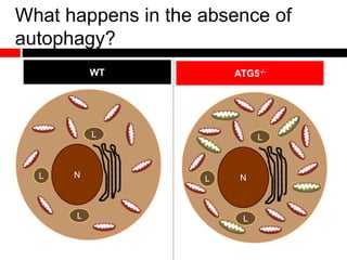
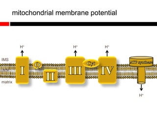
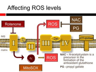
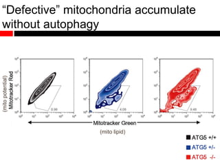
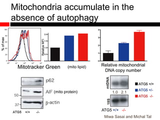
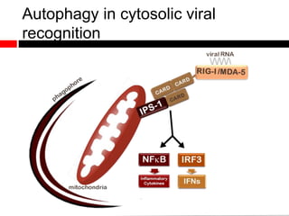
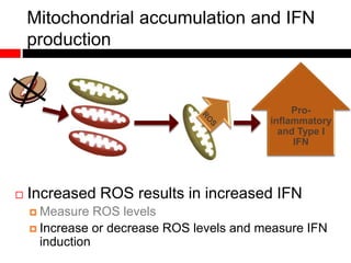
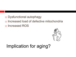
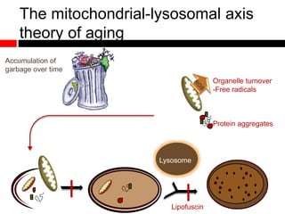
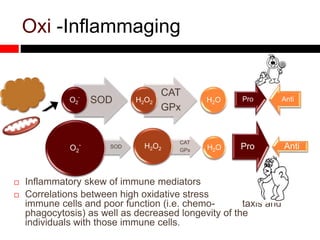
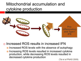
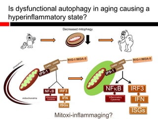
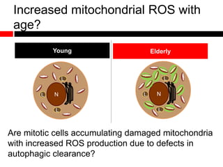
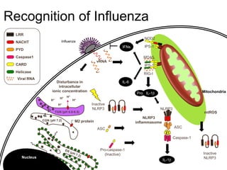
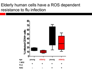
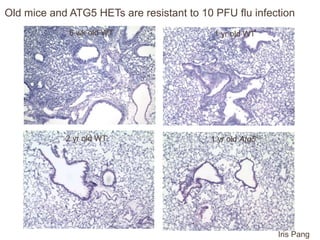
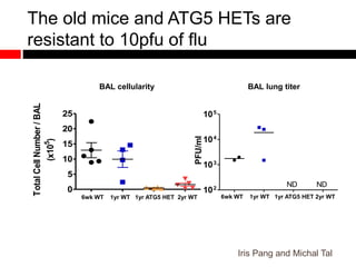
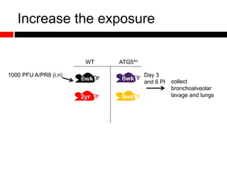
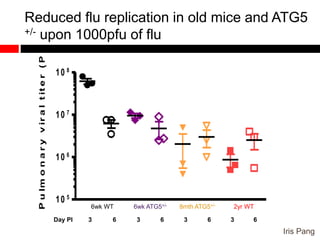
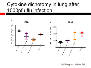
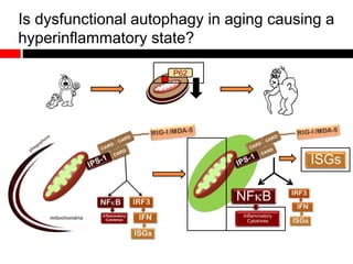
Mitochondrial accumulation and increased ROS levels in the absence of autophagy modulates cytosolic antiviral signaling. Decreased autophagy with age impacts ROS levels and antiviral signaling. Specifically:
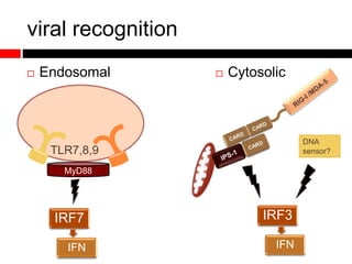
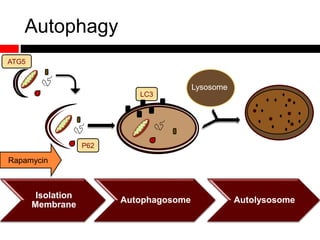
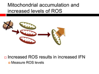
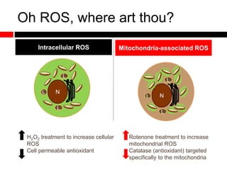
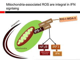
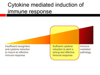
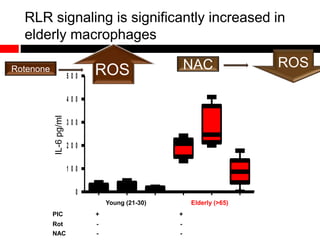
1) In the absence of autophagy, mitochondria accumulate and ROS levels increase. This leads to increased cytosolic antiviral signaling and cytokine production.
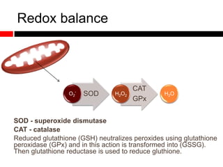
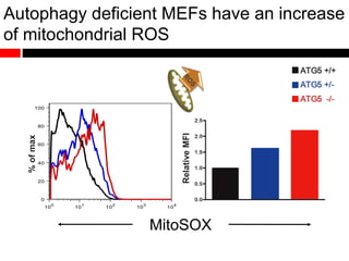
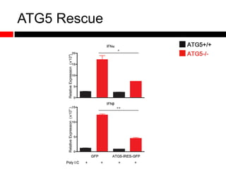
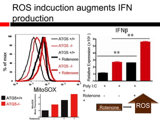
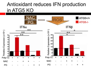
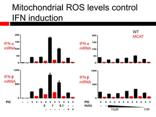
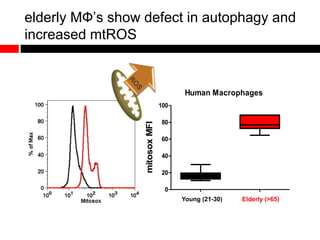
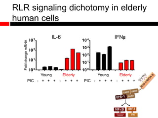
2) Autophagy deficient cells have increased mitochondrial ROS, which modulates cytosolic antiviral signaling pathways and results in increased type I interferon and inflammatory cytokine production.
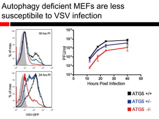
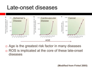
3) Decreased autophagy with age impacts mitochondrial and ROS homeostasis, which affects antiviral signaling. This may contribute to increased susceptibility to viral infection
Autophagy plays multifaceted roles in both the innate and adaptive immune system. In innate immunity, autophagy aids in the recognition of pathogens by pattern recognition receptors like TLRs, NLRs, and RLRs. It also facilitates the handling and destruction of intracellular bacteria by phagocytosis and lysosomal fusion. Autophagy regulates the production of cytokines as well. In adaptive immunity, autophagy contributes to antigen presentation on MHC class I and II molecules and is important for the homeostasis and functions of lymphocytes like T cells and B cells. However, the precise mechanisms in some cases require further clarification.
The document discusses the role of autophagy in the immune system. It describes how autophagy functions in innate immunity through interactions with pattern recognition receptors like TLRs, NLRs, and RLRs. Autophagy helps clear pathogens and regulate cytokine production. The document also examines the role of autophagy in adaptive immunity, particularly antigen presentation through MHC class I and II pathways. Finally, it explores how autophagy impacts T cell and B cell development and homeostasis.
Basophils are granulocytes that develop from bone marrow precursors and make up less than 1% of white blood cells. They contain granules with inflammatory mediators like histamine. Basophils express receptors for IgE (FcεRI) and other ligands that activate inflammatory pathways. Upon activation through IgE-dependent or IgE-independent processes, basophils rapidly degranulate and release mediators such as histamine. They also secrete cytokines like IL-4 and IL-13 over longer periods of time. Basophils play roles in both innate and adaptive immunity through these effector functions.
Neutrophils are specialized white blood cells that play a key role in the innate immune system's defense against pathogens. They are the most abundant type of white blood cell and circulate in the bloodstream, migrating to sites of infection through a multi-step process involving rolling, adhesion, transmigration, and chemotaxis. At the site of infection, neutrophils phagocytose and kill pathogens using granules containing antimicrobial enzymes and reactive oxygen species. They also form neutrophil extracellular traps (NETs) composed of DNA and antimicrobial proteins to ensnare and kill microbes. Uncontrolled activation of neutrophils can cause tissue damage and contribute to chronic inflammatory diseases.
Neutrophils, or polymorphonuclear leukocytes (PMNs), are the most abundant type of white blood cell and play a key role in the innate immune system. PMNs are produced continuously in the bone marrow and circulate in the bloodstream before migrating to sites of infection or inflammation. PMN migration involves rolling, firm adhesion, and transmigration through the endothelium and epithelium guided by cellular adhesion molecules and chemoattractants such as interleukin-8 and leukotriene B4. Upon activation at sites of infection, PMNs phagocytose pathogens and release toxic granule contents and reactive oxygen species to fight infection.
Neutrophils are the most abundant white blood cells, accounting for up to 70% of circulating leukocytes. They are produced in high numbers in the bone marrow to act as sentinels of the innate immune system. Neutrophils have a short lifespan of 10 hours in circulation and up to 48 hours in tissues. They are recruited from blood vessels to sites of infection or damage, where they engulf and kill microbes using oxidative and non-oxidative mechanisms. Neutrophils also release neutrophil extracellular traps that ensnare bacteria. While defending against pathogens, neutrophils must be cleared from tissues to resolve inflammation and prevent tissue damage from their antimicrobial enzymes.
The document discusses apoptosis, or programmed cell death. It defines apoptosis and describes the morphological changes that occur during apoptosis, including cell shrinkage, nuclear fragmentation, and chromatin condensation. It then explains the role of caspases in apoptosis, the intrinsic and extrinsic pathways that initiate apoptosis, and the common pathway involving caspase activation that leads to apoptosis. The document concludes by discussing disorders related to improper apoptosis and references several sources for further information.
This document discusses methods for researching autophagy in mammalian cells. It begins with an introduction to autophagy and the challenges of studying this dynamic process. The authors then provide an overview of techniques for monitoring autophagy at different stages and modulating autophagy to study its functions. They emphasize using multiple techniques together to accurately assess changes in autophagic activity and avoid misinterpreting results.
Neutrophils play an important role in the early stages of Mycobacterial infection by recruiting to sites of infection and phagocytosing bacteria. However, their overall impact is complex, as they may both help control infection initially but also potentially disseminate bacteria as "Trojan horses" to other organs. While neutrophils have antimicrobial mechanisms, it is unclear if they can directly kill all Mycobacterial species. Their interactions with macrophages in clearance of apoptotic neutrophils and antigen presentation also influence acquired immunity. Overall, neutrophils act as a "double-edged sword" in the pathogenesis of Mycobacterial infections.
Apoptosis is the programmed cell death. Aim of cancer therapy is to destroy the invading cells. Cancerous cells can be destroyed by increasing apoptosis.
it can occur in both physiological and pathological conditions. It is different from necrosis. In necrosis, the cell contents leak out and lead to inflammation. But in apoptosis there is no cellular leakage, only apoptotic bodies are formed. They are then engulfed by macrophages.
1. Apoptosis is programmed cell death marked by cell fragmentation, chromatin condensation, and elimination of apoptotic bodies by phagocytosis. 2. Apoptosis can occur physiologically during development and homeostasis or pathologically due to DNA damage, protein misfolding, and viral infections. 3. Apoptosis is initiated through the intrinsic mitochondrial pathway or extrinsic death receptor pathway and executed via caspase activation, resulting in cytoskeletal and DNA breakdown into apoptotic bodies for phagocytic removal.
International Journal of Pharmaceutical Science Invention (IJPSI)inventionjournals
International Journal of Pharmaceutical Science Invention (IJPSI) is an international journal intended for professionals and researchers in all fields of Pahrmaceutical Science. IJPSI publishes research articles and reviews within the whole field Pharmacy and Pharmaceutical Science, new teaching methods, assessment, validation and the impact of new technologies and it will continue to provide information on the latest trends and developments in this ever-expanding subject. The publications of papers are selected through double peer reviewed to ensure originality, relevance, and readability. The articles published in our journal can be accessed online.
This document discusses neutrophil priming and its implications in periodontal disease. It explains that neutrophils can become "primed" by exposure to inflammatory mediators, making them more reactive. In periodontal disease, neutrophils may become primed by substances released in inflamed tissues. Primed neutrophils are hyperactive and release excessive amounts of destructive enzymes and reactive oxygen species that can damage periodontal tissues. This priming of neutrophils may play a role in the pathogenesis and rapid tissue destruction seen in aggressive periodontitis.
Natural killer (NK) cells are cytotoxic lymphocytes that play an important role in the innate immune system analogous to cytotoxic T cells in the adaptive immune system. NK cells differentiate and mature in the bone marrow, lymph nodes, spleen, tonsils and thymus before entering circulation. They recognize and kill stressed, virally infected, or tumor cells through either direct cytolytic granule release or antibody-dependent cellular cytotoxicity. Cytokines released during viral infection activate NK cells to secrete interferon gamma and tumor necrosis factor alpha to help control viral spread and recruit other immune cells.
Cell death, also known as programmed cell death (PCD), is an important process in multicellular organisms whereby cells undergo an regulated death process. There are three main types of cell death - apoptosis, necrosis, and autophagy. Apoptosis is a tightly regulated form of cell death that plays a key role in development and homeostasis. Necrosis is unregulated cell death that results in inflammation. PCD is important in plants for processes like formation of xylem vessels, senescence, and the hypersensitive response to pathogens. Many pathogens have evolved ways to suppress PCD to promote infection.
Apoptosis and necrosis are two types of cell death. Apoptosis is a regulated process where cells actively cause their own death, minimizing harm to surrounding cells and tissue. It occurs normally during development and to remove damaged cells, and is controlled through caspase activation via intrinsic and extrinsic pathways. In contrast, necrosis is unregulated cell death caused by external factors like toxins or trauma. Apoptosis plays important roles in development, tissue homeostasis, and diseases like cancer when its regulation is disrupted. The cell cycle and checkpoints also interact with apoptosis to control cell proliferation and death.
Apoptosis, or programmed cell death, plays an important role in development, immunity, and maintenance of genomic integrity. Disruption of apoptosis can lead to diseases like cancer, autoimmune disorders, and neurodegenerative diseases. The document discusses the key stages and molecular mechanisms of apoptosis, including the roles of caspases, Bcl-2 family proteins, and death receptors. It also covers the importance of apoptosis in processes like immune system development, tissue remodeling, and response to DNA damage. Therapeutic strategies aim to either inhibit inappropriate apoptosis or induce apoptosis in conditions like cancer.
The document discusses different types of cell death, including programmed cell death mechanisms like apoptosis and autophagy. It notes that cell death is tightly regulated and important for development, health, and eliminating damaged or infected cells. The major types of cell death covered are apoptosis (genetically programmed suicide), autophagy (housekeeping role), necrosis (unprogrammed trauma-induced death), and necroptosis (programmed necrosis).
Apoptosis, or programmed cell death, is an important physiological process that eliminates unwanted or damaged cells. There are two main pathways that trigger apoptosis - the extrinsic or death receptor pathway, and the intrinsic or mitochondrial pathway. The extrinsic pathway involves death receptors and ligands that activate caspase enzymes. The intrinsic pathway occurs in response to cellular stress and involves mitochondrial outer membrane permeabilization and the release of proteins like cytochrome c. This forms the apoptosome complex and activates caspase-9 and caspase-3, leading to apoptosis. Apoptosis is a highly regulated process involving Bcl-2 family proteins, caspase enzymes, and characteristic morphological changes including cell shrinkage, nuclear fragmentation, and membrane blebbing. Assays to detect
This document provides an overview and introduction to cell death (apoptosis and necrosis) and cell proliferation. It discusses the differences between necrosis and apoptosis, the major apoptotic pathways involving death receptors, mitochondria and caspases. Different assays for measuring cell death and proliferation are described, along with their advantages and disadvantages. The document aims to help researchers decide which assays are best suited for measuring cell death or proliferation for their particular purposes.
This document summarizes programmed cell death or apoptosis. It discusses the distinct modalities of programmed cell death including apoptosis, autophagy and necroptosis. It covers the role of programmed cell death in disease, the mechanisms and molecular regulators involved in different types of cell death, methods to detect programmed cell death, and the therapeutic potential of modulating programmed cell death pathways.
Apoptosis, or programmed cell death, is an important process in multicellular organisms for maintaining tissue homeostasis. The intrinsic and extrinsic pathways precisely regulate apoptosis through molecular signaling within cells and from other cells. Apoptosis removes infected, damaged, and unnecessary cells, and plays critical roles in development, disease prevention, and cell population control.
This document summarizes various mechanisms of cell death, including apoptosis and necrosis. Apoptosis, or programmed cell death, involves activation of caspases through intrinsic or extrinsic pathways, leading to controlled cell death without inflammation. Necrosis occurs due to external factors causing cell membrane rupture and inflammatory cell death. Other mechanisms discussed include autophagy, which involves lysosomal degradation of cellular components, and entosis, where one cell crawls inside another to die.
This document summarizes information about programmed cell death (PCD) in plants. It discusses how PCD is essential for plant development and defense. There are two main classes of plant PCD - developmental and defensive. Developmental PCD regulates cell division and organ development, while defensive PCD helps destroy infected cells and activate systemic resistance. PCD is controlled by genetically regulated proteases like metacaspases and vacuolar processing enzymes. Hypersensitive response is a form of defensive PCD that rapidly kills cells at infection sites. Necrosis differs from PCD in that it is an unregulated form of cell death caused by injury rather than an active suicide process.
Cell death, or apoptosis, is a tightly regulated process that is essential for development and tissue homeostasis. It occurs through two main pathways: the death receptor pathway and the mitochondrial pathway. The balance between pro-apoptotic and anti-apoptotic BCL-2 family proteins determines whether a cell undergoes apoptosis. Caspases are cysteine proteases that are either initiators or executioners of apoptosis. Impaired apoptosis can lead to diseases like cancer, autoimmunity, and neurodegeneration. Necrosis was traditionally thought to be unregulated cell death, but recent evidence shows some forms of necrosis can be programmed.
Neutrophils play an important role in both oral health and periodontal disease. In oral health, neutrophils help maintain balance through mechanisms like phagocytosis, degranulation, and neutrophil extracellular traps to fight symbiotic bacteria. However, a shift to dysbiotic bacteria due to dental plaque can lead to periodontitis. In periodontitis, dysbiotic bacteria like P. gingivalis and T. denticola interact with neutrophils to inhibit their functions and promote persistent inflammation and tissue damage. This interaction, along with increased recruitment of neutrophils, contributes to chronic inflammation and bone loss in periodontitis. New therapeutic approaches targeting these disease mechanisms show promise for treating periodontitis.
1) Apoptosis is a process of programmed cell death that is important for normal development and physiology, as it helps remove excess, damaged, or dangerous cells.
2) It occurs through intrinsic and extrinsic pathways that involve caspase proteases and results in characteristic cell changes like blebbing and nuclear fragmentation.
3) Between 50-70 billion cells die per day in humans due to apoptosis, which is critical for processes like immune system maturation and tissue remodeling.
This document discusses how cancer cells have altered metabolism that promotes their growth and survival. Oncogenic mutations activate pathways like PI3K/Akt/mTOR that increase nutrient uptake and biosynthesis, putting cancer cells in a constant state of metabolic stress and hunger. This metabolic stress activates autophagy, which recycles cellular components and supports cancer cell metabolism and survival during starvation or chemotherapy. Autophagy degradation provides nutrients and building blocks and removes damaged mitochondria that could increase harmful reactive oxygen species. Cancer cells with oncogenic mutations like in Ras show increased basal autophagy that is essential for their survival. This suggests targeting autophagy may be an effective strategy to treat cancers dependent on it.
Autophagy is a catabolic process in which cells degrade unnecessary or dysfunctional components through lysosomes. It plays an important but complex role in cancer by modulating cell death, quality control, the immune response, and acting as a tumor suppressor. The PI3K/AKT/mTOR pathway is a key regulator of autophagy and is often deregulated in cancer, leading to abnormal autophagy. Recent studies suggest autophagy may be important in cancer development, progression, and response to treatment, though its specific effects depend on the context.
This document describes a study investigating autophagy in Huntington's disease (HD) using mutant huntingtin (mHtt) knock-in striatal cell lines. The study finds that lysosomes accumulate in the perinuclear region of HD model cells but remain evenly distributed in normal cells, indicating impaired vesicular trafficking in HD. Expressing normal huntingtin in HD model cells promotes even lysosomal distribution. Autophagosomal quantities differ between cell lines and in response to starvation, suggesting mHtt causes premature autophagosome-lysosome fusion. Blocking microtubule deacetylation disperses lysosomes in HD model cells, demonstrating a relationship between huntingtin, lysosomal positioning, and autophagy dysfunction in
This document summarizes Luis Emilio Flores Leiva's action research project on implementing an English unit focused on environmental issues with 10th grade students in Chile. The unit utilized various communicative and technology-based activities to develop students' English skills. Assessments included an essay and video interview assignment. While the essay helped develop writing skills, students lacked supporting arguments. The interviews were collaborative and creative but some lower-achieving students did not fully participate. Overall, the unit helped students improve their English communicative competencies, but individual self-evaluations and additional interactive activities could further enhance student learning and participation.
The document summarizes human DNA viruses, including their structure, replication cycles, and associated diseases. It covers adenoviruses, herpesviruses, papovaviruses, parvoviruses, poxviruses, and hepadnaviruses. Adenoviruses use host cell machinery to replicate their DNA and assemble new virus particles that ultimately cause cell lysis. Herpesviruses and papovaviruses replicate through rolling circle mechanisms. Parvoviruses and hepadnaviruses have unique replication features. Poxviruses are the only DNA viruses that replicate in the cytoplasm.
The document discusses viral pathogenesis and genetics. It describes the cycle of viral infection as entry into host cells, primary replication at the infection site, spread within the host, and shedding/transmission. It also discusses the effects of viruses on cells and the host organism. The genetic principles of viruses are explained as mutation, selection, and recombination, which impact viral evolution, management, and experimental study.
This document summarizes an analysis of risk perception and the Risk Priority Number (RPN) index used in Failure Mode and Effects Analysis (FMEA). The study compared how individuals perceive risk from narrative descriptions versus the FMEA method. Results showed individuals did not always assess the same situation similarly depending on how it was presented. Experience level also impacted risk perception. Further research is recommended to improve risk analysis methods and account for cultural differences in risk perception.
Powerpoint presentation M.A. Thesis DefenceCatie Chase
This document summarizes a research study that examined self-determination in post-secondary students with learning disabilities based on whether they were identified as having an LD in primary/secondary school or as an adult. The study found no statistically significant differences in self-determination, as measured by a self-determination scale, between the two groups of students. The discussion considers limitations of the study related to measurement, sample size, and sampling biases. Implications are discussed for further examining the relationship between time of LD identification and self-determination with more reliable measures and larger sample sizes.
This document provides tips for writing a thesis. It discusses starting the writing process early by choosing a title and outline. The outline should summarize the argument in one sentence for each chapter. Material should be collected in a binder as it is researched. Examiners will want to understand the thesis quickly, so the abstract, conclusions, and contents should clearly convey the purpose and findings. Getting feedback from others helps improve the thesis before examination. Regularly interacting with potential examiners also helps them understand and appreciate the research.
This study demonstrated a novel natural transformation mechanism in Actinobacillus actinomycetemcomitans (A.a.) that is independent of uptake signal sequences and the Tfox gene. The study showed that A.a. could be transformed with genomic and plasmid DNA present in microvesicles secreted into the growth medium of donor cells. This transformation occurred both in the presence and absence of components normally required for natural transformation in A.a. The results suggest outer membrane adhesion and fusion of donor microvesicles with recipient cells allows DNA delivery and homologous recombination. This novel mechanism could provide an easier method for genetically transforming A.a. compared to conventional techniques.
This document discusses sepsis, including definitions, pathophysiology, clinical features, diagnosis, and management. It defines sepsis as a life-threatening condition caused by a dysregulated immune response to infection leading to organ dysfunction. The pathophysiology involves a dysregulated inflammatory response and coagulation system. Signs and symptoms may include altered vital signs and organ dysfunction. Diagnosis involves identifying infection source through cultures and biomarkers. Treatment involves prompt antibiotics, fluid resuscitation, and supportive care based on Surviving Sepsis Campaign guidelines.
The document summarizes the key components and mechanisms of innate immunity. It discusses the physical barriers and cells that provide nonspecific immunity, including phagocytes such as macrophages and neutrophils. It describes the effector mechanisms of phagocytes, including oxygen-dependent and -independent intracellular killing using reactive oxygen species, cationic proteins, lysozymes and nitric oxide. It also discusses the roles of natural killer cells, lymphokine-activated killer cells and K cells in nonspecific cytotoxicity against infected or malignant cells.
This document outlines the systemic effects of inflammation, including fever, elevated acute phase proteins, anemia, leukocytosis/leukopenia, sepsis, ARDS, and wasting syndrome. It provides details on the mechanisms and clinical presentations of each effect. For example, it explains that fever occurs due to the effects of pyrogens like prostaglandins on the hypothalamus, while elevated acute phase proteins like C-reactive protein and serum amyloid A are produced in the liver in response to cytokines. Sepsis can lead to disseminated intravascular coagulation, hypoglycemia, and cardiac failure.
Sepsis which is commonly called as septicemia is one of the most dreadful and vulnerable life threatening disease which is quite uncommon in popularity. Around the world 1.8 million cases and in India there is 30% of nosocomial infectious patients get infected per annum accounted epidemiologically. It happens by gradual steps which is based on the incidence of the physiological and biochemical malfunctions from which septic shock is the last severe step. It is not depend on the single specific pathway dysregulation rather regulated by multi biological pathways. Various malfunctions in regulated systems like paralysis of neutrophils, higher production of proinflammatory products, diversion of adaptive immune cells like TH1 and TH2, apoptosis of lymphocytes and dendritic cells leads to sepsis. Complement anaphylatoxin C5a plays a major mediator of inflammatory response and dysregulation of plasmatic cascade. Disruption of any connecting linkers between the coagulation, complement and fibrinolysis leads to inhibition of anticoagulation mechanism. Adrenergic and cholinergic inflammatory pathway which regulates the inflammatory response get altered. In biochemical point of view endothelial dysfunction occurs mainly due to the formation of reactive oxygen species (ROS) and other components, Properdin level also get downregulated. Although much progress has been made in the treatment of inflammatory disease, the continued high mortality in septic shock is a sobering reflection of current therapeutic approaches. Nevertheless, increased understanding the molecular mechanism of various factors, connecting links and the multi effectors of sepsis can provide for better path to conquer sepsis in maximum ways.
This document summarizes neutrophil extracellular traps (NETs). NETs are formed during a process called netosis where neutrophils release chromatin fibers decorated with antimicrobial peptides to trap and kill microbes. NET formation is triggered by various microbes, cytokines, and activated immune cells. The mechanism of netosis involves the release of nuclear and granular contents while keeping the cell membrane intact. NETs play roles in both health and disease, helping to kill pathogens but also potentially contributing to conditions like thrombosis, acute lung injury, and autoimmunity when NET clearance is defective.
Immunosenescence refers to the gradual deterioration of the immune system that occurs with natural aging. As people age, their immune systems become less effective at fighting infections due to atrophy of the thymus gland, changes in the function of B and T cells, and a decline in the production of new immune cells from the bone marrow. This aging of the immune system, known as immunosenescence, is associated with increased risk of infectious diseases and inflammation-related conditions like arthritis. While immunosenescence occurs naturally, certain therapies may help enhance immune function in older individuals.
The document discusses how the immune system deteriorates with age, known as immunosenescence. Key points include:
- Innate immune cells like neutrophils and monocytes/macrophages show functional declines with aging, including altered receptor expression and signaling.
- The adaptive immune system also deteriorates with age. B cell and humoral immunity show reduced costimulatory molecule expression and defects in signaling. T cell immunity declines in naïve cell numbers, cytotoxic function, and proliferation ability. Memory CD8+ T cells lose co-stimulatory molecules like CD28 with age.
- These immune changes contribute to the chronic low-grade inflammation, or "inflamm-aging", seen in older individuals
Antibiotic-induced sepsis occurs when antibiotics cause an excessive release of endotoxins or exotoxins from bacteria, triggering an inflammatory response. Certain antibiotics that target bacterial cell wall synthesis, such as penicillins and cephalosporins, are more likely to cause endotoxin release. The choice of antibiotic class and dose is important in severely ill septic patients to avoid additional toxin release. Aminoglycosides inhibit bacterial protein synthesis with less endotoxin release and can be combined with beta-lactam antibiotics to treat severe sepsis and septic shock while minimizing endotoxin liberation.
This document summarizes various cytokine mediators and their functions. It discusses how cytokines are produced by different cell types and mediate effects through autocrine, paracrine and endocrine signaling. Specific cytokines discussed include interleukins like IL-1, IL-2, IL-4, IL-6, IL-10, IL-12 and IL-18. It also covers interferons, tumor necrosis factor, colony stimulating factors, chemokines and other growth factors. The document discusses the roles of these cytokines in innate immunity, lymphocyte differentiation, and inflammation, as well as their clinical applications.
This document summarizes various cytokine mediators and their functions. It discusses how cytokines are produced by different cell types and mediate effects through autocrine, paracrine and endocrine signaling. Specific cytokines discussed include interleukins (IL), interferons, tumor necrosis factor (TNF), and colony stimulating factors (CSFs). The roles of various cytokines in innate immunity, lymphocyte differentiation, and inflammation are described. Clinical uses of certain cytokines like IL-2, IL-11, anti-TNF antibodies, and GM-CSF are also mentioned.
Cytokines are proteins that are involved in cell signaling and communication during immune responses and inflammation. They modulate processes like immune cell differentiation, activation of lymphocytes and phagocytes, and the development of adaptive immunity. Corticosteroids suppress immunity by blocking cytokine synthesis and release. Cytokines play roles in diseases like cancer, rheumatoid arthritis, and septic shock by regulating immune and inflammatory processes. They can be measured clinically to monitor certain conditions.
Inflammation can cause both local and systemic effects. Systemic effects are mediated by cell-derived cytokines like TNF, IL-1, IL-6 and plasma cell-derived mediators like fibrinogen. Some key systemic effects include fever, elevated acute phase proteins, leukocytosis, anemia, and systemic inflammatory response syndrome (SIRS). The body attempts to counter systemic inflammation through compensatory anti-inflammatory response syndrome (CARS) mediated by cytokines like IL-4 and IL-10. Prolonged systemic inflammation can lead to more severe conditions like sepsis, acute respiratory distress syndrome (ARDS), and wasting syndrome.
Says about most important free radicals and main physiologic roles of free radicals(on transduction and transcriptional gene factors), about stress oxidative , effects of stress oxidative on some common cochlear anthologies and its related processes
The document summarizes the immune system's external and internal defenses against pathogens. It discusses multiple anatomical, cellular, chemical and molecular barriers that prevent pathogen colonization. These include physical barriers like skin and mucous membranes, chemicals in secretions, commensal bacteria, cilia, phagocytes, complement proteins, acute phase proteins and cytokines. The innate immune system provides non-specific protection while the adaptive immune system responds specifically to pathogens.
Cytokines are proteins that mediate communication between cells and help coordinate the body's immune response. They can be divided into groups like lymphokines, monokines, interleukins, and chemokines. Cytokines signal through five main receptor families and activate signaling pathways that induce cellular responses. An imbalance in cytokine signaling has been linked to various diseases. Therapies targeting cytokines and their receptors are used to treat diseases characterized by abnormal cytokine levels like cancer, infections, and autoimmune disorders.
This document summarizes systemic lupus erythematosus (SLE), an autoimmune disease affecting multiple organ systems. SLE has a relapsing and remitting course, and its etiology is unknown. It predominantly affects females aged 16-55. The pathogenesis involves genetic susceptibility, hormones like estrogen increasing autoantibody production, and disturbances in the immune response where self-antigens are mistakenly attacked. Defects in apoptosis, clearance of dead cells, and antibody production against nuclear antigens like DNA contribute to SLE. Main pathology includes overactive B-cells, suppressed T-cell regulation, complement system activation, and DNA being the main antigen attacked by antibodies.
Protozoan parasites cause diseases like malaria, leishmaniasis, and trypanosomiasis. Both the innate and adaptive immune systems play crucial roles in defending against protozoan infections. The innate immune system includes mechanisms like cytokines, complement proteins, macrophages, and neutrophils. The adaptive immune system involves antibody production and T cell responses. Protozoan parasites have evolved ways to evade or subvert the host immune response, such as antigenic variation and inhibiting immune cell function, enabling chronic or recurrent infections. An effective immune response against protozoa involves a balance of pro-inflammatory cytokines, T cell subsets, and effector cells and molecules.
This document summarizes tools and techniques for studying the innate immune system. It describes the differences between innate and adaptive immunity, the phases and cellular components of the innate response, pattern recognition receptors, signaling pathways, and technologies for research including PCR arrays and reporter assays. An example is given of using a PCR array to study the role of TLR3 in the response to Chlamydia infection, finding that TLR3 and IFN-β are major mediators. Another example shows using a reporter array to identify NF-κB and other pathways activated by cytokine stimulation.
5. Mitochondria accumulate and ROS
levels increase in the absence of
autophagy
Mitochondrial ROS modulates cytosolic
antiviral signaling
Decreased autophagy with age impacts
ROS levels and cytosolic antiviral
signaling
Outline
16. Mitochondria accumulate in the
absence of autophagy
Relative mitochondrial
DNA copy number
Mitotracker Green
18SmtDNA
1.0 2.1
ATG5 +/+ -/-
(mito lipid)
(mito protein)
Miwa Sasai and Michal Tal
17. Redox balance
SODO2
-
CAT
GPx
H2O2 H2O
SOD - superoxide dismutase
CAT - catalase
Reduced glutathione (GSH) neutralizes peroxides using glutathione
peroxidase (GPx) and in this action is transformed into (GSSG).
Then glutathione reductase is used to reduce gluthione.
18. Oxidative stress
SODO2
- CAT
GPx
H2O2 H2O
Protein peroxidation
(change in protein activation or multimerization)
Lipid peroxidation
DNA oxidative damage
24. Mitochondrial accumulation and IFN
production
Increased ROS results in increased IFN
Measure ROS levels
Increase or decrease ROS levels and measure IFN
induction
Pro-
inflammatory
and Type I
IFN
30. Oh ROS, where art thou?
Intracellular ROS Mitochondria-associated ROS
NL
L
L
NL
L
L
H2O2 treatment to increase cellular
ROS
Cell permeable antioxidant
Rotenone treatment to increase
mitochondrial ROS
Catalase (antioxidant) targeted
specifically to the mitochondria
33. Cytokine mediated induction of
immune response
Insufficient recognition
and cytokine induction
to mount an effective
immune response
Sufficient cytokine
induction to elicit a
strong and effective
immune response
Immune
mediated
pathology
34. Implication for aging?
Dysfunctional autophagy
Increased load of defective mitochondria
Increased ROS
36. Age is the greatest risk factor in many diseases
ROS is implicated at the core of these late-onset
diseases
Late-onset diseases
AGE
(Modified from Finkel 2005)
Alzheimer’s Cardiovascular Cancer
Disease disease
37. Influenza and pneumonia deaths in 2006
90% of annual Flu related deaths in the USA
are in people over 65
Under1
year1-4
years
5-14
years
15-24
years
25-34
years
35-44
years
45-54
years
55-64
years
65-74
years
75-84
years
85
yearsand
over
0
100
200
300
400
500
600
deathratesper100,000
(based on CDC reported death rates)
38. innate immunity in aging
NKT cells – increased IL-17 in viral infection
DCs and pDCs have decreased TLR
responsiveness in general
Macrophages have decreased TLR4 signaling
and increased TLR3 signaling
Monocytes have decreased TLR signaling with
the exception of increased TLR5 signaling
39. Combining theories of the aging
immune system
Mitochondrial-lysosomal axis theory of aging
Molecular inflammatory theory of aging or
“Inflammaging”
Oxidative stress theory of aging
Oxi-inflammaging to
mitoxi-inflammaging?
41. Inflammaging
A strong immune response that provides
increased fitness will be evolutionarily selected
for, even if it causes pathology in old age.
Insufficient recognition
and cytokine induction
to mount an effective
immune response
Sufficient cytokine
induction to elicit a
strong and effective
immune response
Immune
mediated
pathology
42. Oxi
SODO2
-
CAT
GPx
H2O2 H2O
SODO2
- CAT
GPx
H2O2 H2O
Inflammatory skew of immune mediators
Correlations between high oxidative stress in
immune cells and poor function (i.e. chemo- taxis and
phagocytosis) as well as decreased longevity of the
individuals with those immune cells.
Pro Anti
Pro Anti
-Inflammaging
43. Mitochondrial accumulation and
cytokine production
Increased ROS results in increased IFN
Increased ROS levels with the absence of autophagy
Increasing ROS levels resulted in increased cytokine
production, while decreasing ROS levels resulted in
decreased cytokine production
Pro-
inflammatory
and Type I
IFN
(Tal et al PNAS 2009)
44. Is dysfunctional autophagy in aging causing a
hyperinflammatory state?
NFB
Inflammatory
Cytokines
IRF3
IFN
ISGs
Decreased mitophagy
Mitoxi-inflammaging?
46. Increased mitochondrial ROS with
age?
Young Elderly
NL
L
L
Are mitotic cells accumulating damaged mitochondria
with increased ROS production due to defects in
autophagic clearance?
NL
L
L
47. elderly MФ’s show defect in autophagy and
increased mtROS
Young (21-30) Elderly (>65)
Human Macrophages
0
20
40
60
80
100
mitosoxMFI
51. Why is seasonal Influenza potentially
deadly in the elderly?
Influenza induced primary viral pneumonia or
secondary bacterial pneumonia are one of the
most significant causes of infectious disease
related death in the elderly.
Severe flu infections in the elderly result in
increased lung inflammation reminiscent of the
increased inflammation and “cytokine storm”
induced by highly pathogenic flu strains which
result in inappropriate cellular response,
increased lung damage and inefficient viral
clearance.
52. Increased flu induced IL-6 and IL-1β
production in elderly MФ’s
0
200
400
600
800
1000
IL-6pg/ml
0
10
20
30
40
50
60
70
80
IL-1pg/ml
Young (21-30) Elderly (>65)
5 MOI
FLU
- + + - + +
RAP - - + - - +
Young (21-30) Elderly (>65)
5 MOI
FLU
- + + - + +
RAP - - + - - +
10 individuals per group
PR8 NS-1/GFP virus hMDMs
In vitro infection
53. Increased flu induced IL-6 and IL-1β
production in elderly MФ’s
0
100
200
300
400
IL-6pg/ml
0
10
20
30
40
50
60
70
80
IL-1pg/ml
Young (21-30) Elderly (>65)
1 MOI
FLU
- + + - + +
RAP - - + - - +
Young (21-30) Elderly (>65)
1 MOI
FLU
- + + - + +
RAP - - + - - +
10 individuals per group
PR8 NS-1/GFP virus hMDMs
In vitro infection
54. Elderly human cells have a ROS dependent
resistance to flu infection
0
10
20
30
40
50
60
70
80
%infectedCD14+cells
age young elderly young elderly
1 MOI
FLU
+ + + +
NAC - - + +
55. Decreased IFN-I production and yet
increased resistance to flu
Elderly cells may not be capable of supporting
maximal viral replication.
Increased cell death
Blockage in autophagic clearance
ATP levels
Direct antiviral protection through pro-
inflammatory cytokines?
Increased basal levels of IFNs or ISGs?
Increased production of other types of IFN?
IFNγ has been shown to be increased with age,
correlates with viral titer in humans, and can induce a
subset of ISGs
57. Are the elderly more susceptible to flu
infection?
Increased hospitalizations of flu infected
elderly
When flu infections don’t show increased
morbidity in the elderly, this is attributed to pre-
existing immunity
58. Are the elderly more susceptible to flu
infection?
10 PFU A/PR8 (i.n)
Day 9 PI collect
bronchoalveolar
lavage and lungs
WT
ATG5+/-
59. 6 wk old WT
2 yr old WT
1 yr old WT
1 yr old Atg5+/-
Iris Pang
Old mice and ATG5 HETs are resistant to 10 PFU flu infection
60. Iris Pang and Michal Tal
The old mice and ATG5 HETs are
resistant to 10pfu of flu
102
103
104
105
ND ND
PFU/ml
0
5
10
15
20
25
TotalCellNumber/BAL
(x105
)
6wk WT 1yr WT 1yr ATG5 HET 2yr WT
BAL cellularity BAL lung titer
6wk WT 1yr WT 1yr ATG5 HET 2yr WT
61. Increase the exposure
1000 PFU A/PR8 (i.n) Day 3
and 6 PI collect
bronchoalveolar
lavage and lungs
WT ATG5+/-
62. Reduced flu replication in old mice and ATG5
+/- upon 1000pfu of flu
Iris Pang
10 5
10 6
10 7
10 8
Pulmonaryviraltiter(PFU/ml)
6wk WT 6wk ATG5+/- 8mth ATG5+/- 2yr WT
Day PI 3 6 3 6 3 6 3 6
63. Cytokine dichotomy in lung after
1000pfu flu infection
6w
k
W
T
6w
k
ATG
5
HET
8m
th
ATG
5
HET
2yrW
T
0
2 0
4 0
6 0
8 0
IFNunits/mlBAL
6w
k
W
T
6w
k
ATG
5
HET
8m
th
ATG
5
HET
2yrW
T
0
2 0 0 0
4 0 0 0
6 0 0 0
IL-6pg/mlBAL
IFNα IL-6
Iris Pang and Michal Tal
64. Is dysfunctional autophagy in aging causing a
hyperinflammatory state?
ISGs
P62
NFB
Inflammatory
Cytokines
65. Future directions
Identify the target of oxidation by which
mitochondrial ROS modulates cytosolic
antiviral signaling.
Investigate the IL-6/IFN dichotomy with aging
and long term defects in mitochondrial
clearance.
Assess levels of other IFNs and validate IFITM
expression from elderly cells and old mice.
67. Implications
Autophagic control of mitochondrial number and
integrity modulates cytosolic antiviral responses
Defects in autophagy can lead to a hyper-
inflammatory state and impact many of the chronic
diseases associated with aging
“Feed a cold, starve a fever” – viral anorexia to
induce autophagy?
Modulating autophagy and inflammatory responses
could possibly reduce the morbidity and mortality of
flu infection in the aged population
68. Lung pathology at 6 days post infection
with 1000pfu PR8
peribronchiolar
inflammation
Perivascular
inflammation
alveolitis Lymphoid
aggregates
6wk WT ++ ++ +++ -
6wk ATG5
HET
+ ++ ++ -
8mth ATG5
HET
++ + + -
2yr WT +++ ++ ++ ++++
6wk WT 6wk ATG5 HET 8mth ATG5 HET 2yr WT
uninfected
69. Lung pathology at 6 days post infection
with 1000pfu PR8
peribronchiolar
inflammation
Perivascular
inflammation
alveolitis Lymphoid
aggregates
6wk WT ++ ++ +++ -
6wk ATG5
HET
+ ++ ++ -
8mth ATG5
HET
++ + + -
2yr WT +++ ++ ++ ++++
6wk WT 6wk ATG5 HET 8mth ATG5 HET 2yr WT
uninfected
70. Lessons from in vivo intranasal flu
infections
Decreased mitophagy may contribute to flu
resistance with age, but reduced tolerance to
flu infection with age is likely leading to the
increased morbidity.