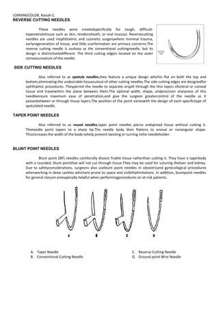
SlideShare a Scribd company logo
1 of 11
LOMANGCOLOB, Raisah C.
The Suture
The word "suture" describes any strand of material used to ligate (tie) blood vessels or approximate
(bring close together) tissues. Sutures are used to close wounds. Sutures and ligatures were used by both the
Egyptians and Syrians as far back as 2,000 B.C. Through the centuries, a wide variety of materials—silk, linen,
cotton, horsehair, animal tendons and intestines, and wire made of precious metals—have been used in
operative procedures. Some of these are still in use today.
The Surgical Needle
THE ANATOMY OF A NEEDLE
Regardless of its intended use, every surgical needle has three basic
components:
The eye.
The body.
The point.
The measurements of these specific components determine, in part, how they will be used most efficiently.
Needle size may be measured in inches or in metric units. The following measurements determine the size of a needle.
CHORD LENGTH—The straight line distance from the point of a curved needle to the swage.
NEEDLE LENGTH—The distance measured along the needle itself from point to end.
RADIUS—The distance from the center of the circle to the body of the needle if the curvature of the needle were
continued to make a full circle.
DIAMETER—The gauge or thickness of the needle wire. Very small needles of fine gauge are needed for
microsurgery. Large, heavy gauge needles are used to penetrate the sternum and to place retention sutures in the
abdominal wall. A broad spectrum of sizes are available between the two extremes.
THE NEEDLE EYE
The eye falls into one of three categories: closed eye, French (split or spring) eye, or swaged (eyeless).
CLOSED EYE
The closed eye is similar to a household sewing needle. The shape of
the eye may be round, oblong, or square.
FRENCH EYE
French eye needles have a slit from inside the eye to the end of the needle with
ridges that catch and hold the suture in place.
LOMANGCOLOB, Raisah C.
SWAGED NEEDLES
Virtually all needles used today are swaged. This configuration joins the
needle and suture together as a continuous unit—one that is convenient to use and
minimizes trauma.
THE NEEDLE BODY
The body of the needle is the portion which is grasped by the needle holder during the surgical procedure. The
body of the needle should be as close as possible to the diameter of the suture material to minimize bleeding and
leakage. This is especially true for cardiovascular, gastrointestinal, and bladder procedures. The curvature of the needle
body may come in a variety of different shapes. Each shape gives the needle different characteristics.
STRAIGHT NEEDLE
This shape may be preferred when suturing easily accessible tissue. Most of these needles are designed
to be used in places where direct finger-held manipulation can easily be performed.
HALF-CURVED NEEDLE
The half-curved or "ski" needle may be used for skin closure or in laparoscopy. Its low profile allows easy
passage down laparoscopic trocars. Its use in skin closure is limited because, while the curved portion passes
through tissue easily, the remaining straight portion of the body is unable to follow the curved path of the
needle without bending or enlarging its path in the tissue.
CURVED NEEDLE
Curved needles allow predictable needle turnout from tissue, and are therefore used most often. This
needle shape requires less space for maneuvering than a straight needle, but the curve necessitates
manipulation with a needle holder. The curvature may be 1/4, 3/8, 1/2, or 5/8 circle.
The most common use for the 3/8 circle is skin closure. The surgeon can easily manipulate this
curvature with slight pronation of the wrist in a relatively large and superficial wound. It is very difficult to use
this needle in a deep body cavity or restricted area because a larger arc of manipulation is required.
LOMANGCOLOB, Raisah C.
The 1/2 circle needle was designed for use in a confined space, although it requires more pronation and
supination of the wrist. But even the tip of this needle may be obscured by tissue deep in the pelvic cavity.
A 5/8 circle needle may be more useful in this situation, especially in some anal, urogenital, intraoral,
and cardiovascular procedures.
COMPOUND CURVED NEEDLE
The compound curved needle was originally developed for anterior segment ophthalmic surgery. It
allows the surgeon to take precise, uniform bites of tissue. The tight 80° curvature of the tip follows into a 45°
curvature throughout the remainder of the body. The initial curve allows reproducible, short, deep bites into the
tissue. The curvature of the remaining portion of the body forces the needle out of the tissue, everting the
wound edges and permitting a view into the wound. This ensures equidistance of the suture material on both
sides of the incision. Equalized pressure on both sides of the comeal-scleral junction minimizes the possibility of
astigmatism following anterior segment surgery.
THE NEEDLE POINT
The point extends from the extreme tip of the needle to the maximum cross-section of the body. Each needle
point is designed and produced to the required degree of sharpness to smoothly penetrate specific types of tissue.
LOMANGCOLOB, Raisah C.
TYPES OF NEEDLES
CUTTING NEEDLES
Cutting needles have at least twoopposing cutting edges. They aresharpened to cut through
tough,difficult-to-penetrate tissue.Cutting needles are ideal for skinsutures that must pass throughdense,
irregular, and relativelythick connective dermal tissue.Because of the sharpness of thecutting edge, care must be
takenin some tissue (tendon sheathor oral mucous membrane) toavoid cutting through more tissuethan
desired.
CONVENTIONAL CUTTING NEEDLES
In addition to the two cutting edges,conventional cutting needles have athird cutting edge on the
insideconcave curvature of the needle.The shape changes from a triangularcutting blade to that of a
flattenedbody on both straight and curvedneedles. This needle type may be prone to cutout of tissue
becausethe inside cutting edge cuts towardthe edges of the incision or wound.
LOMANGCOLOB, Raisah C.
REVERSE CUTTING NEEDLES
These needles were createdspecifically for tough, difficult-
topenetratetissue such as skin, tendonsheath, or oral mucosa. Reversecutting
needles are used inophthalmic and cosmetic surgerywhere minimal trauma,
earlyregeneration of tissue, and little scarformation are primary concerns.The
reverse cutting needle is assharp as the conventional cuttingneedle, but its
design is distinctivelydifferent. The third cutting edgeis located on the outer
convexcurvature of the needle.
SIDE CUTTING NEEDLES
Also referred to as spatula needles,they feature a unique design whichis flat on both the top and
bottom,eliminating the undesirable tissuecutout of other cutting needles.The side-cutting edges are designedfor
ophthalmic procedures. Theypermit the needle to separate orsplit through the thin layers ofscleral or comeal
tissue and travelwithin the plane between them.The optimal width, shape, andprecision sharpness of this
needleensure maximum ease of penetration,and give the surgeon greatercontrol of the needle as it
passesbetween or through tissue layers.The position of the point varieswith the design of each specifictype of
spatulated needle.
TAPER POINT NEEDLES
Also referred to as round needles,taper point needles pierce andspread tissue without cutting it.
Theneedle point tapers to a sharp tip.The needle body then flattens to anoval or rectangular shape.
Thisincreases the width of the body tohelp prevent twisting or turning inthe needleholder.
BLUNT POINT NEEDLES
Blunt point (BP) needles canliterally dissect friable tissue ratherthan cutting it. They have a taperbody
with a rounded, blunt pointthat will not cut through tissue.They may be used for suturing theliver and kidney.
Due to safetyconsiderations, surgeons also useblunt point needles in obstetricand gynecological procedures
whenworking in deep cavities whichare prone to space and visibilitylimitations. In addition, bluntpoint needles
for general closure areespecially helpful when performingprocedures on at-risk patients.
A. Taper Needle
B. Conventional Cutting Needle
C. Reverse Cutting Needle
D. Ground point Wire Needle
LOMANGCOLOB, Raisah C.
The Surgical Suture
SUTURE CHARACTERISTICS
The choice of suture materials generally depends on whether the wound closure occurs in one or
more layers. In selecting the most appropriate sutures, the surgeon takes into account the amount of
tension on the wound, the number of layers of closure, depth of suture placement, anticipated amount of
edema, and anticipated timing of suture removal.
Optimal suture qualities include:
High uniform tensile strength, permitting use of finer sizes.
High tensile strength retention in vivo, holding the wound securely throughout the critical
healing period, followed by rapid absorption.
Consistent uniform diameter.
Sterile.
Pliable for ease of handling and knot security.
Freedom from irritating substances or impurities for optimum tissue acceptance.
Predictable performance.
SIZE AND TENSILE STRENGTH
Sizedenotes the diameter of the suture material. The accepted surgical practice is to use the
smallest diameter suture that will adequately hold the mending wounded tissue. This practice minimizes
trauma as the suture is passed through the tissue to effect closure. It also ensures that the minimum mass
of foreign material is left in the body. Suture size is stated numerically; as the number of 0s in the suture
size increases, the diameter of the strand decreases. For example, size 5-0, or 00000, is smaller in diameter
than size 4-0, or 0000. The smaller the size, the less tensile strength the suture will have.
Knot tensile strengthis measured by the force, in pounds, which the suture strand can withstand
before it breaks when knotted. The tensile strength of the tissue to be mended (its ability to withstand
stress) determines the size and tensile strength of the suturing material the surgeon selects. The accepted
rule is that the tensile strength of the suture need never exceed the tensile strength of the tissue. However,
sutures should be at least as strong as normal tissue through which they are being placed.
MONOFILAMENT VS. MULTIFILAMENT STRANDS
Sutures are classified according to the number of strands of which they are comprised.
MONOFILAMENT SUTURES
Monofilament suturesare made of a single strand of material. Because of their simplified
structure, they encounter less resistance as they pass through tissue than multifilament suture
material. They also resist harboring organisms which may cause infection. These characteristics
make monofilament sutures well-suited to vascular surgery. Monofilament sutures tie down easily.
However, because of their construction, extreme care must be taken when handling and tying these
sutures. Crushing or crimping of this suture type can nick or create a weak spot in the strand. This
may result in suture breakage.
MULTIFILAMENT SUTURES
Multifilament suturesconsist of several filaments, or strands, twisted or braided together.
This affords greater tensile strength, pliability, and flexibility. Multifilament sutures may also be
coated to help them pass relatively smoothly through tissue and enhance handling characteristics.
Coated multifilament sutures are well-suited to intestinal procedures.
LOMANGCOLOB, Raisah C.
ABSORBABLE VS. NONABSORBABLE SUTURES
Sutures are classified according to their degradation properties.Sutures that undergo rapid
degradation in tissues, losing their tensile strength within 60 days, are considered absorbable sutures.
Sutures that generally maintain their tensile strength for longer than 60 days are nonabsorbablesutures.
ABSORBABLE SUTURES
Absorbable sutures may be used to hold wound edges in approximation temporarily, until
they have healed sufficiently to withstand normal stress. These sutures are prepared either from the
collagen of healthy mammals or from synthetic polymers. Some are absorbed rapidly, while others
are treated or chemically structured to lengthen absorption time. They may also be impregnated or
coated with agents that improve their handling properties, and colored with an FDA-approved dye
to increase visibility in tissue. Natural absorbable sutures are digested by body enzymes which attack
and break down the suture strand.
NONABSORBABLE SUTURES
Nonabsorbable sutures are those which are not digested by body enzymes or hydrolyzed in
body tissue. They are made from a variety of nonbiodegradable materials and are ultimately
encapsulated or walled off by the body’s fibroblasts. Nonabsorbable sutures ordinarily remain
where they are buried within the tissues. When used forskin closure, they must be removed
postoperatively.
Nonabsorbable sutures may be used in a variety of applications:
Exterior skin closure, to be removed after sufficient healing has occurred.
Within the body cavity, where they will remain permanently encapsulated in tissue.
Patient history of reaction to absorbable sutures, keloidal tendency, or possible tissue
hypertrophy.
Prosthesis attachment (i.e., defibrillators, pacemakers, drug delivery mechanisms).
LOMANGCOLOB, Raisah C.
Common Suturing Techniques
LIGATURES
A suture tied around a vessel to occlude the lumen is called a ligature or
tie. It may be used to affect hemostasis or to close off a structure to prevent
leakage. There are two primary types of ligatures.
Free tieor freehand ligaturesare single strands of suture material used to
ligate a vessel, duct, or other structure. After a hemostat or other similar type of
surgical clamp has been placed on the end of the structure, the suture strand is
tied around the vessel under the tip of the hemostat. The hemostat is removed
after the first throw and the surgeon tightens the knot using his or her
fingertips, taking care to avoid instrument damage to the suture. Additional
throws are added as needed to square and secure the knot.
Stick tie, suture ligature, or transfixion sutureis a strand of suture
material attached to a needle to ligate a vessel, duct, or other structure. This
technique is used on deep structures where placement of a hemostat is
difficult or on vessels of large diameter. The needle is passed through the
structure or adjacent tissue first to anchor the suture, and then tied around
the structure. Additional throws are used as needed to secure the knot.
THE PRIMARY SUTURE LINE
The primary suture lineis the line of sutures that holds the wound edges in approximation during
healing by first intention. It may consist of a continuous strand of material or a series of interrupted suture
strands. Other types of primary sutures, such as deep sutures, buried sutures, purse-string sutures, and
LOMANGCOLOB, Raisah C.
subcuticular sutures, are used for specific indications. Regardless of technique, a surgical needle is attached
to the suture strand to permit repeated passes through tissue.
CONTINUOUS SUTURES
Also referred to as running stitches, continuous sutures are a series of stitches taken with
one strand of material. The strand may be tied to itself at each end, or looped, with both cut ends of
the strand tied together. A continuous suture line can be placed rapidly. It derives its strength from
tension distributed evenly along the full length of the suture strand. However, care must be taken to
apply firm tension, rather than tight tension, to avoid tissue strangulation. Excessive tension and
instrument damage should be avoided to prevent suture breakage which could disrupt the entire
line of a continuous suture. Continuous suturing leaves less foreign body mass in the wound. In the
presence of infection, it may be desirable to use a monofilament suture material because it has no
interstices which can harbor microorganisms. This is especially critical as a continuous suture line
can transmit infection along the entire length of the strand. A continuous one layer mass closure
may be used on peritoneum and/orfascial layers of the abdominal wall to provide a temporary seal
during the healing process.
CONTINUOUS SUTURING TECHNIQUES
INTERRUPTED SUTURES
Interrupted suturesuse a number of strands to close the wound.
Each strand is tied and cut after insertion. This provides a more secure
closure, because if one suture breaks, the remaining sutures will hold
the wound edges in approximation. Interrupted sutures may be used if
a wound is infected, because microorganisms may be less likely to
travel along a series of interrupted stitches.
DEEP SUTURES
Deep suturesare placed completely under the epidermal skin layer.
They may be placed as continuous or interrupted sutures and are not removed
postoperatively.
LOMANGCOLOB, Raisah C.
BURIED SUTURES
Buried suturesare placed so that the knot protrudes to the inside, under the layer to be
closed. This technique is useful when using large diameter permanent sutures on deeper layers in
thin patients who may be able to feel large knots that are not buried.
PURSE-STRING SUTURES
Purse-string sutures are continuous sutures placed around a
lumen and tightened like a drawstring to invert the opening. They
may be placed around the stump of the appendix, in the bowel to
secure an intestinal stapling device, or in an organ prior to insertion of
a tube (such as the aorta, to hold the cannulation tube in place during
an open heart procedure).
SUBCUTICULAR SUTURES
Subcuticular suturesare continuous or interrupted sutures
placed in the dermis, beneath the epithelial layer. Continuous
subcuticular sutures are placed in a line parallel to the wound. This
technique involves taking short, lateral stitches the full length of the
wound. After the suture has been drawn taut, the distal end is anchored
in the same manner as the proximal end. This may involve tying or any
of a variety of anchoring devices. Subcuticular suturing may be
performed with absorbable suture which does not require removal, or
with monofilament nonabsorbable suture that is later removed by
simply removing the anchoring device at one end and pulling the
opposite end.
THE SECONDARY SUTURE LINE
A secondary line of sutures may be used:
To reinforce and support the primary suture line, eliminate dead space, and prevent fluid
accumulation in an abdominal wound during healing by first intention. When used for this purpose,
they may also be called retention, stay, or tension sutures.
To support wounds for healing by second intention.
For secondary closure following wound disruption when healing by third intention.
NOTE: If secondary sutures are used in cases of nonhealing, they should be placed in opposite
fashion from the primary sutures (i.e., interrupted if the primary sutures were continuous, continuous if the
primary sutures were interrupted).
Retention suturesare placed approximately 2 inches from each edge of the wound. The tension
exerted lateral to the primary suture line contributes to the tensile
strength of the wound. Through-and-through suturesare placed from
inside the peritoneal cavity through all layers of the abdominal wall,
including the peritoneum. They should be inserted before the
peritoneum is closed using a simple interrupted stitch. The wound may
be closed in layers for a distance of approximately three-fourths its
length. Then the retention sutures in this area may be drawn together
and tied. It is important that a finger be placed within the abdominal
cavity to prevent strangulation of the viscera in the closure. The
remainder of the wound may then be closed. Prior to tightening and
tying the final retention sutures, it is important to explore the
abdomen
LOMANGCOLOB, Raisah C.
again with a finger to prevent strangulation of viscera in the closure. The remainder of the wound may then
be closed.Retention sutures utilize
nonabsorbable suture material. They should therefore be removed as soon as the danger of
suddenincreases in intra-abdominal pressure is over— usually 2 to 6 weeks, with an average of 3 weeks.
STITCH PLACEMENT
Many types of stitches are used for both continuous and interrupted suturing. In every case, equal
"bites" of tissue should be taken on each side of the wound. The needle should be inserted from 1 to 3
centimeters from the edge of the wound, depending upon the type and condition of the tissue being
sutured.
Principles of Suturing
1. The completed knot must be tight, firm, and tied so that slippage will not occur.
2. To avoid wicking of bacteria, knot should not be placed in incision lines.
3. Knots should be small and the ends cut short (2-3mm).
4. Avoid excessive tension to finer gauge materials as breakage may occur.
5. Avoid using a jerking motion, which may break the suture.
6. Avoid crushing or crimping of suture materials by not using hemostats or needle holders on them except
on the free end for tying.
7. Do not tie suture too tightly as tissue necrosis may occur. Knot tension should not produce tissue
blanching.
8. Maintain adequate traction on one end while trying to avoid losing the first loop.
Principles of Suture Removal
1. The area should be swabbed with hydrogen peroxide for removal of encrusted necrotic debris, blood,
and serum from about the sutures.
2. A sharp suture scissors should be used to cut the loops of individual or continuous sutures about the
teeth.
3. It is often helpful to use a No. 23 explorer to help lift the sutures if they are within the sulcus or in close
opposition to the tissue.
4. A cotton pliers is used to remove the suture. The location of the knots should be noted so that they can
be removed first. This will prevent unnecessary entrapment under the flap.
5. Suture should be removed in 7 to 10 days to prevent epithelialization or wicking about the suture.

More Related Content

What's hot

Principles of use and abuse of suture 1
Principles of use and abuse of suture 1Principles of use and abuse of suture 1
Principles of use and abuse of suture 1Drkabiru2012
 
Surgical Sutures and Suturing Techniques
Surgical Sutures and Suturing TechniquesSurgical Sutures and Suturing Techniques
Surgical Sutures and Suturing TechniquesDene W. Daugherty
 
Needles & sutures (A quick Review)
Needles & sutures (A quick Review)Needles & sutures (A quick Review)
Needles & sutures (A quick Review)Shady Nafie
 
Hemostasis and methods to control bleeding
Hemostasis and methods to control bleedingHemostasis and methods to control bleeding
Hemostasis and methods to control bleedingAbdul Rahman Ibrahim
 
SUTURES AND SUTURING
SUTURES AND SUTURINGSUTURES AND SUTURING
SUTURES AND SUTURINGShilpa Shiv
 
Instruments SURGERY updated PPT
Instruments SURGERY updated PPT Instruments SURGERY updated PPT
Instruments SURGERY updated PPT TONY SCARIA
 
Surgical Sutures and Needles
Surgical Sutures and NeedlesSurgical Sutures and Needles
Surgical Sutures and NeedlesTega Ejeheri
 
sutures and suturing technique and knots
sutures and suturing technique and knotssutures and suturing technique and knots
sutures and suturing technique and knotsAvinash Rathore
 
Sutures Materials
Sutures MaterialsSutures Materials
Sutures MaterialsIAU Dent
 
Surgical instruments I
Surgical instruments ISurgical instruments I
Surgical instruments IAnkita Singh
 
Surgical instrument ppt
Surgical instrument ppt Surgical instrument ppt
Surgical instrument ppt PoonamPurkar
 
Suturing 101 - Basic Surgical Skills for Medical Students and Junior Doctors
Suturing 101 - Basic Surgical Skills for Medical Students and Junior DoctorsSuturing 101 - Basic Surgical Skills for Medical Students and Junior Doctors
Suturing 101 - Basic Surgical Skills for Medical Students and Junior DoctorsAaron Sparshott
 
Suture materials and techniques
Suture materials and techniquesSuture materials and techniques
Suture materials and techniquesmemoalawad
 

What's hot (20)

Principles of use and abuse of suture 1
Principles of use and abuse of suture 1Principles of use and abuse of suture 1
Principles of use and abuse of suture 1
 
Surgical Sutures and Suturing Techniques
Surgical Sutures and Suturing TechniquesSurgical Sutures and Suturing Techniques
Surgical Sutures and Suturing Techniques
 
Needles & sutures (A quick Review)
Needles & sutures (A quick Review)Needles & sutures (A quick Review)
Needles & sutures (A quick Review)
 
Hemostasis and methods to control bleeding
Hemostasis and methods to control bleedingHemostasis and methods to control bleeding
Hemostasis and methods to control bleeding
 
SUTURES AND SUTURING
SUTURES AND SUTURINGSUTURES AND SUTURING
SUTURES AND SUTURING
 
Instruments SURGERY updated PPT
Instruments SURGERY updated PPT Instruments SURGERY updated PPT
Instruments SURGERY updated PPT
 
Surgical instruments
Surgical instrumentsSurgical instruments
Surgical instruments
 
Suture materials
Suture materialsSuture materials
Suture materials
 
Cardiac procedure tray
Cardiac procedure trayCardiac procedure tray
Cardiac procedure tray
 
Surgery 6th year, Tutorial (Dr. AbdulWahid)
Surgery 6th year, Tutorial (Dr. AbdulWahid)Surgery 6th year, Tutorial (Dr. AbdulWahid)
Surgery 6th year, Tutorial (Dr. AbdulWahid)
 
Surgical Sutures and Needles
Surgical Sutures and NeedlesSurgical Sutures and Needles
Surgical Sutures and Needles
 
Suture materials and techniques
Suture materials and techniquesSuture materials and techniques
Suture materials and techniques
 
sutures and suturing technique and knots
sutures and suturing technique and knotssutures and suturing technique and knots
sutures and suturing technique and knots
 
Sutures Materials
Sutures MaterialsSutures Materials
Sutures Materials
 
Surgical instruments I
Surgical instruments ISurgical instruments I
Surgical instruments I
 
Surgical suture basics
Surgical suture basicsSurgical suture basics
Surgical suture basics
 
Surgical instrument ppt
Surgical instrument ppt Surgical instrument ppt
Surgical instrument ppt
 
Suturing 101 - Basic Surgical Skills for Medical Students and Junior Doctors
Suturing 101 - Basic Surgical Skills for Medical Students and Junior DoctorsSuturing 101 - Basic Surgical Skills for Medical Students and Junior Doctors
Suturing 101 - Basic Surgical Skills for Medical Students and Junior Doctors
 
Wound management
Wound managementWound management
Wound management
 
Suture materials and techniques
Suture materials and techniquesSuture materials and techniques
Suture materials and techniques
 

Similar to The Suture

Practical skills in medical practice
Practical skills in medical practicePractical skills in medical practice
Practical skills in medical practiceAvinash Bhondwe
 
common_surgical_instruments_module.pdf
common_surgical_instruments_module.pdfcommon_surgical_instruments_module.pdf
common_surgical_instruments_module.pdfPRINCEBALHARA101
 
Principles of suturing in surgery
Principles of suturing in surgeryPrinciples of suturing in surgery
Principles of suturing in surgeryJamil Kifayatullah
 
Instrumentation for Basic Oral Surgery Dr.Ali Mohammed AbuTrab
Instrumentation for Basic Oral Surgery Dr.Ali Mohammed AbuTrabInstrumentation for Basic Oral Surgery Dr.Ali Mohammed AbuTrab
Instrumentation for Basic Oral Surgery Dr.Ali Mohammed AbuTrabAli Mohammed AbuTrab
 
sutures and suturing technique
sutures and suturing techniquesutures and suturing technique
sutures and suturing techniqueMangeshAndhare2
 
suture material and suturing
suture material and suturingsuture material and suturing
suture material and suturingJoel D'silva
 
General principles of arthroscopy kle, belgaum, dr utkarsh dwivedi
General principles of arthroscopy kle, belgaum, dr utkarsh dwivediGeneral principles of arthroscopy kle, belgaum, dr utkarsh dwivedi
General principles of arthroscopy kle, belgaum, dr utkarsh dwivediUtkarsh Dwivedi
 
Surgical Instruments.pdf
Surgical Instruments.pdfSurgical Instruments.pdf
Surgical Instruments.pdfPPALIVE
 
General instruments by Prashant Kumar
General instruments by Prashant KumarGeneral instruments by Prashant Kumar
General instruments by Prashant KumarKGMU Lucknow
 
Incision, suture & suture meteri al
Incision, suture & suture meteri alIncision, suture & suture meteri al
Incision, suture & suture meteri alSanjukta Saha
 
Basic armamentarium for minor oral surgery
Basic armamentarium for minor oral surgeryBasic armamentarium for minor oral surgery
Basic armamentarium for minor oral surgeryIyad Abou Rabii
 
Basic Surgical Orthopedic Instruments
Basic Surgical Orthopedic InstrumentsBasic Surgical Orthopedic Instruments
Basic Surgical Orthopedic InstrumentsDr. Zubair Younis
 
Basic armamentarium for minor oral surgery
Basic armamentarium for minor oral surgeryBasic armamentarium for minor oral surgery
Basic armamentarium for minor oral surgeryBabajide Sokoya
 
Operative instruments in Craniotomy
 Operative instruments in Craniotomy Operative instruments in Craniotomy
Operative instruments in CraniotomyBipul Thakur
 

Similar to The Suture (20)

Practical skills in medical practice
Practical skills in medical practicePractical skills in medical practice
Practical skills in medical practice
 
common_surgical_instruments_module.pdf
common_surgical_instruments_module.pdfcommon_surgical_instruments_module.pdf
common_surgical_instruments_module.pdf
 
Principles of suturing in surgery
Principles of suturing in surgeryPrinciples of suturing in surgery
Principles of suturing in surgery
 
Instrumentation for Basic Oral Surgery Dr.Ali Mohammed AbuTrab
Instrumentation for Basic Oral Surgery Dr.Ali Mohammed AbuTrabInstrumentation for Basic Oral Surgery Dr.Ali Mohammed AbuTrab
Instrumentation for Basic Oral Surgery Dr.Ali Mohammed AbuTrab
 
instruments (1).pptx
instruments (1).pptxinstruments (1).pptx
instruments (1).pptx
 
Basics of arthroscopy ppt
Basics of arthroscopy pptBasics of arthroscopy ppt
Basics of arthroscopy ppt
 
Periodontal microsurgery
Periodontal microsurgeryPeriodontal microsurgery
Periodontal microsurgery
 
sutures and suturing technique
sutures and suturing techniquesutures and suturing technique
sutures and suturing technique
 
suture material and suturing
suture material and suturingsuture material and suturing
suture material and suturing
 
General instruments
General instrumentsGeneral instruments
General instruments
 
General principles of arthroscopy kle, belgaum, dr utkarsh dwivedi
General principles of arthroscopy kle, belgaum, dr utkarsh dwivediGeneral principles of arthroscopy kle, belgaum, dr utkarsh dwivedi
General principles of arthroscopy kle, belgaum, dr utkarsh dwivedi
 
Surgical Instruments.pdf
Surgical Instruments.pdfSurgical Instruments.pdf
Surgical Instruments.pdf
 
surgical instruments
surgical instrumentssurgical instruments
surgical instruments
 
Sutures & Drainage
Sutures & DrainageSutures & Drainage
Sutures & Drainage
 
General instruments by Prashant Kumar
General instruments by Prashant KumarGeneral instruments by Prashant Kumar
General instruments by Prashant Kumar
 
Incision, suture & suture meteri al
Incision, suture & suture meteri alIncision, suture & suture meteri al
Incision, suture & suture meteri al
 
Basic armamentarium for minor oral surgery
Basic armamentarium for minor oral surgeryBasic armamentarium for minor oral surgery
Basic armamentarium for minor oral surgery
 
Basic Surgical Orthopedic Instruments
Basic Surgical Orthopedic InstrumentsBasic Surgical Orthopedic Instruments
Basic Surgical Orthopedic Instruments
 
Basic armamentarium for minor oral surgery
Basic armamentarium for minor oral surgeryBasic armamentarium for minor oral surgery
Basic armamentarium for minor oral surgery
 
Operative instruments in Craniotomy
 Operative instruments in Craniotomy Operative instruments in Craniotomy
Operative instruments in Craniotomy
 

Recently uploaded

History Class XII Ch. 3 Kinship, Caste and Class (1).pptx
History Class XII Ch. 3 Kinship, Caste and Class (1).pptxHistory Class XII Ch. 3 Kinship, Caste and Class (1).pptx
History Class XII Ch. 3 Kinship, Caste and Class (1).pptxsocialsciencegdgrohi
 
SOCIAL AND HISTORICAL CONTEXT - LFTVD.pptx
SOCIAL AND HISTORICAL CONTEXT - LFTVD.pptxSOCIAL AND HISTORICAL CONTEXT - LFTVD.pptx
SOCIAL AND HISTORICAL CONTEXT - LFTVD.pptxiammrhaywood
 
Final demo Grade 9 for demo Plan dessert.pptx
Final demo Grade 9 for demo Plan dessert.pptxFinal demo Grade 9 for demo Plan dessert.pptx
Final demo Grade 9 for demo Plan dessert.pptxAvyJaneVismanos
 
Solving Puzzles Benefits Everyone (English).pptx
Solving Puzzles Benefits Everyone (English).pptxSolving Puzzles Benefits Everyone (English).pptx
Solving Puzzles Benefits Everyone (English).pptxOH TEIK BIN
 
ENGLISH5 QUARTER4 MODULE1 WEEK1-3 How Visual and Multimedia Elements.pptx
ENGLISH5 QUARTER4 MODULE1 WEEK1-3 How Visual and Multimedia Elements.pptxENGLISH5 QUARTER4 MODULE1 WEEK1-3 How Visual and Multimedia Elements.pptx
ENGLISH5 QUARTER4 MODULE1 WEEK1-3 How Visual and Multimedia Elements.pptxAnaBeatriceAblay2
 
Introduction to AI in Higher Education_draft.pptx
Introduction to AI in Higher Education_draft.pptxIntroduction to AI in Higher Education_draft.pptx
Introduction to AI in Higher Education_draft.pptxpboyjonauth
 
Proudly South Africa powerpoint Thorisha.pptx
Proudly South Africa powerpoint Thorisha.pptxProudly South Africa powerpoint Thorisha.pptx
Proudly South Africa powerpoint Thorisha.pptxthorishapillay1
 
भारत-रोम व्यापार.pptx, Indo-Roman Trade,
भारत-रोम व्यापार.pptx, Indo-Roman Trade,भारत-रोम व्यापार.pptx, Indo-Roman Trade,
भारत-रोम व्यापार.pptx, Indo-Roman Trade,Virag Sontakke
 
Presiding Officer Training module 2024 lok sabha elections
Presiding Officer Training module 2024 lok sabha electionsPresiding Officer Training module 2024 lok sabha elections
Presiding Officer Training module 2024 lok sabha electionsanshu789521
 
Alper Gobel In Media Res Media Component
Alper Gobel In Media Res Media ComponentAlper Gobel In Media Res Media Component
Alper Gobel In Media Res Media ComponentInMediaRes1
 
Introduction to ArtificiaI Intelligence in Higher Education
Introduction to ArtificiaI Intelligence in Higher EducationIntroduction to ArtificiaI Intelligence in Higher Education
Introduction to ArtificiaI Intelligence in Higher Educationpboyjonauth
 
Software Engineering Methodologies (overview)
Software Engineering Methodologies (overview)Software Engineering Methodologies (overview)
Software Engineering Methodologies (overview)eniolaolutunde
 
Science lesson Moon for 4th quarter lesson
Science lesson Moon for 4th quarter lessonScience lesson Moon for 4th quarter lesson
Science lesson Moon for 4th quarter lessonJericReyAuditor
 
Class 11 Legal Studies Ch-1 Concept of State .pdf
Class 11 Legal Studies Ch-1 Concept of State .pdfClass 11 Legal Studies Ch-1 Concept of State .pdf
Class 11 Legal Studies Ch-1 Concept of State .pdfakmcokerachita
 
Enzyme, Pharmaceutical Aids, Miscellaneous Last Part of Chapter no 5th.pdf
Enzyme, Pharmaceutical Aids, Miscellaneous Last Part of Chapter no 5th.pdfEnzyme, Pharmaceutical Aids, Miscellaneous Last Part of Chapter no 5th.pdf
Enzyme, Pharmaceutical Aids, Miscellaneous Last Part of Chapter no 5th.pdfSumit Tiwari
 
Employee wellbeing at the workplace.pptx
Employee wellbeing at the workplace.pptxEmployee wellbeing at the workplace.pptx
Employee wellbeing at the workplace.pptxNirmalaLoungPoorunde1
 
Biting mechanism of poisonous snakes.pdf
Biting mechanism of poisonous snakes.pdfBiting mechanism of poisonous snakes.pdf
Biting mechanism of poisonous snakes.pdfadityarao40181
 

Recently uploaded (20)

History Class XII Ch. 3 Kinship, Caste and Class (1).pptx
History Class XII Ch. 3 Kinship, Caste and Class (1).pptxHistory Class XII Ch. 3 Kinship, Caste and Class (1).pptx
History Class XII Ch. 3 Kinship, Caste and Class (1).pptx
 
SOCIAL AND HISTORICAL CONTEXT - LFTVD.pptx
SOCIAL AND HISTORICAL CONTEXT - LFTVD.pptxSOCIAL AND HISTORICAL CONTEXT - LFTVD.pptx
SOCIAL AND HISTORICAL CONTEXT - LFTVD.pptx
 
Model Call Girl in Bikash Puri Delhi reach out to us at 🔝9953056974🔝
Model Call Girl in Bikash Puri  Delhi reach out to us at 🔝9953056974🔝Model Call Girl in Bikash Puri  Delhi reach out to us at 🔝9953056974🔝
Model Call Girl in Bikash Puri Delhi reach out to us at 🔝9953056974🔝
 
Final demo Grade 9 for demo Plan dessert.pptx
Final demo Grade 9 for demo Plan dessert.pptxFinal demo Grade 9 for demo Plan dessert.pptx
Final demo Grade 9 for demo Plan dessert.pptx
 
Solving Puzzles Benefits Everyone (English).pptx
Solving Puzzles Benefits Everyone (English).pptxSolving Puzzles Benefits Everyone (English).pptx
Solving Puzzles Benefits Everyone (English).pptx
 
ENGLISH5 QUARTER4 MODULE1 WEEK1-3 How Visual and Multimedia Elements.pptx
ENGLISH5 QUARTER4 MODULE1 WEEK1-3 How Visual and Multimedia Elements.pptxENGLISH5 QUARTER4 MODULE1 WEEK1-3 How Visual and Multimedia Elements.pptx
ENGLISH5 QUARTER4 MODULE1 WEEK1-3 How Visual and Multimedia Elements.pptx
 
Introduction to AI in Higher Education_draft.pptx
Introduction to AI in Higher Education_draft.pptxIntroduction to AI in Higher Education_draft.pptx
Introduction to AI in Higher Education_draft.pptx
 
Proudly South Africa powerpoint Thorisha.pptx
Proudly South Africa powerpoint Thorisha.pptxProudly South Africa powerpoint Thorisha.pptx
Proudly South Africa powerpoint Thorisha.pptx
 
भारत-रोम व्यापार.pptx, Indo-Roman Trade,
भारत-रोम व्यापार.pptx, Indo-Roman Trade,भारत-रोम व्यापार.pptx, Indo-Roman Trade,
भारत-रोम व्यापार.pptx, Indo-Roman Trade,
 
Presiding Officer Training module 2024 lok sabha elections
Presiding Officer Training module 2024 lok sabha electionsPresiding Officer Training module 2024 lok sabha elections
Presiding Officer Training module 2024 lok sabha elections
 
Alper Gobel In Media Res Media Component
Alper Gobel In Media Res Media ComponentAlper Gobel In Media Res Media Component
Alper Gobel In Media Res Media Component
 
Introduction to ArtificiaI Intelligence in Higher Education
Introduction to ArtificiaI Intelligence in Higher EducationIntroduction to ArtificiaI Intelligence in Higher Education
Introduction to ArtificiaI Intelligence in Higher Education
 
TataKelola dan KamSiber Kecerdasan Buatan v022.pdf
TataKelola dan KamSiber Kecerdasan Buatan v022.pdfTataKelola dan KamSiber Kecerdasan Buatan v022.pdf
TataKelola dan KamSiber Kecerdasan Buatan v022.pdf
 
Software Engineering Methodologies (overview)
Software Engineering Methodologies (overview)Software Engineering Methodologies (overview)
Software Engineering Methodologies (overview)
 
Science lesson Moon for 4th quarter lesson
Science lesson Moon for 4th quarter lessonScience lesson Moon for 4th quarter lesson
Science lesson Moon for 4th quarter lesson
 
Class 11 Legal Studies Ch-1 Concept of State .pdf
Class 11 Legal Studies Ch-1 Concept of State .pdfClass 11 Legal Studies Ch-1 Concept of State .pdf
Class 11 Legal Studies Ch-1 Concept of State .pdf
 
Enzyme, Pharmaceutical Aids, Miscellaneous Last Part of Chapter no 5th.pdf
Enzyme, Pharmaceutical Aids, Miscellaneous Last Part of Chapter no 5th.pdfEnzyme, Pharmaceutical Aids, Miscellaneous Last Part of Chapter no 5th.pdf
Enzyme, Pharmaceutical Aids, Miscellaneous Last Part of Chapter no 5th.pdf
 
Employee wellbeing at the workplace.pptx
Employee wellbeing at the workplace.pptxEmployee wellbeing at the workplace.pptx
Employee wellbeing at the workplace.pptx
 
9953330565 Low Rate Call Girls In Rohini Delhi NCR
9953330565 Low Rate Call Girls In Rohini  Delhi NCR9953330565 Low Rate Call Girls In Rohini  Delhi NCR
9953330565 Low Rate Call Girls In Rohini Delhi NCR
 
Biting mechanism of poisonous snakes.pdf
Biting mechanism of poisonous snakes.pdfBiting mechanism of poisonous snakes.pdf
Biting mechanism of poisonous snakes.pdf
 

The Suture

  • 1. LOMANGCOLOB, Raisah C. The Suture The word "suture" describes any strand of material used to ligate (tie) blood vessels or approximate (bring close together) tissues. Sutures are used to close wounds. Sutures and ligatures were used by both the Egyptians and Syrians as far back as 2,000 B.C. Through the centuries, a wide variety of materials—silk, linen, cotton, horsehair, animal tendons and intestines, and wire made of precious metals—have been used in operative procedures. Some of these are still in use today. The Surgical Needle THE ANATOMY OF A NEEDLE Regardless of its intended use, every surgical needle has three basic components: The eye. The body. The point. The measurements of these specific components determine, in part, how they will be used most efficiently. Needle size may be measured in inches or in metric units. The following measurements determine the size of a needle. CHORD LENGTH—The straight line distance from the point of a curved needle to the swage. NEEDLE LENGTH—The distance measured along the needle itself from point to end. RADIUS—The distance from the center of the circle to the body of the needle if the curvature of the needle were continued to make a full circle. DIAMETER—The gauge or thickness of the needle wire. Very small needles of fine gauge are needed for microsurgery. Large, heavy gauge needles are used to penetrate the sternum and to place retention sutures in the abdominal wall. A broad spectrum of sizes are available between the two extremes. THE NEEDLE EYE The eye falls into one of three categories: closed eye, French (split or spring) eye, or swaged (eyeless). CLOSED EYE The closed eye is similar to a household sewing needle. The shape of the eye may be round, oblong, or square. FRENCH EYE French eye needles have a slit from inside the eye to the end of the needle with ridges that catch and hold the suture in place.
  • 2. LOMANGCOLOB, Raisah C. SWAGED NEEDLES Virtually all needles used today are swaged. This configuration joins the needle and suture together as a continuous unit—one that is convenient to use and minimizes trauma. THE NEEDLE BODY The body of the needle is the portion which is grasped by the needle holder during the surgical procedure. The body of the needle should be as close as possible to the diameter of the suture material to minimize bleeding and leakage. This is especially true for cardiovascular, gastrointestinal, and bladder procedures. The curvature of the needle body may come in a variety of different shapes. Each shape gives the needle different characteristics. STRAIGHT NEEDLE This shape may be preferred when suturing easily accessible tissue. Most of these needles are designed to be used in places where direct finger-held manipulation can easily be performed. HALF-CURVED NEEDLE The half-curved or "ski" needle may be used for skin closure or in laparoscopy. Its low profile allows easy passage down laparoscopic trocars. Its use in skin closure is limited because, while the curved portion passes through tissue easily, the remaining straight portion of the body is unable to follow the curved path of the needle without bending or enlarging its path in the tissue. CURVED NEEDLE Curved needles allow predictable needle turnout from tissue, and are therefore used most often. This needle shape requires less space for maneuvering than a straight needle, but the curve necessitates manipulation with a needle holder. The curvature may be 1/4, 3/8, 1/2, or 5/8 circle. The most common use for the 3/8 circle is skin closure. The surgeon can easily manipulate this curvature with slight pronation of the wrist in a relatively large and superficial wound. It is very difficult to use this needle in a deep body cavity or restricted area because a larger arc of manipulation is required.
  • 3. LOMANGCOLOB, Raisah C. The 1/2 circle needle was designed for use in a confined space, although it requires more pronation and supination of the wrist. But even the tip of this needle may be obscured by tissue deep in the pelvic cavity. A 5/8 circle needle may be more useful in this situation, especially in some anal, urogenital, intraoral, and cardiovascular procedures. COMPOUND CURVED NEEDLE The compound curved needle was originally developed for anterior segment ophthalmic surgery. It allows the surgeon to take precise, uniform bites of tissue. The tight 80° curvature of the tip follows into a 45° curvature throughout the remainder of the body. The initial curve allows reproducible, short, deep bites into the tissue. The curvature of the remaining portion of the body forces the needle out of the tissue, everting the wound edges and permitting a view into the wound. This ensures equidistance of the suture material on both sides of the incision. Equalized pressure on both sides of the comeal-scleral junction minimizes the possibility of astigmatism following anterior segment surgery. THE NEEDLE POINT The point extends from the extreme tip of the needle to the maximum cross-section of the body. Each needle point is designed and produced to the required degree of sharpness to smoothly penetrate specific types of tissue.
  • 4. LOMANGCOLOB, Raisah C. TYPES OF NEEDLES CUTTING NEEDLES Cutting needles have at least twoopposing cutting edges. They aresharpened to cut through tough,difficult-to-penetrate tissue.Cutting needles are ideal for skinsutures that must pass throughdense, irregular, and relativelythick connective dermal tissue.Because of the sharpness of thecutting edge, care must be takenin some tissue (tendon sheathor oral mucous membrane) toavoid cutting through more tissuethan desired. CONVENTIONAL CUTTING NEEDLES In addition to the two cutting edges,conventional cutting needles have athird cutting edge on the insideconcave curvature of the needle.The shape changes from a triangularcutting blade to that of a flattenedbody on both straight and curvedneedles. This needle type may be prone to cutout of tissue becausethe inside cutting edge cuts towardthe edges of the incision or wound.
  • 5. LOMANGCOLOB, Raisah C. REVERSE CUTTING NEEDLES These needles were createdspecifically for tough, difficult- topenetratetissue such as skin, tendonsheath, or oral mucosa. Reversecutting needles are used inophthalmic and cosmetic surgerywhere minimal trauma, earlyregeneration of tissue, and little scarformation are primary concerns.The reverse cutting needle is assharp as the conventional cuttingneedle, but its design is distinctivelydifferent. The third cutting edgeis located on the outer convexcurvature of the needle. SIDE CUTTING NEEDLES Also referred to as spatula needles,they feature a unique design whichis flat on both the top and bottom,eliminating the undesirable tissuecutout of other cutting needles.The side-cutting edges are designedfor ophthalmic procedures. Theypermit the needle to separate orsplit through the thin layers ofscleral or comeal tissue and travelwithin the plane between them.The optimal width, shape, andprecision sharpness of this needleensure maximum ease of penetration,and give the surgeon greatercontrol of the needle as it passesbetween or through tissue layers.The position of the point varieswith the design of each specifictype of spatulated needle. TAPER POINT NEEDLES Also referred to as round needles,taper point needles pierce andspread tissue without cutting it. Theneedle point tapers to a sharp tip.The needle body then flattens to anoval or rectangular shape. Thisincreases the width of the body tohelp prevent twisting or turning inthe needleholder. BLUNT POINT NEEDLES Blunt point (BP) needles canliterally dissect friable tissue ratherthan cutting it. They have a taperbody with a rounded, blunt pointthat will not cut through tissue.They may be used for suturing theliver and kidney. Due to safetyconsiderations, surgeons also useblunt point needles in obstetricand gynecological procedures whenworking in deep cavities whichare prone to space and visibilitylimitations. In addition, bluntpoint needles for general closure areespecially helpful when performingprocedures on at-risk patients. A. Taper Needle B. Conventional Cutting Needle C. Reverse Cutting Needle D. Ground point Wire Needle
  • 6. LOMANGCOLOB, Raisah C. The Surgical Suture SUTURE CHARACTERISTICS The choice of suture materials generally depends on whether the wound closure occurs in one or more layers. In selecting the most appropriate sutures, the surgeon takes into account the amount of tension on the wound, the number of layers of closure, depth of suture placement, anticipated amount of edema, and anticipated timing of suture removal. Optimal suture qualities include: High uniform tensile strength, permitting use of finer sizes. High tensile strength retention in vivo, holding the wound securely throughout the critical healing period, followed by rapid absorption. Consistent uniform diameter. Sterile. Pliable for ease of handling and knot security. Freedom from irritating substances or impurities for optimum tissue acceptance. Predictable performance. SIZE AND TENSILE STRENGTH Sizedenotes the diameter of the suture material. The accepted surgical practice is to use the smallest diameter suture that will adequately hold the mending wounded tissue. This practice minimizes trauma as the suture is passed through the tissue to effect closure. It also ensures that the minimum mass of foreign material is left in the body. Suture size is stated numerically; as the number of 0s in the suture size increases, the diameter of the strand decreases. For example, size 5-0, or 00000, is smaller in diameter than size 4-0, or 0000. The smaller the size, the less tensile strength the suture will have. Knot tensile strengthis measured by the force, in pounds, which the suture strand can withstand before it breaks when knotted. The tensile strength of the tissue to be mended (its ability to withstand stress) determines the size and tensile strength of the suturing material the surgeon selects. The accepted rule is that the tensile strength of the suture need never exceed the tensile strength of the tissue. However, sutures should be at least as strong as normal tissue through which they are being placed. MONOFILAMENT VS. MULTIFILAMENT STRANDS Sutures are classified according to the number of strands of which they are comprised. MONOFILAMENT SUTURES Monofilament suturesare made of a single strand of material. Because of their simplified structure, they encounter less resistance as they pass through tissue than multifilament suture material. They also resist harboring organisms which may cause infection. These characteristics make monofilament sutures well-suited to vascular surgery. Monofilament sutures tie down easily. However, because of their construction, extreme care must be taken when handling and tying these sutures. Crushing or crimping of this suture type can nick or create a weak spot in the strand. This may result in suture breakage. MULTIFILAMENT SUTURES Multifilament suturesconsist of several filaments, or strands, twisted or braided together. This affords greater tensile strength, pliability, and flexibility. Multifilament sutures may also be coated to help them pass relatively smoothly through tissue and enhance handling characteristics. Coated multifilament sutures are well-suited to intestinal procedures.
  • 7. LOMANGCOLOB, Raisah C. ABSORBABLE VS. NONABSORBABLE SUTURES Sutures are classified according to their degradation properties.Sutures that undergo rapid degradation in tissues, losing their tensile strength within 60 days, are considered absorbable sutures. Sutures that generally maintain their tensile strength for longer than 60 days are nonabsorbablesutures. ABSORBABLE SUTURES Absorbable sutures may be used to hold wound edges in approximation temporarily, until they have healed sufficiently to withstand normal stress. These sutures are prepared either from the collagen of healthy mammals or from synthetic polymers. Some are absorbed rapidly, while others are treated or chemically structured to lengthen absorption time. They may also be impregnated or coated with agents that improve their handling properties, and colored with an FDA-approved dye to increase visibility in tissue. Natural absorbable sutures are digested by body enzymes which attack and break down the suture strand. NONABSORBABLE SUTURES Nonabsorbable sutures are those which are not digested by body enzymes or hydrolyzed in body tissue. They are made from a variety of nonbiodegradable materials and are ultimately encapsulated or walled off by the body’s fibroblasts. Nonabsorbable sutures ordinarily remain where they are buried within the tissues. When used forskin closure, they must be removed postoperatively. Nonabsorbable sutures may be used in a variety of applications: Exterior skin closure, to be removed after sufficient healing has occurred. Within the body cavity, where they will remain permanently encapsulated in tissue. Patient history of reaction to absorbable sutures, keloidal tendency, or possible tissue hypertrophy. Prosthesis attachment (i.e., defibrillators, pacemakers, drug delivery mechanisms).
  • 8. LOMANGCOLOB, Raisah C. Common Suturing Techniques LIGATURES A suture tied around a vessel to occlude the lumen is called a ligature or tie. It may be used to affect hemostasis or to close off a structure to prevent leakage. There are two primary types of ligatures. Free tieor freehand ligaturesare single strands of suture material used to ligate a vessel, duct, or other structure. After a hemostat or other similar type of surgical clamp has been placed on the end of the structure, the suture strand is tied around the vessel under the tip of the hemostat. The hemostat is removed after the first throw and the surgeon tightens the knot using his or her fingertips, taking care to avoid instrument damage to the suture. Additional throws are added as needed to square and secure the knot. Stick tie, suture ligature, or transfixion sutureis a strand of suture material attached to a needle to ligate a vessel, duct, or other structure. This technique is used on deep structures where placement of a hemostat is difficult or on vessels of large diameter. The needle is passed through the structure or adjacent tissue first to anchor the suture, and then tied around the structure. Additional throws are used as needed to secure the knot. THE PRIMARY SUTURE LINE The primary suture lineis the line of sutures that holds the wound edges in approximation during healing by first intention. It may consist of a continuous strand of material or a series of interrupted suture strands. Other types of primary sutures, such as deep sutures, buried sutures, purse-string sutures, and
  • 9. LOMANGCOLOB, Raisah C. subcuticular sutures, are used for specific indications. Regardless of technique, a surgical needle is attached to the suture strand to permit repeated passes through tissue. CONTINUOUS SUTURES Also referred to as running stitches, continuous sutures are a series of stitches taken with one strand of material. The strand may be tied to itself at each end, or looped, with both cut ends of the strand tied together. A continuous suture line can be placed rapidly. It derives its strength from tension distributed evenly along the full length of the suture strand. However, care must be taken to apply firm tension, rather than tight tension, to avoid tissue strangulation. Excessive tension and instrument damage should be avoided to prevent suture breakage which could disrupt the entire line of a continuous suture. Continuous suturing leaves less foreign body mass in the wound. In the presence of infection, it may be desirable to use a monofilament suture material because it has no interstices which can harbor microorganisms. This is especially critical as a continuous suture line can transmit infection along the entire length of the strand. A continuous one layer mass closure may be used on peritoneum and/orfascial layers of the abdominal wall to provide a temporary seal during the healing process. CONTINUOUS SUTURING TECHNIQUES INTERRUPTED SUTURES Interrupted suturesuse a number of strands to close the wound. Each strand is tied and cut after insertion. This provides a more secure closure, because if one suture breaks, the remaining sutures will hold the wound edges in approximation. Interrupted sutures may be used if a wound is infected, because microorganisms may be less likely to travel along a series of interrupted stitches. DEEP SUTURES Deep suturesare placed completely under the epidermal skin layer. They may be placed as continuous or interrupted sutures and are not removed postoperatively.
  • 10. LOMANGCOLOB, Raisah C. BURIED SUTURES Buried suturesare placed so that the knot protrudes to the inside, under the layer to be closed. This technique is useful when using large diameter permanent sutures on deeper layers in thin patients who may be able to feel large knots that are not buried. PURSE-STRING SUTURES Purse-string sutures are continuous sutures placed around a lumen and tightened like a drawstring to invert the opening. They may be placed around the stump of the appendix, in the bowel to secure an intestinal stapling device, or in an organ prior to insertion of a tube (such as the aorta, to hold the cannulation tube in place during an open heart procedure). SUBCUTICULAR SUTURES Subcuticular suturesare continuous or interrupted sutures placed in the dermis, beneath the epithelial layer. Continuous subcuticular sutures are placed in a line parallel to the wound. This technique involves taking short, lateral stitches the full length of the wound. After the suture has been drawn taut, the distal end is anchored in the same manner as the proximal end. This may involve tying or any of a variety of anchoring devices. Subcuticular suturing may be performed with absorbable suture which does not require removal, or with monofilament nonabsorbable suture that is later removed by simply removing the anchoring device at one end and pulling the opposite end. THE SECONDARY SUTURE LINE A secondary line of sutures may be used: To reinforce and support the primary suture line, eliminate dead space, and prevent fluid accumulation in an abdominal wound during healing by first intention. When used for this purpose, they may also be called retention, stay, or tension sutures. To support wounds for healing by second intention. For secondary closure following wound disruption when healing by third intention. NOTE: If secondary sutures are used in cases of nonhealing, they should be placed in opposite fashion from the primary sutures (i.e., interrupted if the primary sutures were continuous, continuous if the primary sutures were interrupted). Retention suturesare placed approximately 2 inches from each edge of the wound. The tension exerted lateral to the primary suture line contributes to the tensile strength of the wound. Through-and-through suturesare placed from inside the peritoneal cavity through all layers of the abdominal wall, including the peritoneum. They should be inserted before the peritoneum is closed using a simple interrupted stitch. The wound may be closed in layers for a distance of approximately three-fourths its length. Then the retention sutures in this area may be drawn together and tied. It is important that a finger be placed within the abdominal cavity to prevent strangulation of the viscera in the closure. The remainder of the wound may then be closed. Prior to tightening and tying the final retention sutures, it is important to explore the abdomen
  • 11. LOMANGCOLOB, Raisah C. again with a finger to prevent strangulation of viscera in the closure. The remainder of the wound may then be closed.Retention sutures utilize nonabsorbable suture material. They should therefore be removed as soon as the danger of suddenincreases in intra-abdominal pressure is over— usually 2 to 6 weeks, with an average of 3 weeks. STITCH PLACEMENT Many types of stitches are used for both continuous and interrupted suturing. In every case, equal "bites" of tissue should be taken on each side of the wound. The needle should be inserted from 1 to 3 centimeters from the edge of the wound, depending upon the type and condition of the tissue being sutured. Principles of Suturing 1. The completed knot must be tight, firm, and tied so that slippage will not occur. 2. To avoid wicking of bacteria, knot should not be placed in incision lines. 3. Knots should be small and the ends cut short (2-3mm). 4. Avoid excessive tension to finer gauge materials as breakage may occur. 5. Avoid using a jerking motion, which may break the suture. 6. Avoid crushing or crimping of suture materials by not using hemostats or needle holders on them except on the free end for tying. 7. Do not tie suture too tightly as tissue necrosis may occur. Knot tension should not produce tissue blanching. 8. Maintain adequate traction on one end while trying to avoid losing the first loop. Principles of Suture Removal 1. The area should be swabbed with hydrogen peroxide for removal of encrusted necrotic debris, blood, and serum from about the sutures. 2. A sharp suture scissors should be used to cut the loops of individual or continuous sutures about the teeth. 3. It is often helpful to use a No. 23 explorer to help lift the sutures if they are within the sulcus or in close opposition to the tissue. 4. A cotton pliers is used to remove the suture. The location of the knots should be noted so that they can be removed first. This will prevent unnecessary entrapment under the flap. 5. Suture should be removed in 7 to 10 days to prevent epithelialization or wicking about the suture.