The ribosome, which constitutes one of the most com
plex and sophisticated macromolecules in the bacterial
cell, lies at the centre of translation. In bacteria, the small
30S ribosomal subunit associates with the large 50S sub
unit to form a functional 70S ribosome. The 30S subunit
consists of the 16S ribosomal RNA (rRNA) and 21 pro
teins (denoted S1–S21; prefix S for ‘small’), whereas the
50S subunit contains two rRNAs (the 23S and 5S rRNAs)
and 33 different proteins (known as L proteins; prefix L
for ‘large’)1. All components are present in one copy, with
the exception of L7/L12, which is present in four or six
copies per ribosome in bacteria2,3 and archaea4,5 (L7 is the
Nacetylated form of L12). These proteins are the only
ribosomal proteins that do not directly interact with
rRNA; their binding is mediated by L10, and together
they form a stable pentameric or heptameric complex 6
known as the L7/L12 stalk (referred to hereafter as the L12
stalk). This stalk is an essential component of the docking
site for the translational guanosinenucleotidebinding
proteins (G proteins), which assist the ribosome at vari
ous stages of translation. Despite the large number of
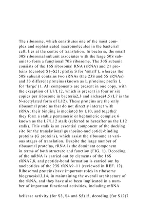
ribosomal proteins, rRNA is the dominant component
in terms of both structure and function (FIG. 1). Decoding
of the mRNA is carried out by elements of the 16S
rRNA7,8, and peptidebond formation is carried out by
nucleotides of the 23S rRNA9–11 (reviewed in REF. 12).
Ribosomal proteins have important roles in ribosome
biogenesis13,14, in maintaining the overall architecture of
the rRNA, and they have also been implicated in a num
ber of important functional activities, including mRNA
helicase activity (for S3, S4 and S5)15, decoding (for S12)7
and peptidyltransferase activity (for L27 (REF. 16) and
L2 (REF. 17)).
The ribosome passes through four functional phases
for the synthesis of a single protein: initiation, elonga
tion, termination and recycling (FIG. 2). All phases are
mediated by specific factors, some of which are bacteria
specific, whereas others (such as the elongation factors
EFTu and EFG) are universally conserved. The amino
acid substrates that are attached to tRNAs (known as
aminoacyltRNAs (aatRNAs)) are delivered to the ribo
some in a ternary complex with EFTu and GTP, and
the tRNAs move through three distinct binding sites
(the aminoacyl (A), peptidyl (P) and exit (E) sites)
located at the interface of the 30S and 50S subunits.
After initiation — which involves placement of the
mRNA start codon and the specific initiator tRNA
(formyl methionine tRNA; fMettRNA) at the Psite
of the 30S subunit, followed by association of the 50S
sub unit — the elongation cycle ensues. The ribo
some moves along an mRNA in the 5ʹ to 3ʹ direction
and decodes each consecutive codon with the help of
the incoming aatRNAs. After successful decoding, the
aatRNA swings fully into the Asite (in a process that is
known ...
Differences in translation and transcription in prokaryotes and e.pdfmanjan6
Differences in translation and transcription in prokaryotes and eukaryotes?
Differences in translation and transcription in prokaryotes and eukaryotes?
Solution
Transcription is the generation of RNA molecules from DNA and Translation is the generation
of protein molecules from RNA. In this way the information from DNA is passed for synthesis
of new proteins or enzymes. Although the basic concepts of transcription and translation are
same into prokaryotes and eukaryotes but due to organizational differences between the two cell
types some differences are there in their transcription and translation.
Because in prokaryotes there are no nucleus both processes here take place in cytoplasm. But in
eukaryotes RNA transcript generation and post transcriptional processing occurs in nucleus.
Apart from that following differences are present in transcription for the two cell types.
In transcription RNA polymerase is responsible for reading the codes of DNA. Three types of
RNA molecules are there: rRNA for ribosomal RNA, mRNA or messenger RNA for all RNA
except ribosomal and tRNA and tRNA or transfer RNA required during translation or transfer of
information from RNA to protein. In prokaryotes all the three types of RNA are produced by
single type of RNA polymerase and the polymerase is composed of five polypeptides. In
eukaryotes there are three types of polymerases namely rRNA is transcriped by RNA Pol I,
mRNA by Pol II and tRNA by Pol III. Each type is composed of 10-15 polypeptides.
Transcription has three phases: Initiation, elongation and termination. Seperate enzymes and
protein factors are required during each phase. In prokaryotes no initiation factors are there &
number of elongation factors are much less than prokaryotes. In eukaryotes initiation factors are
TFIIA, TFIIB, TFIID, TFIIE, TFIIF and TFIIH.
Another major difference is the polycistronic nature of mRNA in prokaryotes i.e genes more
than one present on a single mRNA transcript. But eukaryotic mRNA is monocistronic i.e each
mRNA contains a single gene.
In prokaryotes termination is of 2 types rho factor dependent and rho factor independent. But in
eukaryotes transcripts are vary long and actual process is still unknown.
After generation of primary transcript the post transcriptional processing of the RNAs are less
complex in prokaryotes in comparison to eukaryotes. In prokaryotes translation begins
immediately following transcription. But in eukaryotes all the three types of RNAs undergo
many post transcriptional modifications where the unnecessary sequences are cut off and some
sequences are also added up. For example 5\' capping, addition of the poly A tail, and splicing.
The 5\' capping reaction replaces the triphosphate group at the 5\' end of the RNA chain with a
special nucleotide that is referred to as the 5\' cap. It is thought to help with mRNA recognition
by the ribosome during translation. A modification also takes place at the opposite end of the
RNA transcript. To the 3\.
Protein synthesis and processing: Ribosome, formation of initiation complex, initiation factors and their regulation, elongation and elongation factors, termination, genetic code, aminoacylation of tRNA, tRNA-identity, aminoacyl tRNA synthetase, and translational proof-reading, translational inhibitors, Post Translational modification of proteins. Protein targeting.
This is a process by which the genetic code contained within a messenger RNA (mRNA) molecule is decoded to produce a specific sequence of amino acids in a polypeptide chain.
Translation involves translating the sequence of a messenger RNA (mRNA) molecule to a sequence of amino acids during protein synthesis. It is the process in which ribosomes in the cytoplasm or ER synthesize proteins after the process of transcription of DNA to RNA.
All scientific theories must be able to make testable predictions. S.docxoreo10
All scientific theories must be able to make testable predictions. Such predictions are based on observations. Experiments can then be conducted to verify (or falsify) such predictions. Darwin theorized that evolution occurred through natural selection; however, this may not have occurred in a smooth process. Some evolutionary theorists suggest that evolution by natural selection occurred in step-wise fashion.
Assignment
Write 3–4 pages on the following (not including the title and reference pages):
Explain the concepts of phyletic gradualism and punctuated equilibrium.
What predictions about the fossil record does punctuated equilibrium make?
In this model, what are the processes that produce rapid evolution? Which evolutionary factors are responsible for the periods of relative stasis?
Patterns of punctuated equilibrium have been observed in some cases, but the debate between punctuated equilibrium and phyletic gradualism continues and provides interesting areas of research. Based on your research into the scientific process, what evidence do we see today that supports a long history of life on the planet?
What evidence do we see that supports evolution by gradual change?
What evidence do we see that supports the concept of punctuated equilibrium?
.
All I wnat is to write a reflection paper on my project which is hac.docxoreo10
All I wnat is to write a reflection paper on my project which is hacking tools
My project is about using those 5 tools :
1-
Ice Hole for
Phishing
2-
SocialKlepto for
Social
3-
SmartphonePF and
Mactans
for Mobile
4-
Hping and
Yersinia for networks
5-
LCP and
Cain and Abel for
PasswordCracking
.
More Related Content
Similar to The ribosome, which constitutes one of the most complex and.docx
Differences in translation and transcription in prokaryotes and e.pdfmanjan6
Differences in translation and transcription in prokaryotes and eukaryotes?
Differences in translation and transcription in prokaryotes and eukaryotes?
Solution
Transcription is the generation of RNA molecules from DNA and Translation is the generation
of protein molecules from RNA. In this way the information from DNA is passed for synthesis
of new proteins or enzymes. Although the basic concepts of transcription and translation are
same into prokaryotes and eukaryotes but due to organizational differences between the two cell
types some differences are there in their transcription and translation.
Because in prokaryotes there are no nucleus both processes here take place in cytoplasm. But in
eukaryotes RNA transcript generation and post transcriptional processing occurs in nucleus.
Apart from that following differences are present in transcription for the two cell types.
In transcription RNA polymerase is responsible for reading the codes of DNA. Three types of
RNA molecules are there: rRNA for ribosomal RNA, mRNA or messenger RNA for all RNA
except ribosomal and tRNA and tRNA or transfer RNA required during translation or transfer of
information from RNA to protein. In prokaryotes all the three types of RNA are produced by
single type of RNA polymerase and the polymerase is composed of five polypeptides. In
eukaryotes there are three types of polymerases namely rRNA is transcriped by RNA Pol I,
mRNA by Pol II and tRNA by Pol III. Each type is composed of 10-15 polypeptides.
Transcription has three phases: Initiation, elongation and termination. Seperate enzymes and
protein factors are required during each phase. In prokaryotes no initiation factors are there &
number of elongation factors are much less than prokaryotes. In eukaryotes initiation factors are
TFIIA, TFIIB, TFIID, TFIIE, TFIIF and TFIIH.
Another major difference is the polycistronic nature of mRNA in prokaryotes i.e genes more
than one present on a single mRNA transcript. But eukaryotic mRNA is monocistronic i.e each
mRNA contains a single gene.
In prokaryotes termination is of 2 types rho factor dependent and rho factor independent. But in
eukaryotes transcripts are vary long and actual process is still unknown.
After generation of primary transcript the post transcriptional processing of the RNAs are less
complex in prokaryotes in comparison to eukaryotes. In prokaryotes translation begins
immediately following transcription. But in eukaryotes all the three types of RNAs undergo
many post transcriptional modifications where the unnecessary sequences are cut off and some
sequences are also added up. For example 5\' capping, addition of the poly A tail, and splicing.
The 5\' capping reaction replaces the triphosphate group at the 5\' end of the RNA chain with a
special nucleotide that is referred to as the 5\' cap. It is thought to help with mRNA recognition
by the ribosome during translation. A modification also takes place at the opposite end of the
RNA transcript. To the 3\.
Protein synthesis and processing: Ribosome, formation of initiation complex, initiation factors and their regulation, elongation and elongation factors, termination, genetic code, aminoacylation of tRNA, tRNA-identity, aminoacyl tRNA synthetase, and translational proof-reading, translational inhibitors, Post Translational modification of proteins. Protein targeting.
This is a process by which the genetic code contained within a messenger RNA (mRNA) molecule is decoded to produce a specific sequence of amino acids in a polypeptide chain.
Translation involves translating the sequence of a messenger RNA (mRNA) molecule to a sequence of amino acids during protein synthesis. It is the process in which ribosomes in the cytoplasm or ER synthesize proteins after the process of transcription of DNA to RNA.
Similar to The ribosome, which constitutes one of the most complex and.docx (20)
All scientific theories must be able to make testable predictions. S.docxoreo10
All scientific theories must be able to make testable predictions. Such predictions are based on observations. Experiments can then be conducted to verify (or falsify) such predictions. Darwin theorized that evolution occurred through natural selection; however, this may not have occurred in a smooth process. Some evolutionary theorists suggest that evolution by natural selection occurred in step-wise fashion.
Assignment
Write 3–4 pages on the following (not including the title and reference pages):
Explain the concepts of phyletic gradualism and punctuated equilibrium.
What predictions about the fossil record does punctuated equilibrium make?
In this model, what are the processes that produce rapid evolution? Which evolutionary factors are responsible for the periods of relative stasis?
Patterns of punctuated equilibrium have been observed in some cases, but the debate between punctuated equilibrium and phyletic gradualism continues and provides interesting areas of research. Based on your research into the scientific process, what evidence do we see today that supports a long history of life on the planet?
What evidence do we see that supports evolution by gradual change?
What evidence do we see that supports the concept of punctuated equilibrium?
.
All I wnat is to write a reflection paper on my project which is hac.docxoreo10
All I wnat is to write a reflection paper on my project which is hacking tools
My project is about using those 5 tools :
1-
Ice Hole for
Phishing
2-
SocialKlepto for
Social
3-
SmartphonePF and
Mactans
for Mobile
4-
Hping and
Yersinia for networks
5-
LCP and
Cain and Abel for
PasswordCracking
.
Alice,Betty, and Carol are playing a game with 48 marbles in a circl.docxoreo10
Alice,Betty, and Carol are playing a game with 48 marbles in a circle. Alice takes 2 marbles. Betty takes 4 marbles and Carol takes 6 marbles. One of them (not saying which one) now takes as many marbles as she did the first time. Another girl takes twice as many as she had before and the remaining girl takes 4 times as many as before. There are now 10 marbles left in the circle. Which girl took the same amount as the first time?
.
All healthcare organizations must convert to an Electronic Health Re.docxoreo10
All healthcare organizations must convert to an Electronic Health Record (EHR) system by 2014. This is a major expense that will cost many healthcare organizations millions of dollars. Purchasing an EHR system will undoubtedly require the acquisition and use of long-term assets. Prepare a proposal, addressed to the hospital's management, for financing a 1.5 million dollar EHR system for your local hospital. Be sure to include a strategic plan for financing, administering, operating and marketing the new system.
.
All round writer onlyThis is an individual Mediation assignment..docxoreo10
All round writer only
This is an individual Mediation assignment.
Read the attached information.
Assume the first role, that of Samantha (Sam) Pinder,
Executive V.P. of Finance, a mediator/facilitator . Two other roles are also printed out to assist you in understanding how the parties view the dispute.
Resolve
this dispute using the Steps in the Mediation Process and the Mediator's guide which follow the role information.
Describe the
outcome
for all parties.
.
Alice was wondering whether it was a good idea to invest her money i.docxoreo10
Alice was wondering whether it was a good idea to invest her money in treasury bonds. Discuss with Alice the pros and cons of that type of investment, contrasting U.S. treasuries to other sovereign debt. Be sure to include a discussion of risk. Also, show her the different types of bonds that exist in the corporate world.
-400-600 words
- Please include references
.
All organisms have DNA, which differs only in the number and order o.docxoreo10
All organisms have DNA, which differs only in the number and order of each type of nucleotide. This suggests that all organisms have
Answers available in ...
10
Take this time to do your
best on this question.
Eliminate
Reactivate
Eliminate
Reactivate
Eliminate
Reactivate
.
All literature involves some kind of performance which is intended f.docxoreo10
All literature involves some kind of performance which is intended for an audience. Sometimes, however, the performative quality of a work (i.e., the fact that it is being presented to an audience) is more obvious than at others. Drama and poetry, for example, tend to emphasize overt performance more than do short stories, which more often are read silently and in solitude.
How is the more direct performative aspect of drama and/or poetry reflected in these forms? (Consider for example, each genre’s uses of literary structure, language, technique, and style.) How do these literary elements affect your reading experience?
In your post, identify key qualities of drama and poetry which emphasize their performative qualities. Discuss how these characteristics shape your reading response. Support your views with
at least one example of a dramatic text and one example of a poem
.
.
All key elements of the assignment are covered in a substantiv.docxoreo10
All key elements of the assignment are covered in a substantive way.
·
Presentation follows the timeline of the evolution of business.
·
Presentation provides information on the different stages of business evolution, including:
o
Feudalism
o
Mercantilism
o
Capitalism
o
Commerce
o
Property rights
o
The Industrial Revolution
·
Presentation consists of 10 to 15 slides appropriate for the speaker’s audience.
·
Speaker notes are included for each slide.
·
Title and APA reference slide are included.
.
Alice, Betty and Carol are playing a game with 48 marbles in a circl.docxoreo10
Alice, Betty and Carol are playing a game with 48 marbles in a circle. Alice takes 2 marbles, Betty takes 4 marbles and Carol takes 6 marbles. One of them (not saying which one) now takes as many marbles as she did the first time. Another girl takes twice as many as she had taken before and the remaining girl takes 4 times as many as before. There are now 10 marbles left in the circle. Which girl took the same amount as the first time?
.
Alice Jones was employed as a clerk-typist by a company. She request.docxoreo10
Alice Jones was employed as a clerk-typist by a company. She requested and was refused a vacation day. The employer's refusal was based on her failure to submit the request at least two weeks in advance as required by company policy. She announced that she would take the day anyway, and when she subsequently failed to report for work, was fired for insubordination, plus the unexcused absence. Jones claimed that the company's real reason for firing her was a complaint that she had made to her state's department of labor concerning elimination of employee rest breaks. Explain and evaluate the possible causes of action available to Jones, and identify and explain the possible defenses available to Jones’ employer with regard to each cause of action. Integrate case law and statutory support into your response. (Points : 30)
.
Air and Water Pollution PaperAir and water pollutants exist in m.docxoreo10
Air and Water Pollution Paper
Air and water pollutants exist in many forms. Understanding what they are and where they come from better equips you to address the issues of air and water pollution.
Select
two types of air pollutants and two types of water pollutants.
Write
a 950 paper in which you analyze your selected pollutants and their effect on the environment. In your analysis, include the following items:
·
Indicate whether the selected air pollutants are considered primary or secondary pollutants. Explain why they are considered to be primary or secondary and discuss the sources of these pollutants.
·
Describe how the selected air pollutants affect the different layers of the atmosphere. In looking at this interaction, how do greenhouse gases influence Earth’s climate? Discuss how these air pollutants and greenhouse gases affect human, plant, and animal life.
·
Examine the selected water pollutants. Discuss the sources of these pollutants and indicate their effects on water resources and aquatic life.
·
Discuss the effect of poor water quality on humans and the environment. What are some solutions for reducing poor water quality?
Cite
at least two references.
Format
your paper consistent with APA guidelines.
.
Air pollution is an environmental health problem in many cities thro.docxoreo10
Air pollution is an environmental health problem in many cities throughout the world. Residents in an urban community through which a major freeway transportation route runs are suffering from a number of health effects. The local organization arranges for you, a health educator, to consult with and assist a nurse from a local community clinic in
planning and implementing
a program that will address reduction in exposure of the community, particularly children, to air pollution and thereby reducing the impact of air pollution in the community.
Write a 2-3 page paper in which you do the following:
1. Describe the common health problems associated with
indoor and outdoor air pollution
in urban settings.
2. Describe why children are more vulnerable to the effects of air pollutants.
3. Describe how you (as a Health Educator and consultant in this multi-disciplinary team), would assist the nurse to
plan and implement
a program that will reduce the exposure of this community to air pollution as well as reduce the impact of air pollution on the health of children. In your response, make sure to include preventive steps that can be taken by the community (home and school, for example) to reduce the exposure of children to air pollutants.
.
After your topic has been approved, the next step is to research.docxoreo10
After your topic has been approved, the next step is to research current articles and publications (within one year) and write an outline. You will need to use at least six credible sources that provide objective, authoritative, and accurate descriptions of your selected microeconomics topic. Two of your sources must be from academic journals. Your sources can include the following:
Academic journals
Financial and economic publications like the Wall Street Journal, the Economist, and industry-specific publications
Newspapers such as the New York Times and the Washington Post
Research databases like ProQuest
Use the
Hunt Library (Links to an external site.)
to conduct your research. Do not use Investopedia or Wikipedia.
You may write a formal outline or present your information in paragraph form using current APA formatting. Regardless of the outline format, you must provide the following:
Details that align with your microeconomics topic
List of your sources
Two of the sources must be from academic journals.
.
After watching three of the five movie clips listed in the Multime.docxoreo10
After watching three of the five movie clips listed in the
Multimedia
section, above, describe how they fit into a specific genre (or subgenre) as explained in the text. What elements of the film are characteristic of that genre? How does it fulfill the expectations of that genre? How does it play against these expectations?
.
Aging and Disability WorksheetPart IIdentify 2 or .docxoreo10
Aging and Disability Worksheet
Part I
Identify 2 or 3 issues faced by the aging population.
1.
2.
3.
Answer the following questions in 100 to 200 words each
.
Provide citations for all
the
sources
you use
.
·
What is ageism? How does ageism influence the presence of diversity in society?
·
What is the
Age
Dis
criminitation in Employment
Act (AD
E
A)? How does the AD
E
A address issues for the aging population?
·
What is being done to address the issues you identified?
·
Is the number of aging population expected to rise in numbers or decrease?
·
What types of legislation may or may not be affected by the aging population?
·
How does poverty affect the aging population?
Part II
Answer the following questions in 100 to 200 words each
.
Provide citations for all
the
sources
you use
.
·
What does the ADA provide for people with disabilities?
·
How have people with disabilities been treated in the past?
·
How has the attitude toward people with disabilities changed over time?
·
What are some unique circumstances or issues encountered by people with disabilities?
·
What is being done to address those issues?
·
What types of legislation have been introduced to address issues faced by people with disabilities?
.
After watching the video and reading the Web Resource, CDC Autism .docxoreo10
After watching the video and reading the Web Resource, "CDC: Autism Spectrum Disorders: Signs and Symptoms," discuss how you as an early childhood professional can use this information to increase awareness and early identification of autism, specifically discussing assessment tools that can be used with children with autism.
.
AI Artificial Intelligence1Reading responsePeter .docxoreo10
AI: Artificial Intelligence
1
Reading response
Peter Dormer, “Craft and the Turing Test for Practical Thinking,” in The Challenge of Technology.
What is personal know-how? What is distributed knowledge?
How do they relate to the Turing test?
Give one example of your own how these concepts matter today to artists and makers, or better yet, in your own experience?
Journal homework
Keep a record (text and drawings) of events in daily life where human and machine intersect and interact. Fill at least two pages with your observations.
Mary Shelley, Frankenstein, or The Modern Prometheus, 1818
Boris Karloff in Frankenstein in 1931 directed by James Whale
Mary Shelley first published Frankenstein, or the Modern Prometheus 1818. the novel allegorizes the Romantic obsession with discovering the power or principle of life. Ideas about a life power were consistent with the scientific understanding of the day. Darwin himself spoke of an organizing “spirit of animation” in his Zoonomia; or, The Laws of Organic Life, in which he stated “the world itself might have been generated, rather than created.”
Dr. Frankenstein picked all the parts for his monster based on their beauty, but when it comes to life, the monster is unbearably ugly. “I had worked hard for nearly two years, for the sole purpose of infusing life into an inanimate body…the beauty of the dream vanished, and breathless horror and disgust filled my heart. Unable to endure the aspect of the being I had created, I rushed out of the room”.
4
Two definitions of AI:
“The use of computer programs and programming techniques to cast light on the principles of intelligence in general and human thought in particular.
--Margaret Boden
“The science of making machines do things that would require intelligence if done by humans.”
-Marvin Minsky
BOTH OF THESE STATEMENTS ORIGINATE IN ALAN TURING’S FIRST COMPUTER SCIENCE ARTICLE
Working assumption: all cognition is computable
Question:
Is what’s not yet known to be computable actually computable?
if so, then what?
if not, why not, and what does that tell us about cognition?
7
Who was Alan Turing?
B. 1912 London, attended King’s College, Cambridge and Princeton University. He studied mathematics and logic (he hadn’t invented computer science yet)
At 23, he invented the “Turing machine” and published “On Computable Numbers in 1936, the first and most important paper in comp. sci.
During WWII, solved the German Enigma code by use of electromechanical devices—a precursor to the computer
Laid the foundation for major subfields of comp sci: theory of computation, design of hardware and software, and the study of artificial intelligence
“The Imitation Game,”
aka
“The Turing Test”
In 1950, Turing posited a way to test machine intelligence: a person in a room before a screen. S/he would correspond with two agents and based on their responses, decide which was a machine and which was human. If the machine can pass fo.
Agree or disagree with, and discuss the following statement Corp.docxoreo10
Agree or disagree with, and discuss the following statement: "Corporate intelligence is not corporate espionage because 95 percent of the information a company needs to make strategic decisions is available and accessible to the public." Explain your rationale.
Your response should be at least 200 words in length. All sources used, including the textbook, must be referenced; paraphrased and quoted material must have accompanying citations.
David, F. (2011). 1.
Strategic management: concepts & cases
(Custom Edition ed., pp. 72-74). New York: McGraw-Hill Irwin.
No Wiki, Dictionary.com or Plagiarism
.
After watching Reactions to an Impending Death Sentence and Ti.docxoreo10
After watching
Reactions to an Impending Death Sentence
and
Ties That Bind
, discuss the impact of our various relationships with those who are dying on how death impacts us. For example, how would you relate to a dying child, adult or older adult when they are dying? How would you relate to those left behind: parents, siblings, children, grandchildren, spouse? How do you feel about your own death? Discuss these questions in 250 – 300 words
.
Synthetic Fiber Construction in lab .pptxPavel ( NSTU)
Synthetic fiber production is a fascinating and complex field that blends chemistry, engineering, and environmental science. By understanding these aspects, students can gain a comprehensive view of synthetic fiber production, its impact on society and the environment, and the potential for future innovations. Synthetic fibers play a crucial role in modern society, impacting various aspects of daily life, industry, and the environment. ynthetic fibers are integral to modern life, offering a range of benefits from cost-effectiveness and versatility to innovative applications and performance characteristics. While they pose environmental challenges, ongoing research and development aim to create more sustainable and eco-friendly alternatives. Understanding the importance of synthetic fibers helps in appreciating their role in the economy, industry, and daily life, while also emphasizing the need for sustainable practices and innovation.
How to Make a Field invisible in Odoo 17Celine George
It is possible to hide or invisible some fields in odoo. Commonly using “invisible” attribute in the field definition to invisible the fields. This slide will show how to make a field invisible in odoo 17.
Unit 8 - Information and Communication Technology (Paper I).pdfThiyagu K
This slides describes the basic concepts of ICT, basics of Email, Emerging Technology and Digital Initiatives in Education. This presentations aligns with the UGC Paper I syllabus.
Read| The latest issue of The Challenger is here! We are thrilled to announce that our school paper has qualified for the NATIONAL SCHOOLS PRESS CONFERENCE (NSPC) 2024. Thank you for your unwavering support and trust. Dive into the stories that made us stand out!
Students, digital devices and success - Andreas Schleicher - 27 May 2024..pptxEduSkills OECD
Andreas Schleicher presents at the OECD webinar ‘Digital devices in schools: detrimental distraction or secret to success?’ on 27 May 2024. The presentation was based on findings from PISA 2022 results and the webinar helped launch the PISA in Focus ‘Managing screen time: How to protect and equip students against distraction’ https://www.oecd-ilibrary.org/education/managing-screen-time_7c225af4-en and the OECD Education Policy Perspective ‘Students, digital devices and success’ can be found here - https://oe.cd/il/5yV
Welcome to TechSoup New Member Orientation and Q&A (May 2024).pdfTechSoup
In this webinar you will learn how your organization can access TechSoup's wide variety of product discount and donation programs. From hardware to software, we'll give you a tour of the tools available to help your nonprofit with productivity, collaboration, financial management, donor tracking, security, and more.
The Art Pastor's Guide to Sabbath | Steve ThomasonSteve Thomason
What is the purpose of the Sabbath Law in the Torah. It is interesting to compare how the context of the law shifts from Exodus to Deuteronomy. Who gets to rest, and why?
The Roman Empire A Historical Colossus.pdfkaushalkr1407
The Roman Empire, a vast and enduring power, stands as one of history's most remarkable civilizations, leaving an indelible imprint on the world. It emerged from the Roman Republic, transitioning into an imperial powerhouse under the leadership of Augustus Caesar in 27 BCE. This transformation marked the beginning of an era defined by unprecedented territorial expansion, architectural marvels, and profound cultural influence.
The empire's roots lie in the city of Rome, founded, according to legend, by Romulus in 753 BCE. Over centuries, Rome evolved from a small settlement to a formidable republic, characterized by a complex political system with elected officials and checks on power. However, internal strife, class conflicts, and military ambitions paved the way for the end of the Republic. Julius Caesar’s dictatorship and subsequent assassination in 44 BCE created a power vacuum, leading to a civil war. Octavian, later Augustus, emerged victorious, heralding the Roman Empire’s birth.
Under Augustus, the empire experienced the Pax Romana, a 200-year period of relative peace and stability. Augustus reformed the military, established efficient administrative systems, and initiated grand construction projects. The empire's borders expanded, encompassing territories from Britain to Egypt and from Spain to the Euphrates. Roman legions, renowned for their discipline and engineering prowess, secured and maintained these vast territories, building roads, fortifications, and cities that facilitated control and integration.
The Roman Empire’s society was hierarchical, with a rigid class system. At the top were the patricians, wealthy elites who held significant political power. Below them were the plebeians, free citizens with limited political influence, and the vast numbers of slaves who formed the backbone of the economy. The family unit was central, governed by the paterfamilias, the male head who held absolute authority.
Culturally, the Romans were eclectic, absorbing and adapting elements from the civilizations they encountered, particularly the Greeks. Roman art, literature, and philosophy reflected this synthesis, creating a rich cultural tapestry. Latin, the Roman language, became the lingua franca of the Western world, influencing numerous modern languages.
Roman architecture and engineering achievements were monumental. They perfected the arch, vault, and dome, constructing enduring structures like the Colosseum, Pantheon, and aqueducts. These engineering marvels not only showcased Roman ingenuity but also served practical purposes, from public entertainment to water supply.
This is a presentation by Dada Robert in a Your Skill Boost masterclass organised by the Excellence Foundation for South Sudan (EFSS) on Saturday, the 25th and Sunday, the 26th of May 2024.
He discussed the concept of quality improvement, emphasizing its applicability to various aspects of life, including personal, project, and program improvements. He defined quality as doing the right thing at the right time in the right way to achieve the best possible results and discussed the concept of the "gap" between what we know and what we do, and how this gap represents the areas we need to improve. He explained the scientific approach to quality improvement, which involves systematic performance analysis, testing and learning, and implementing change ideas. He also highlighted the importance of client focus and a team approach to quality improvement.
Cambridge International AS A Level Biology Coursebook - EBook (MaryFosbery J...
The ribosome, which constitutes one of the most complex and.docx
1. The ribosome, which constitutes one of the most com-
plex and sophisticated macromolecules in the bacterial
cell, lies at the centre of translation. In bacteria, the small
30S ribosomal subunit associates with the large 50S sub-
unit to form a functional 70S ribosome. The 30S subunit
consists of the 16S ribosomal RNA (rRNA) and 21 pro-
teins (denoted S1–S21; prefix S for ‘small’), whereas the
50S subunit contains two rRNAs (the 23S and 5S rRNAs)
and 33 different proteins (known as L proteins; prefix L
for ‘large’)1. All components are present in one copy, with
the exception of L7/L12, which is present in four or six
copies per ribosome in bacteria2,3 and archaea4,5 (L7 is the
N-acetylated form of L12). These proteins are the only
ribosomal proteins that do not directly interact with
rRNA; their binding is mediated by L10, and together
they form a stable pentameric or heptameric complex 6
known as the L7/L12 stalk (referred to hereafter as the L12
stalk). This stalk is an essential component of the docking
site for the translational guanosine-nucleotide-binding
proteins (G proteins), which assist the ribosome at vari-
ous stages of translation. Despite the large number of
ribosomal proteins, rRNA is the dominant component
in terms of both structure and function (FIG. 1). Decoding
of the mRNA is carried out by elements of the 16S
rRNA7,8, and peptide-bond formation is carried out by
nucleotides of the 23S rRNA9–11 (reviewed in REF. 12).
Ribosomal proteins have important roles in ribosome
biogenesis13,14, in maintaining the overall architecture of
the rRNA, and they have also been implicated in a num-
ber of important functional activities, including mRNA
helicase activity (for S3, S4 and S5)15, decoding (for S12)7
2. and peptidyltransferase activity (for L27 (REF. 16) and
L2 (REF. 17)).
The ribosome passes through four functional phases
for the synthesis of a single protein: initiation, elonga-
tion, termination and recycling (FIG. 2). All phases are
mediated by specific factors, some of which are bacteria-
specific, whereas others (such as the elongation factors
EF-Tu and EF-G) are universally conserved. The amino
acid substrates that are attached to tRNAs (known as
aminoacyl-tRNAs (aa-tRNAs)) are delivered to the ribo-
some in a ternary complex with EF-Tu and GTP, and
the tRNAs move through three distinct binding sites
(the aminoacyl- (A-), peptidyl- (P-) and exit- (E-) sites)
located at the interface of the 30S and 50S subunits.
After initiation — which involves placement of the
mRNA start codon and the specific initiator tRNA
(formyl methionine tRNA; fMet-tRNA) at the P-site
of the 30S subunit, followed by association of the 50S
sub unit — the elongation cycle ensues. The ribo-
some moves along an mRNA in the 5ʹ to 3ʹ direction
and decodes each consecutive codon with the help of
the incoming aa-tRNAs. After successful decoding, the
aa-tRNA swings fully into the A-site (in a process that is
known as accommodation). Decoding and accommo-
dation are often collectively referred to as ‘Asite occu
pation’. The swing docks the aminoacyl residue into the
peptidyltransferase centre, resulting in rapid peptide
bond formation. The nascent chain is transferred from
the peptidyl-tRNA at the P-site to the charged tRNA at
Decoding
Selection of the cognate
ternary complex of aminoacyl-
tRNA–EF-Tu–GTP on the basis
3. of correct codon-anticodon
interactions between the
mRNA and tRNA, respectively.
EF‑G and EF4: translocation and
back‑translocation on the bacterial
ribosome
Hiroshi Yamamoto1*, Yan Qin2*, John Achenbach3*,
Chengmin Li2, Jaroslaw Kijek4,
Christian M. T. Spahn1 and Knud H. Nierhaus1,4
Abstract | Ribosomes translate the codon sequence of an mRNA
into the amino acid
sequence of the corresponding protein. One of the most crucial
events is the translocation
reaction, which involves movement of both the mRNA and the
attached tRNAs by one codon
length and is catalysed by the GTPase elongation factor G
(EF‑G). Interestingly, recent
studies have identified a structurally related GTPase, EF4, that
catalyses movement of the
tRNA
2
–mRNA complex in the opposite direction when the ribosome
stalls, which is known as
back‑translocation. In this Review, we describe recent insights
into the mechanistic basis of
both translocation and back‑translocation.
1Institut für Medizinische
Physik und Biophysik, Charité
– Universitätsmedizin Berlin,
Charitéplatz 1,10117 Berlin,
Germany.
5. after peptide bond formation and before translocation
as pre-translocational states (PRE-states). To accom-
modate the next incoming aa-tRNA, the peptidyl-tRNA
at the A-site and the deacylated tRNA at the P-site are
translocated to the P- and E-sites, respectively, and this
is catalysed by EF-G–GTP. The resulting state, in which
the P- and E-sites are occupied and the A-site is vacant,
is called the post-translocational state (POST-state)
(reviewed in REF. 18). The release of the deacylated tRNA
from the E-site is thought to occur after trans location19,20
or, alternatively, on occupation of the A-site with the
next aa-tRNA21–23.
It is possible that ribosomes mistranslocate, which
leads to an arrest in protein synthesis as the ribosome
stalls and thereby blocks the progression of other ribo-
somes on the same mRNA. Recent studies suggest that
such stalled ribosomes can be rescued by a GTPase
known as EF4, which is structurally related to EF-G. This
factor recognizes stalled ribosomes that have a deacylated
tRNA in the E-site and a peptidyl-tRNA in the P-site
(the POST-state) and catalyses a back-translocation
reaction (FIG. 2). The tRNAs are dragged back into the
P- and A-sites, thereby giving the ribosome a second
chance to properly translocate24–26. Other studies suggest
that EF4 can also bind to and mobilize ribosomes that
are stalled in the PRE-state27 (see below). Translation is
terminated when a ribosome encounters a stop codon on
the mRNA, which is recognized by a release factor that
triggers release of the nascent polypeptide. During the
final phase of translation, which is known as recycling,
the 70S ribosome is thought to dissociate into its 30S and
50S subunits, which are re-used for subsequent rounds
of initiation (reviewed in REF. 18).
6. In this Review, we discuss a number of recent struc-
tural and biochemical studies in bacteria, primarily
Escherichia coli and Thermus thermophilus, that have
enhanced our understanding of the mechanisms of bac-
terial translocation and back-translocation. The binding
modes and functional roles of EF-G and EF4 are dis-
cussed, as well as the proposed physiological relevance
of back-translocation.
EF‑G and EF4
Structural similarities. EF-G and EF-Tu are universal
translation factors, whereas EF4 is found in almost all
bacteria, in mitochondria and chloroplasts, but is absent
in archaea and the cytoplasm of eukaryotes. EF4 is the
third most highly conserved bacterial protein after
EF-Tu and EF-G, with a 55–68% amino acid identity
between different bacterial species24.
The three-dimensional structures of EF-G and the
ternary complex (aa-tRNA–EF-Tu–GTP) are highly
similar (FIG. 3a,b). The five structural domains of EF-G
(FIG. 3a) fold into a structure that resembles the ternary
complex, and domain IV of EF-G corresponds to the
anticodon stem–loop of the tRNA within the ternary
Nature Reviews | Microbiology
50S subunit
L12
L12 stalk
L1
L1 stalk
7. CP
PTC
Head
Body
Platform
S13
S12E P
A
30S subunit
E
P
A
L11
L10
SRL
23S rRNA 16S rRNA
5S rRNA
Figure 1 | Overall architecture of the large and small subunits of
the bacterial ribosome. Both subunits are shown
from the interface side. The large 50S subunit contains the 23S
ribosomal RNA (rRNA) and 5S rRNA (light grey and dark
9. ribosome) only undergoes a
single round of catalysis.
complex (FIG. 3b). This is probably the most famous
example of molecular mimicry, which highlights the
need for both EF-G and the ternary complex to occupy
a similar site at the interface of the ribosomal subunits.
Similarly, the domain structure of EF4 is highly related
to that of EF-G (FIG. 3a). Both factors share domains I
(known as the G domain), II, III and V, which are
responsible for ribosome binding and GTPase activ-
ity. In addition, both factors have specific domains:
EFG contains Gʹ (which is a subdomain of domain I)
and domain IV, whereas EF4 has a unique carboxy-
terminal domain (CTD)24. Domain IV of EF-G and the
CTD of EF4 are responsible for mediating the opposing
roles of these two factors in translation (FIG. 3c).
First contacts with the ribosome. The first contacts of
EF-G and EF4 with the ribosome involve the L12 stalk
and seem to follow the same pathway. The substrate for
EF-G is the 70S ribosome in the PRE-state, whereas the
substrate for EF4 is still unclear. One study suggests that
EF4 preferentially binds to the POST-state ribosome,
owing to observations that EF4 binds to the POST-state
with higher affinity than to the PRE-state, and that
EF4-dependent GTP hydrolysis has a higher turnover
rate with POST-state ribosomes than with PRE-state
ribosomes28. However, single-turnover experiments and
single-molecule FRET (Förster resonance energy trans-
fer) measurements suggest that the PRE-state is the
preferential but not the exclusive target of EF4. In this
study, EF4 could compete with EF-G for binding to the
PRE-state27. Thus, EF-G recognizes a specific functional
state, whereas EF4 seems to be more promiscuous in its
10. specificity.
It is thought that EF-G makes its first ribosomal con-
tact with the CTD of L12 using the Gʹ domain3. The next
step might be shared by other factors (such as EF-Tu and
EF4) and involves contact with the base of the L12 stalk,
resulting in interactions between the L12 CTD and the
amino-terminal domain (NTD) of L11, as demonstrated
by cryo-electron microscopy(cryo-EM)29,30 and X-ray
crystallography31,32. This interaction is controlled by the
universally conserved Pro22 residue of L11, which is in
a trans-configuration when the ribosome is free of GTP-
binding proteins or when a non-GTPase factor is bound
(Supplementary information S1 (figure)). However, when
a G-protein factor such as EF-G, EF-Tu or EF4 binds to
the ribosome, Pro22 adopts the cis-configuration, which
facilitates the L11–L12 interaction. Interestingly, the
trans–cis transition is catalysed by a peptidyl-prolyl cis–trans
isomerase (PPIase) centre, comprising amino acyl residues
that reside mainly in the G domain of translational fac-
tors. Before the factor dissociates from the ribosome after
GTP hydrolysis and inorganic phosphate (Pi) release, the
PPIase activity of the factor stimulates reversion of Pro22
to the trans-configuration33,34.
The early contacts of EF-G with the ribosome pre-
sent a conundrum: EF-G triggers the movement of the
tRNA2–mRNA complex from a PRE-state to the POST-
state, but the initial EF-G contacts with the ribosome
that are essential for activating the ribosome and setting
the tRNA2–mRNA complex in motion are currently
unknown. When EF-G is added to a PRE-state ribo-
some and its dissociation from the ribosome is inhibited
(using the antibiotic fusidic acid or the non-cleavable
GTP analogues GDPNP (guanosine 5ʹtetrahydro
gen triphosphate) or GDPCP (5ʹguanosylmethylene
11. triphosphate), X-ray and cryo-EM structures have dem-
onstrated that the peptidyl-tRNA has left the A-site
and approaches the P-site, and domain IV of EF-G is
flipped into the A-site, where it functions as a doorstop
to prevent back-translocation of the tRNA2–mRNA
Nature Reviews | Microbiology
aa-tRNA–EF-Tu–GTP
EF-Tu–GDP + P
i
A-site occupationTranslocation
Peptidyl transfer
EF-G–GTP
E-tRNA
EF-G–GDP + P
i
Elongation
cycle
Initiation
Termination
Recycling
70S
initiation complex
12. mRNA
fMet-tRNA
50S
30S
APE
APE
EF4–GDP + P
i
EF4–GTP
APE
APE
APE APE
Figure 2 | The functional phases of the ribosome during
translation. The 70S
initiation complex contains the initiator tRNA
(formylmethionine tRNA (fMet‑tRNA)) at
the ribosomal P‑site, which interacts with the start codon
(typically AUG) of the mRNA
via the formation of a codon–anticodon duplex. The 70S
initiation complex enters the
elongation cycle on binding the ternary complex
aminoacyl‑tRNA–elongation factor
Tu–GTP (aa‑tRNA–EF‑Tu–GTP). After successful decoding,
GTP is hydrolysed, EF‑Tu–GDP
and inorganic phosphate (P
14. Single‑molecule FRET
(Single-molecule Förster
resonance energy transfer). A
phenomenon in which energy
induced by light excitation is
transferred from one
fluorophore to another in a
distance-dependent manner,
observed on a single complex
or molecule.
complex 31,35–39 (FIG. 3c; Supplementary information S2
(figure)). In other words, in all previous ribosome struc-
tures with EF-G, the factor has already triggered a first
step of translocation. However, a recent report describes
the structure of a pre-translocational EF-G—ribosome
complex with two tRNAs in hybrid positions. The com-
plex was prepared in the presence of GTP; EF-G disso-
ciation was blocked with the antibiotic fusidic acid and
translocation of the tRNA2–mRNA complex was inhib-
ited with the antibiotic viomycin115. In this PRE-state, the
tip of EF-G domain IV makes strong contacts with the
anticodon loop of the A-site tRNA. A comparison of
the EF-G structure in the POST state31 revealed that
EF-G undergoes a ~20° rotation around the sarcin–ricin
loop (SRL) of the 23S rRNA. This rotation results in
a movement of the tip of domain IV by 20 Å into the
decoding centre during the transition from the PRE- to
the POST-state. Although this study reveals important
insights, it is still unclear what triggers the dramatic
conformational change of EF-G and which contacts
between EF-G and the ribosome (or its ligands) set the
tRNA2-mRNA in motion.
15. When EF4 is added to POST-state ribosomes, the
structures that are available show the peptidyl-tRNA in
a back-translocated position, having established either
an intermediate state (possibly identical with a trans-
location intermediate25) or a PRE-state28. Thus, a struc-
ture in which EF4 is bound to the POST-state before the
onset of back-translocation is currently lacking.
The specific domains of EF‑G and EF4. Both factors
reduce the activation-energy barrier between PRE- and
POST-states, but the binding of each factor induces
one distinct state of the tRNA2–mRNA complex; EF-G
favours the POST-state and EF4 favours the PRE-state.
EF-G flips domain IV into the A-site, resulting in a door-
stop effect that stabilizes the POST-state. This suggests
that domain IV is essential for translocation. Indeed,
Thermus thermophilus EF-G fragments that lack this
domain are unable to translocate, but they retain GTPase
activity and are able to bind to the ribosome40. As men-
tioned above, EF4 lacks domain IV of EF-G and, as such,
lacks the doorstop function, which is considered to be a
prerequisite to allow for the back-movement of tRNAs
from the POST-state to the PRE-state. This is clearly seen
in the cryo-EM structure28 (Supplementary information S2
(figure), left panel), in which the back-translocated
peptidyl-tRNA in the A-site is attached to the unique
CTD of EF4, whereas domain IV of EF-G would prevent
movement into this position. After movement back into
the A-site, the CTD of EF4 halts the peptidyl-tRNA in
this position, thereby re-establishing the PRE-state. This
halting effect is caused by surface patches of strong posi-
tive charges on EF4 that attract the negative charges of
the A-site tRNA28,41. The CTD of EF4 contacts the inner
side of the elbow and the acceptor-stem down to the
CCA end of the A-site tRNA (Supplementary information
S2 (figure), right panel).
16. To preserve the reading frame during back-translocation,
maintenance of codon–anticodon interactions is essen-
tial. The presence of a cognate E-site tRNA is crucial
for EF4-mediated back-translocation24 because a back-
translocated tRNA in the P-site must sustain codon–
anticodon interactions; without such interactions, a P-site
tRNA cannot be fixed on the 30S subunit42.
Mechanism of translocation
A wealth of recent structural data describing the dynam-
ics and structural transitions of the ribosome during
translocation now allows for a comprehensive overview
of the mechanisms involved. In this section, we describe
Nature Reviews | Microbiology
EF4
a
b
P/P
P/P
E/E
A/L
EF4
EF-G
EF-Tu
17. G Gʹ G II III IV V CTD
c
1–158 159–253 254–289 290–404 405–482 483–603
1– –212 213–313 314–405 tRNA
604–691
1– –188 189–281 291–371 398–486 487–599
EF-G
EF4
G
IIIII
VV
CTD
EF-Tu
G
II
III
A/T-tRNA
EF-G
G
18. II
III
IV
Gʹ
Common domainsSpecific domains
Backwards
Forwards
Figure 3 | Structure, binding sites and functions of the
elongation factors. a | Domain
organization of elongation factor G (EF‑G), EF4 and EF‑Tu. b |
EF‑G, EF4 and EF‑Tu have a
highly similar domain organization and fold into similar
three‑dimensional structures
(EF‑G, Protein Data Bank (PDB) accession 2WRI31; EF4, PDB
accession 3DEG28; and the
ternary complex aminoacyl‑tRNA−EF‑Tu−GTP, PDB accession
2WRN70). c | EF‑G and EF4
bind to a similar site on the ribosome, but their specific
domains promote opposing effects.
EF‑G catalyses forward movement of the tRNAs from the A/A
and P/P sites to the P/P and
E/E sites, whereas EF4 can reverse this reaction to promote
back translocation, moving
the tRNAs from E/E to P/P and from P/P even beyond the A/A
site toward the L12 stalk.
The latter position is only seen in the presence of EF4 and is
referred to as the A/L position.
20. the N-glycosidase ricin or
cleaving the 23S rRNA after
G2661 by the RNase α-sarcin
impairs the binding and GTPase
activity of both elongation
factor Tu (EF-Tu) and EF-G,
thereby blocking translation.
Activation‑energy barrier
The energy barrier that
separates reactants and
products in a chemical
reaction.
the role of intersubunit rotation (formerly called ‘ratch
eting’43) and swivelling of the head of the 30S subunit in
translocation, as well as recent insights into the role of
GTP hydrolysis.
The PRE‑states. After peptidebond formation, the ribo-
some can adopt at least three PRE-states; in each state,
both the A- and P-sites on the 30S subunit are occupied
by a tRNA-anticodon stem, whereas the CCA ends of
the tRNAs on the 50S subunit can vary in their location.
In the classical PRE-state, the anticodon stem and the
CCA end of the two tRNAs are positioned in the same
site on each ribosomal subunit (known as A/A for the
A-site tRNA and P/P for the P-site tRNA). The ribosome
spontaneously fluctuates between this classical state and
a rotated state44. Rotation involves a 4–7 ° anticlockwise
rotation of the 30S subunit relative to the 50S subunit,
around a pivot axis close to the middle of helix 44 (h44)43
(FIG. 4a). The intersubunit rotation is coupled to a move-
ment of the CCA end of the P-site tRNA on the 50S sub-
unit to the E-site; simultaneous movement of the CCA
end of the A-site tRNA into the 50S P-site may occur
21. but is not strictly coupled. The tRNA positions within
the 30S subunit remain unchanged, giving rise to hybrid
sites45. The functional state of a ribosome with a tRNA
in an A/P hybrid site (anticodon stem in the A-site on
the 30S subunit and the CCA end in the P-site on the
50S subunit), and a deacylated tRNA in a P/E hybrid site
(anticodon stem in the P-site of the 30S and the CCA
end in the E-site of the 50S) is known as hybrid state 1
(H1). The third PRE-state (A/A and P/E), which corre-
sponds to movement of the P-site tRNA only, is known
as hybrid state 2 (H2)44,46 (FIG. 4b). Back-rotation of the
30S subunit re-establishes the tRNAs in the classical A/A
and P/P binding positions.
These fluctuations between the various PRE-states
only occur in the absence of EF-G47. All three PRE-
states are substrates for EF-G; EF-G can enter the
sequence of PRE-states (classical, H2 and H1) at any
stage in order to move the tRNA2–mRNA complex to the
POST-state, although EF-G–GTP seems to favour
the 30S rotated state with tRNAs in hybrid positions48,49.
In other words, this sequence of PRE-states is the
only route to the transition state and is thus essential
Nature Reviews | Microbiology
4–7°
Non-rotated
30S head
30S head
Rotated
22. Classical
H1 H2
18°
P/P A/A
P/E A/P P/E A/A
30S body
50S subunit
L1 stalk
L12 stalk
L12 stalk
L1 stalk
a
b
Swivelled
Non-swivelled
c
30S body
50S subunit
24. for translocation47. Inhibition of intersubunit rotation
by crosslinking the 30S and 50S subunits blocks trans-
location50, which shows that this is an essential step in
translocation. Single-molecule FRET measurements have
revealed that there are two populations of pre-translocation
complexes: one in which the ribosome rapidly fluctu-
ates between classical and hybrid states, and another
in which the tRNA positions are long-lived in either
the classical or hybrid state configuration. Following the
addition of EF-G, both populations of pre-translocation
complexes are translocated47, but it is currently unclear
whether only one or both populations exist in vivo.
The transition from PRE‑states to the POST‑state. After
binding to the A-site, a tRNA must translocate twice
(from the A-site to the P-site and from the P-site to the
E-site) during the course of translation, which involves
five distinct combinations of tRNA binding sites:
A/A, A/P, P/P, P/E and E/E. Analyses of ribosomes in
polysomes51,52 or during poly(Phe) synthesis53 have
revealed that at least two tRNAs are always present
on the ribosome during the elongation cycle; in the
PRE-state this corresponds to either the classical state
(A/A and P/P) or the hybrid states (H1 or H2). By con-
trast, only one POST-state exists, which is characterized
by a peptidyl-tRNA in the P/P site and a deacylated tRNA
in the E/E site (Supplementary information S3 (figure)).
A transition intermediate between the PRE- and POST-
states is observed when EF-G is trapped on the ribosome
either by using GDPNP or fusidic acid. This intermedi-
ate is characterized by another large-scale movement of
the ribosome, this time exclusively within the small sub-
unit. It involves an anticlockwise rotation of the 30S head
relative to the 30S body, termed swivelling, which turns
the head by about 18 ° towards the E-site35–39,54–56
25. (FIG. 4c).
In agreement with measurements of head rotation and
mRNA movement 57, structural data show an almost
complete translocation of the tRNA2–mRNA complex in
the POST-state transition intermediate (TIPOST)35,58. EF-G
dependent GTP hydrolysis is not required for translo-
cation, however, it must occur to ensure that EF-G is
released from the ribosome. A reversal of the head swivel
and 30S back-rotation ensues, thereby establishing the
stable POST-state, in which the tRNAs fully occupy
the P/P and E/E sites.
It is important to note that during translocation of
the tRNA2–mRNA complex, it is the tRNAs that are
physically moved by the ribosome, whereas the mRNA
co-migrates with the tRNAs, mainly owing to codon–
anticodon interactions. This conclusion is supported
by the observation that the main physical contacts
between the mRNA and the ribosome during elongation
are mediated by the codon–anticodon interactions59.
This highlights the importance of codon–anticodon
inter actions not only during decoding at the A-site but
also at the P-site31,60,61 and the E-site22,32,62.
Activation‑energy barrier between PRE‑ and POST‑states.
The PRE-states are separated from the POST-state by
a high activation-energy barrier of 90 kJ mol–1 (REF. 63).
EF-G reduces this barrier by establishing the TIPOST state
and accelerates the translocation rate by 104- to 106-fold
compared with spontaneous translocation (reviewed
in REF. 64). Structures that possibly have a role in estab-
lishing the energy barrier are the bridges that connect
the 30S and 50S subunits at the intersubunit face and the
ribosomal proteins S12 and S13 (REF. 65), which are
located close to the A-site and P-site tRNAs. However,
26. studies have shown that disruption of some of the
bridges66 or removal of S12 and S13 (REF. 65) only con-
fer a modest increase in the rates of both spontaneous
translocation and back-translocation, which indicates
that they have only a marginal role in establishing the
energy barrier.
By contrast, it has been proposed that a structural
element of the 16S rRNA might have a decisive role in
creating the activation-energy barrier. A ridge of four
bases, G1338-A-N-U1341 (where N represents any
base), in the 30S head and the nucleotide A790 of the 30S
platform form a gate that blocks movement of the tRNA
anticodon stem between the P- and E-sites67 (FIG. 5a,b).
Four of the five nucleotides of this gate, which is referred
to as the A790 gate, are universally conserved in all three
domains of life. The A790 gate is 13.8 Å in width in the
absence of EF-G (closed gate), which is too narrow to
allow the passage of an RNA duplex, such as the anti-
codon stem of the P-site tRNA (which has a diameter
of 20 Å). Therefore, this gate needs to open in order to
enable movement of a P-site tRNA to the E-site. A series
of published functional complexes in the absence and
presence of EF-G have been analysed, which suggest that
the A790 gate is closed in the absence of EF-G and in the
POST-state31,46, but that it opens to a width of approxi-
mately 24 Å exclusively in the intermediate TIPOST state35.
These findings are in clear agreement with a recent crys-
tal structure of translocation intermediates of bacterial
ribosomes68 as well as with a first cryo-EM structure of
a TIPOST ribosome containing two tRNAs116. Opening of
the gate is accompanied and probably caused by the 18 °
swivel of the 30S head68, as the gate is closed in the non-
swivelled PRE-states (FIG. 5b). Swivelling of the 30S head
not only opens the A790 gate, but also induces move-
ment of the tRNA2–mRNA complex on the 30S subunit
29. PRE-states
POSTClassical
a
b
c
H1 TI POST
Closed Intermediate Intermediate
Figure 5 | Ribosomal conformational changes during
translocation.
a | After peptidyl ‑transfer, the tRNAs are in the classical state
(A/A and
P/P), which establishes an equilibrium with the hybrid states H1
and H2
(H2 not shown) owing to intersubunit rotation. When elongation
factor G
(EF‑G) binds to one of these three PRE‑states, swivelling of the
30S head
is induced, leading to the formation of the translocation
intermediate
TIPOST, which later resolves into the post‑translocational state
(POST‑state)
after a reversal of the head swivel and 30S back‑rotation. Top
row, view of
the 70S ribosome from the 30S solvent side showing the
intersubunit
movements. Bottom row, view from above the 70S ribosome
showing the
tRNA positions. b | Positions of the 16S rRNA base A790,
which forms an
32. L1 positions are observed31,35,37,46,69,70 (FIG. 5c): it adopts
an open position during decoding and in the classical
PRE-state; a closed position in the hybrid PRE-states
(H1 and H2); and an intermediate position in the
TIPOST and POST-state. Thus, the L1 stalk is proposed
to function as a gate for the deacylated E-site tRNA,
blocking release of the tRNA when it is in the closed
position, but enabling free dissociation when it is in the
open position71. This hypothesis is consistent with the
allosteric three-site model for the elongation cycle72,
which posits that the E-site tRNA is only released
when the A-site becomes occupied with the next
aa-tRNA21–23,73, coinciding with opening of the L1 stalk
during decoding. The coupling of different transloca-
tional states to distinct positions of the L1 stalk is clearly
visible in X-ray and cryo-EM structures46, whereas FRET
measurements have indicated that, at least under the
in vitro conditions that were used, anticlockwise subunit
rotation and L1 closure are only loosely coupled74,75.
As the L1 stalk is in contact with the deacylated
tRNA in the H1, TIPOST and the POST-states (FIG. 5c), it
has been suggested that it might carry the tRNA from
the P-site to the E-site during translocation37,69. However,
L1 is not an essential protein and its removal only leads
to a 50% reduction in the growth rate of E. coli, which
corresponds to a 50% reduction in poly(Phe) synthesis
in vitro76. Furthermore, deletion of the L1 gene has no
effect on EF-G-dependent translocation77, which sug-
gests that the L1 protein is unlikely to have an active role
in tRNA transport from the P-site to the E-site. However,
the importance of the L1 rRNA-binding site, which also
makes contact with the tRNA, is unknown.
GTP hydrolysis. GTP hydrolysis on EF-G and EF4 is
33. mediated by domains that are shared by both factors
(FIG. 3c) and therefore probably follows identical path-
ways. GTP cleavage is not essential for tRNA movement,
although EF-G-mediated translocation occurs at least
fourfold faster with GTP compared with GDPNP78–80.
How this acceleration is achieved is unclear, but it is
modest, considering that EF-G-dependent transloca-
tion (with or without GTP hydrolysis) is at least four
orders of magnitude faster than spontaneous transloca-
tion64 (BOX 1). GTP hydrolysis is primarily thought to be
important for fast and efficient release of EF-G, which
is required to enable the incoming ternary complex to
bind to the ribosome. Although EF-G dependent GTP
cleavage can precede translocation78, GTP hydrolysis and
Pi release are not strictly coupled to the movement of the
tRNA2–mRNA complex81.
Residues in the SRL of the 50S sub unit are impor-
tant for factor binding and are involved in trig-
gering GTP cleavage36,38,39,82,83. The SRL comprises
the 2660 loop of H95 of the 23S rRNA, which contains the
longest universally conserved stretch of 12 RNA nucleo-
tides82,84. Ribosomes in which the SRL is cleaved by the
RNase toxin αsarcin, as well as studies of SRL mutants,
have revealed that the SRL is important for EF-Tu binding
and essential for anchoring EF-G to the ribosome during
the various conformational changes of the translocation
process82,85,86. It has been shown that the exocyclic group
of A2660, rather than the actual chemistry of this base,
is crucial for GTP hydrolysis87, although the effects are
indirect, as A2660 points away from the GTPase centre.
Our current understanding for the mechanism that
triggers GTPase activity involves the hydrophobic resi-
dues Ile19 and Ile61 (E. coli nomenclature) of EF-G. These
two amino acids are proposed to form a hydrophobic gate,
34. which needs to open to enable His92 to approach GTP.
Box 1 | Spontaneous translocation and back‑translocation
in vitro
Spontaneous translocation has been observed by several
groups101,102, but it occurs at a
rate that is at least four orders of magnitude slower than
translocation catalysed by
elongation factor G (EF‑G)–GTP (reviewed in REF. 64).
Thiol‑modifying reagents, such as
p‑chloromercuribenzoate103, or the absence of the ribosomal
proteins S12 and S13 from
the small ribosomal subunit65 accelerate the rate of
spontaneous translocation, but the
rate is still orders of magnitude slower than translocation
catalysed by EF‑G–GTP.
Addition of deacylated tRNAs cognate to the codon at the E‑site
can induce
back‑translocation of ribosomes from the post‑translocational
state (POST‑state) to a
pre‑translocational state (PRE‑state)104,105. However, direct
binding of a deacylated
tRNA to the E‑site does not occur in vivo because deacylated
tRNAs are always
complexed with components of the translational machinery,
such as the ribosomes or
tRNA synthetases106. This is true despite the large fraction
(30%) of deacylated tRNAs
that are observed in minimal media107; in rich media, the
percentage might be
substantially lower. Thus, there is almost no pool of free
deacylated tRNA under
non‑starvation conditions because most of the tRNAs that are
not bound to ribosomes
35. or synthetases are fully charged with amino acids106,108.
Interestingly, when EF‑G is removed from a population of
ribosomes in the
post‑translocational state (POST‑state), the ribosomes partially
fall back into the pre‑
translocational state (PRE‑state)95,104. This suggests that the
energetic levels of PRE‑
and POST‑states are very similar, and that, in some cases, the
PRE‑state might be
slightly thermodynamically favoured over the POST‑state. The
rates of spontaneous
forward and reverse translocation are similar (about 0.5 to 2 ×
10–3 s–1), which suggests
that even small energetic increments could shift the equilibrium
to either side. Such
shifts are observed with antibiotics, which was first noted with
sparsomycin‑triggered
translocation109. Other examples are streptomycin, neomycin,
paromomycin and
viomycin, which shift the ribosome from the POST‑state to a
PRE‑state, whereas
hygromycin favours the POST‑state95,104.
The induction of back translocation by the addition of
deacylated tRNAs to the
POST‑state has been analysed in a time‑resolved cryo‑electron
microscopy study, and
the observed structures have been used to describe the
conformational changes that
occur during canonical forward translocation110. However, the
validity of these
interpretations is questionable for two main reasons. First, the
induced back translocation
is more than four orders of magnitude slower than an enzymatic
translocation. Second,
37. follows the same mechanism in EF-G and EF-Tu.
Because the ‘active’ orientation of His92 is only
observed in three translocation intermediates38,39,88 and
the essential residues of the GTPase centre are positioned
so that they are ready to cleave GTP, the time of GTP
cleavage can now be identified: it occurs just before, or
during, the formation of TIPOST (REF. 35), before the A790
gate fully opens39. Interestingly, His92 occupies a dif-
ferent orientation in one of the recent structures of the
transition intermediates68: it is located 9 Å away from
the γphosphate and points away from the bound nucleo
tide, which indicates an inactive GTPase centre (FIG. 6b),
similar to two unrotated states with an inactive GTPase
centre, the POST-state31 and the EF-Tu–70S complex70
after GTP cleavage. The observation of an open A790
gate in the translocation intermediate38,39,88 and an inac-
tive GTPase centre (which occurs in the POST-state31)
suggests that this structure represents a late transition
intermediate just before arriving at the POST-state.
EF4 and back‑translocation
The data available on 70S–EF4 complexes and the mecha-
nism of EF4 dependent back-translocation are still insuf-
ficient to provide a detailed description of the structural
transitions that occur during this reaction. For example,
the molecular basis by which EF4 might open the A790
gate to facilitate a reversal of the E-site tRNA to the
P-site is unknown. However, a model for EF4-mediated
back-translocation has been proposed28. By examining
EF4-mediated back-translocation of POST-state ribo-
somes, the tRNAs were observed in a PRE-state that
was unique to back-translocation. In this state, a
deacylated tRNA was found in the P/P site, whereas the
peptidyl-tRNA had moved beyond the A/A site to a posi-
38. tion known as the A/L site (L for LepA, the original name
of EF4 (REF. 28)). In this position, the elbow of the A-site
tRNA is displaced by ~14 Å towards the L12 stalk (FIG. 3c).
When EF4 is released, the peptidyl-tRNA is predicted to fall
back into the A/A position, which might be an important
step for the re-mobilization of a stalled ribosome. These
data indicate that EF4-dependent back-translocation
is not a simple reversal of translocation; this view is also
supported by FRET analysis of back -translocation25.
Nature Reviews | Microbiology
SRL
His92
Ile61
P-loop
a b
GTPase
centre
of EF-G
GDPCP
His18
Ile19
Asp20
Inactive His92
Active GTPase
conformation
39. Inactive GTPase
conformation
Active His92
Active His92
SW II
γ-Ph
A2662G2661
γ-Ph
SW I
Figure 6 | Mechanism of GTP hydrolysis on EF-G. a | The active
GTPase centre of EF‑G in complex with a translocation
intermediate in the presence of the non‑cleavable GTP analogue
GDPCP (5ʹ‑guanosyl‑methylene‑triphosphate). The
functional motifs of EF‑G are shown, namely the P‑loop, switch
I (SW I) and switch II (SW II), together with a portion of the
ribosomal sarcin–ricin loop (SRL). Interactions of His18 and
the ‘catalytic’ His92 (Escherichia coli nomenclature) with
nucleotides of the SRL are shown as dashed lines. In the active
GTPase state, the catalytic His92 is oriented towards the
γ‑phosphate (γ‑Ph) of GDPCP (distance 3 Å). Note that His18
and His92 interact with the backbone of the SRL
(phosphate‑OH groups of G2661 and A2662, respectively;
Protein Data Bank (PDB) accessions 4BTC and 4BTD32).
b | Left panel, His92 from three crystal structures of the
translocation intermediate38,39,88 have been aligned according
to
the bound nucleotide; His92 occupies an almost identical
position in all three structures, which corresponds to an active
41. that EF4 could have an important physiological role
at high ionic strength, which could be caused by high
intracellular levels of K+ and Mg2+. For example, under
hyperosmotic conditions, the intracellular concentra-
tions of Mg2+ and K+ (together with glutamate) increase
three- to sevenfold92,93. A change in K+ concentration
over a wide range has only a marginal effect on protein
synthesis in vitro. By contrast, an increase in Mg2+ leads
to the ribosome becoming more compact and less flex-
ible, resulting in an increase in error rate and a decrease
in translation rate owing to both decelerated ribosome
movement and an increase in the number of stalled
ribosomes on mRNAs94,95.
A recent analysis showed that EF4 has no effect on the
rate of elongation under physiological Mg2+ concentra-
tions (4.5 mM), whereas it accelerates protein synthesis by
about fivefold when the Mg2+ concentration is increased
threefold in vitro26. These data suggest that EF4 might
function in recognizing ribosomes that are stalled either
in the PRE- or the POST-state, and that it re-mobilizes
them, thus recycling both the mRNA and the associ-
ated ribosomes of the polysome. It was shown in vivo
and in vitro that EF4 does not reduce misincorporation
errors26,96, whereas a previous study 24 showed that EF4
increases the fraction of functional proteins produced in
the cell (which could be due to a reduction in misincor-
poration rate). However, this effect was only observed at
increased Mg2+ concentrations, but not in the presence
of aminoglycosides, which are known to increase the
misincorporation rate97. A possible explanation is that
EF4 indirectly leads to increased synthesis of functional
proteins by preventing the misfolding of proteins (rather
than counteracting misincorporations). Consistent with
this hypothesis, protein misfolding is known to occur
when the ribosome is subject to unscheduled stalls98.
42. The relationship between increased Mg2+ concentra-
tion and EF4 activity is consistent with the pheno type
that is associated with LepAdepleted (ΔlepA) E. coli
mutants grown in competition with wild-type cells in
media containing 100 mM Mg 2+ at pH 6. Wild-type
cells show a strong growth advantage under these con-
ditions, whereas there was no substantial difference
between wildtype and ΔlepA mutants in medium that
contains 1 mM Mg2+ at pH 7 (REF. 26). Surprisingly, the
intracellular concentration of EF4 in vivo is the same
during growth under physiological and hyperosmotic
conditions. However, during physiological growth con-
ditions, almost all EF4 proteins are associated with the
membrane, whereas the majority of EF4 is found in
the cytoplasm under hyperosmotic conditions26. This sug-
gests that the membrane is a storage vessel for EF4 under
optimal growth conditions and that EF4 is liberated when
the Mg2+ concentration rises to unfavourable levels.
The lack of EF4 orthologues in archaea and the cyto-
plasm of eukaryotes might be related to the fact that
hyperosmotic conditions generally leave the intra cellular
concentrations of K+ and Mg 2+ largely unchanged99.
However, the EF4 orthologue in mitochondria and
chloro plasts might have the same function as EF4 in
bacteria. Depending on the rates of respiration and
photo synthesis, the inner membrane potential of these
organelles can change sharply, which affects the pH of the
cytosol close to the membrane where protein synthesis
occurs. Similarly to E. coli EF4, the mitochondrial homo-
logue Guf1 is found at the inner membrane. A Δguf1
yeast strain has a reduced growth rate under suboptimal
temperatures and starvation conditions. Protein synthe-
sis is only marginally perturbed in the knockout strain,
43. but the production of functional proteins is reduced100.
Similarly to bacterial EF4 (REF. 98), this would suggest that
Guf1 might also reactivate stalled ribosomes and thereby
enhance the production of functional proteins. The pro-
posed ability of EF4 to resolve stalled ribosomes when the
pH and Mg2+ concentrations are unfavourable has two
important consequences: it could accelerate protein syn-
thesis by mobilizing stalled ribosomes and it could also
prevent co-translational misfolding. However, it should
be noted that the evidence of a role for EF4 in rescuing
stalled ribosomes is suggestive rather than direct, thus
further studies are required to confirm this potential role.
Summary and outlook
The opposing functions of EF-G and EF4, which trig-
ger translocation and back-translocation, respectively,
are mediated by their specific domains (domain IV
of EF-G and the CTD of EF4 (FIG. 3)). During trans-
location, EF-G reduces the activation-energy barrier
between the PRE- and POST-states, probably by open-
ing of the A790 gate during swivelling (FIG. 5B), which
enables the tRNAs to translocate to the POST-state.
Domain IV of EF-G enters the A-site as soon as the
tRNAs have moved from the PRE- to the POST-state
and thereby blocks back translocation. The exact details
of the mechanism of EF4-mediated back-transloca-
tion of the tRNA2–mRNA complex have not yet been
resolved. Deacylated tRNA and peptidyl-tRNA in the
E- and P-sites are moved to the P- and A-sites, respec-
tively, and it seems as though the CTD of EF4 halts the
peptidyl-tRNA at the A-site and drags the elbow of
the peptidyl-tRNA beyond the A-site to the A/L posi-
tion (FIG. 3c; Supplementary information S2 (figure)).
The data suggest that EF4-triggered back translocation
is not a simple reversal of translocation. However, we
have much to learn about the structural transitions
45. 5. Naganuma, T. et al. Structural basis for translation
factor recruitment to the eukaryotic/archaeal
ribosomes. J. Biol. Chem. 285, 4747–4756 (2010).
6. Pettersson, I., Hardy, S. J. S. & Liljas, A. The ribosomal
protein L8 is a complex of L7/L12 and L10. FEBS Lett.
6, 135–138 (1976).
7. Ogle, J. M. et al. Recognition of cognate transfer RNA
by the 30S ribosomal subunit. Science 292, 897–902
(2001).
8. Demeshkina, N., Jenner, L., Westhof, E., Yusupov, M.
& Yusupova, G. A new understanding of the decoding
principle on the ribosome. Nature 484, 256–259
(2012).
9. Nissen, P., Hansen, J., Ban, N., Moore, P. B. &
Steitz, T. A. The structural basis of ribosome activity in
peptide bond synthesis. Science 289, 920–930
(2000).
10. Voorhees, R. M., Weixlbaumer, A., Loakes, D.,
Kelley, A. C. & Ramakrishnan, V. Insights into
substrate stabilization from snapshots of the peptidyl
transferase center of the intact 70S ribosome. Nature
Struct. Mol. Biol. 16, 528–533 (2009).
11. Hiller, D. A., Singh, V., Zhong, M. & Strobel, S. A. A
two‑step chemical mechanism for ribosome‑catalysed
peptide bond formation. Nature 476, 236–239
(2011).
12. Pech, M. & Nierhaus, K. H. The thorny way to the
mechanism of ribosomal peptide‑bond formation.
Chembiochem 13, 189–192 (2012).
46. 13. Nomura, M. Assembly of bacterial ribosomes. Science
179, 864–873 (1973).
14. Nierhaus, K. H. The assembly of prokaryotic
ribosomes. Biochimie 73, 739–755 (1991).
15. Takyar, S., Hickerson, R. P. & Noller, H. F. mRNA
helicase activity of the ribosome. Cell 120, 49–58
(2005).
16. Wower, I., Wower, J. & Zimmermann, R. Ribosomal
protein L27 participates in both 50S subunit
assembly and the peptidyl transferase reaction.
J. Biol. Chem. 273, 19847–19852 (1998).
17. Diedrich, G. et al. Ribosomal protein L2 is involved in
the association of the ribosomal subunits, tRNA
binding to A and P sites and peptidyl transfer. EMBO J.
19, 5241–5250 (2000).
18. Schmeing, T. & Ramakrishnan, V. What recent
ribosome structures have revealed about the
mechanism of translation. Nature 461, 1234–1242
(2009).
19. Robertson, J. M. & Wintermeyer, W. Mechanism of
ribosomal translocation. tRNA binds transiently to an
exit site before leaving the ribosome during
translocation. J. Mol. Biol. 196, 525–540 (1987).
20. Uemura, S. et al. Real‑time tRNA transit on single
translating ribosomes at codon resolution. Nature
464, 1012–1017 (2010).
21. Chen, C. et al. Allosteric versus spontaneous exit‑site
47. (E‑site) tRNA dissociation early in protein synthesis.
Proc. Natl Acad. Sci. USA 108, 16980–16985
(2011).
22. Triana‑Alonso, F. J., Chakraburtty, K. & Nierhaus, K. H.
The elongation factor 3 unique in higher fungi and
essential for protein biosynthesis is an E site factor.
J. Biol. Chem. 270, 20473–20478 (1995).
23. Dinos, G., Kalpaxis, D. L., Wilson, D. N. &
Nierhaus, K. H. Deacylated tRNA is released from the
E site upon A site occupation but before GTP is
hydrolyzed by EF‑Tu. Nucleic Acids Res. 33,
5291–5296 (2005).
24. Qin, Y. et al. The highly conserved LepA is a ribosomal
elongation factor that back‑translocates the ribosome.
Cell 127, 721–733 (2006).
This paper reports the discovery that EF4 is a
ribosomal back-translocase.
25. Liu, H., Pan, D., Pech, M. & Cooperman, B. S.
Interrupted catalysis: the EF4 (LepA) effect on back‑
translocation. J. Mol. Biol. 396, 1043–1052 (2010).
26. Pech, M. et al. Elongation factor 4 (EF4/LepA)
accelerates protein synthesis at increased Mg2+
concentrations. Proc. Natl Acad. Sci. USA 108,
3199–3203 (2011).
An in vivo and in vitro study suggesting that EF4
rescues stalled ribosomes.
27. Liu, H. et al. The conserved protein EF4 (LepA)
modulates the elongation cycle of protein synthesis.
Proc. Natl Acad. Sci. USA 108, 16223–16228 (2011).
This study suggests that the PRE-state is the
48. preferential target for EF4.
28. Connell, S. R. et al. A new tRNA intermediate revealed
on the ribosome during EF4‑mediated back‑
translocation. Nature Struct. Mol. Biol. 15, 910–915
(2008).
This study shows the cryo-EM structure of EF4 in
action.
29. Datta, P. P., Sharma, M. R., Qi, L., Frank, J. &
Agrawal, R. K. Interaction of the Gʹ domain of
elongation factor G and the C‑terminal domain of
ribosomal protein L7/L12 during translocation as
revealed by cryo‑EM. Mol. Cell 20, 723–731 (2005).
30. Yokoyama, T. et al. Structural insights into initial and
intermediate steps of the ribosome‑recycling process.
EMBO J. 31, 1836–1846 (2012).
31. Gao, Y. G. et al. The structure of the ribosome with
elongation factor G trapped in the posttranslocational
state. Science 326, 694–699 (2009).
This paper shows the crystal structure of an
EF-G–70S complex in the POST-state.
32. Feng, S., Chen, Y. & Gao, Y. G. Crystal structure of 70S
ribosome with both cognate tRNAs in the E and P
sites representing an authentic elongation complex.
PLoS ONE 8, e58829 (2013).
33. Wang, L. et al. A conserved proline switch on the
ribosome facilitates the recruitment and binding of
trGTPases. Nature Struct. Mol. Biol. 19, 403–410
(2012).
34. Zhang, D. et al. Common chaperone activity in the
49. G‑domain of trGTPase protects L11‑L12 interaction on
the ribosome. Nucleic Acids Res. 40, 10851–10865
(2012).
35. Ratje, A. H. et al. Head swivel on the ribosome
facilitates translocation by means of intra‑subunit
tRNA hybrid sites. Nature 468, 713–716 (2010).
This paper reports the first visualization of distinct
structural intermediates during translocation.
36. Connell, S. R. et al. Structural basis for interaction of
the ribosome with the switch regions of GTP‑bound
elongation factors. Mol. Cell 25, 751–764 (2007).
37. Khade, P. K., Shi, X. & Joseph, S. Steric
complementarity in the decoding center Is Important
for tRNA selection by the ribosome. J. Mol. Biol. (2013).
38. Chen, Y., Feng, S., Kumar, V. & Gao, Y.‑G. Structure of
EF‑G–ribosome complex in a pre‑translocation state.
Nature Struct. Mol. Biol. 20, 1077–1084 (2013).
This paper (along with references 39, 68 and 88) is
one of four simultaneously published crystal
structures of translocation intermediates.
39. Tourigny, D. S., Fernandez, I. S., Kelley, A. C. &
Ramakrishnan, V. Elongation factor G bound to the
ribosome in an intermediate state of translocation.
Science 340, 1235490 (2013).
40. Martemyanov, K. A. & Gudkov, A. T. Domain IV of
elongation factor G from Thermus thermophilus is
strictly required for translocation. FEBS Lett. 452,
155–159 (1999).
41. Evans, R. N., Blaha, G., Bailey, S. & Steitz, T. A.
50. The structure of LepA, the ribosomal back translocase.
Proc. Natl Acad. Sci. USA 105, 4673–4678 (2008).
This paper shows the crystal structure of EF4.
42. Schäfer, M. A. et al. Codon–anticodon interaction at
the P site is a prerequisite for tRNA interaction with
the small ribosomal subunit. J. Biol. Chem. 277,
19095–19105 (2002).
43. Frank, J. & Agrawal, R. K. A ratchet‑like inter‑subunit
reorganization of the ribosome during translocation.
Nature 406, 318–322 (2000).
44. Munro, J. B., Altman, R. B., O’Connor, N. &
Blanchard, S. C. Identification of two distinct hybrid
state intermediates on the ribosome. Mol. Cell 25,
505–517 (2007).
This study is the first demonstration of the two
ribosomal hybrid states, H1 and H2.
45. Moazed, D. & Noller, H. F. Intermediate states in the
movement of transfer RNA in the ribosome. Nature
342, 142–148 (1989).
46. Agirrezabala, X. et al. Structural characterization of
mRNA–tRNA translocation intermediates. Proc. Natl
Acad. Sci. USA 109, 6094–6099 (2012).
47. Chen, C. et al. Single‑molecule fluorescence
measurements of ribosomal translocation dynamics.
Mol. Cell 42, 367–377 (2011).
48. Cornish, P. V., Ermolenko, D. N., Noller, H. F. & Ha, T.
Spontaneous intersubunit rotation in single
ribosomes. Mol. Cell 30, 578–588 (2008).
51. 49. Munro, J. B., Altman, R. B., Tung, C. S.,
Sanbonmatsu, K. Y. & Blanchard, S. C. A fast dynamic
mode of the EF‑G‑bound ribosome. EMBO J. 29,
770–781 (2010).
50. Horan, L. H. & Noller, H. F. Intersubunit movement is
required for ribosomal translocation. Proc. Natl Acad.
Sci. USA 104, 4881–4885 (2007).
51. Warner, J. R. & Rich, A. The number of soluble RNA
molecules on reticulocyte polyribosomes. Proc. Natl
Acad. Sci. USA 51, 1134–1141 (1964).
52. Remme, J., Margus, T., Villems, R. & Nierhaus, K. H.
The third ribosomal tRNA‑binding site, the E site, is
occupied in native polysomes. Eur. J. Biochem. 183,
281–284 (1989).
53. Rheinberger, H.‑J. & Nierhaus, K. H. Testing an
alternative model for the ribosomal peptide elongation
cycle. Proc. Natl Acad. Sci. USA 80,
4213–4217 (1983).
54. Spahn, C. M. et al. Localization of the ribosomal
protection protein Tet(O) on the ribosome and the
mechanism of tetracycline resistance. Mol. Cell. 7,
1037–1045 (2001).
55. Spahn, C. M. et al. Domain movements of elongation
factor eEF2 and the eukaryotic 80S ribosome facilitate
tRNA translocation. EMBO J. 23, 1008–1019 (2004).
Although we now know a great deal about the
mechanistic details of translocation, there is one major
step about which we still know very little, although it is
crucial for a complete understanding of both translo-
53. Natl Acad. Sci. USA 109, 20391–22034 (2012).
58. Ramrath, D. J. et al. The complex of tmRNA‑SmpB
and EF‑G on translocating ribosomes. Nature 485,
526–529 (2012).
59. Alexeeva, E. V., Shpanchenko, O. V., Dontsova, O. A.,
Bogdanov, A. A. & Nierhaus, K. H. Interaction of
mRNA with the Escherichia coli ribosome: accessibility
of phosphorothioate‑containing mRNA bound to
ribosomes for iodine cleavage. Nucleic Acids Res. 24,
2228–2235 (1996).
60. Wurmbach, P. & Nierhaus, K. H. Codon–anticodon
interaction at the ribosomal P (peptidyl‑tRNA) site.
Proc. Natl Acad. Sci. USA 76, 2143–2147 (1979).
61. Lührmann, R., Eckhardt, H. & Stöffler, G. Codon–
anticodon interaction at the ribosomal peptidyl‑site.
Nature 280, 423–425 (1979).
62. Jenner, L., Rees, B., Yusupov, M. & Yusupova, G.
Messenger RNA conformations in the ribosomal E site
revealed by X‑ray crystallography. EMBO Rep. 8,
846–850 (2007).
63. Schilling‑Bartetzko, S., Bartetzko, A. & Nierhaus, K. H.
Kinetic and thermodynamic parameters for transfer
RNA binding to the ribosome and for the translocation
reaction. J. Biol. Chem. 267, 4703–4712 (1992).
64. Shoji, S., Walker, S. E. & Fredrick, K. Ribosomal
translocation: one step closer to the molecular
mechanism. ACS Chem. Biol. 20, 93–107 (2009).
65. Cukras, A. R., Southworth, D. R., Brunelle, J. L.,
54. Culver, G. M. & Green, R. Ribosomal proteins S12 and
S13 function as control elements for translocation of
the mRNA:tRNA complex. Mol. Cell 12, 321–328
(2003).
66. Liu, Q. & Fredrick, K. Contribution of intersubunit
bridges to the energy barrier of ribosomal translocation.
Nucleic Acids Res. 41, 565–574 (2013).
67. Schuwirth, B. S. et al. Structures of the bacterial
ribosome at 3.5 Å resolution. Science 310, 827–834
(2005).
This paper reports the first crystal structure of an
E. coli ribosome and also demonstrates the closure
of the A790 gate.
68. Zhou, J., Lancaster, L., Donohue, J. P. & Noller, H. F.
Crystal structures of EF‑G‑ribosome complexes
trapped in intermediate states of translocation.
Science 340, 1236086 (2013).
This paper reports the crystal structure of a
translocation intermediate showing 16S rRNA
bases C1397 and A1503 intercalating with the
mRNA, thereby preventing back-translocation.
69. Valle, M. et al. Locking and unlocking of ribosomal
motions. Cell 114, 123–134 (2003).
70. Schmeing, T. M. et al. The crystal structure of the
ribosome bound to EF‑Tu and aminoacyl‑tRNA.
Science 326, 688–694 (2009).
71. Trabuco, L. G. et al. The role of L1 stalk‑tRNA
interaction in the ribosome elongation cycle. J. Mol.
Biol. 402, 741–760 (2010).
55. 72. Nierhaus, K. H. The allosteric three‑site model for the
ribosomal elongation cycle: features and future.
Biochemistry 29, 4997–5008 (1990).
73. Rheinberger, H.‑J. & Nierhaus, K. H. Allosteric
interactions between the ribosomal transfer RNA‑
binding sites A and E. J. Biol. Chem. 261, 9133–9139
(1986).
74. Cornish, P. V. et al. Following movement of the L1
stalk between three functional states in single
ribosomes. Proc. Natl Acad. Sci. USA 106,
2571–2576 (2009).
75. Munro, J. B. et al. Spontaneous formation of the
unlocked state of the ribosome is a multistep process.
Proc. Natl Acad. Sci. USA 107, 709–714 (2010).
76. Subramanian, A. R. & Dabbs, E. R. Functional studies
on ribosomes lacking protein L1 from mutant
Escherichia coli. Eur. J. Biochem. 112, 425–430
(1980).
77. Sander, G. Ribosomal protein L1 from Escherichia coli.
Its role in the binding of tRNA to the ribosome and in
elongation factor G‑dependent GTP hydrolysis. J. Biol.
Chem. 258, 10098–10103 (1983).
78. Rodnina, M. V., Savelsbergh, A., Katunin, V. I. &
Wintermeyer, W. Hydrolysis of GTP by elongation
factor G drives tRNA movement on the ribosome.
Nature 385, 37–41 (1997).
79. Zavialov, A. V. & Ehrenberg, M. Peptidyl‑tRNA
regulates the GTPase activity of translational factors.
Cell 114, 113–122 (2003).
56. 80. Pan, D., Kirillov, S. V. & Cooperman, B. S. Kinetically
competent intermediates in the translocation step of
protein synthesis. Mol. Cell 25, 519–529 (2007).
81. Peske, F., Savelsbergh, A., Katunin, V. I.,
Rodnina, M. V. & Wintermeyer, W. Conformational
changes of the small ribosomal subunit during
elongation factor G‑dependent tRNA–mRNA
translocation. J. Mol. Biol. 343, 1183–1194 (2004).
82. Hausner, T. P., Atmadja, J. & Nierhaus, K. H. Evidence
that the G2661 region of 23S rRNA is located at the
ribosomal binding sites of both elongation factors.
Biochimie 69, 911–923 (1987).
83. Clementi, N. & Polacek, N. Ribosome‑associated
GTPases: the role of RNA for GTPase activation.
RNA Biol. 7, 521–527 (2010).
84. Endo, Y. & Wool, I. G. The site of action of α‑sarcin on
eukaryotic ribosomes. The sequence at the α‑sarcin
cleavage site in 28S rRNA. J. Biol. Chem. 257,
9054–9060 (1982).
85. Garcia‑Ortega, L., Alvarez‑Garcia, E., Gavilanes, J. G.,
Martinez‑del‑Pozo, A. & Joseph, S. Cleavage of the
sarcin‑ricin loop of 23S rRNA differentially affects
EF‑G and EF‑Tu binding. Nucleic Acids Res. 38,
4108–4119 (2010).
86. Shi, X., Khade, P. K., Sanbonmatsu, K. Y. & Joseph, S.
Functional role of the sarcin–ricin loop of the 23S
rRNA in the elongation cycle of protein synthesis.
J. Mol. Biol. 419, 125–138 (2012).
57. 87. Clementi, N., Chirkova, A., Puffer, B., Micura, R. &
Polacek, N. Atomic mutagenesis reveals A2660 of 23S
ribosomal RNA as key to EF‑G GTPase activation.
Nature Chem. Biol. 6, 344–351 (2010).
88. Pulk, A. & Cate, J. H. Control of ribosomal subunit
rotation by elongation factor G. Science 340,
1235970 (2013).
89. Berchtold, H. et al. Crystal structure of active
elongation factor Tu reveals major domain
rearrangements. Nature 365, 126–132 (1993).
90. Dibb, N. J. & Wolfe, P. B. lep operon proximal gene is
not required for growth or secretion by Escherichia
coli. J. Bacteriol. 166, 83–87 (1986).
91. Bijlsma, J. J., Lie, A. L. M., Nootenboom, I. C.,
Vandenbroucke‑Grauls, C. M. & Kusters, J. G.
Identification of loci essential for the growth of
Helicobacter pylori under acidic conditions. J. Infect.
Dis. 182, 1566–1569 (2000).
92. Froschauer, E. M., Kolisek, M., Dieterich, F.,
Schweigel, M. & Schweyen, R. J. Fluorescence
measurements of free [Mg2+] by use of mag‑fura 2 in
Salmonella enterica. FEMS Microbiol. Lett. 237,
49–55 (2004).
93. Richey, B. et al. Variability of intracellular ionic
environment of Escherichia coli. J. Biol. Chem. 262,
7157–7164 (1987).
94. Bartetzko, A. & Nierhaus, K. H. A simple Mg2+/NH
4
58. +/
polyamine system for poly(U) dependent poly(Phe)
synthesis with near in vivo characteristics. Methods
Enzymol. 164, 650–658 (1988).
95. Szaflarski, W. et al. New features of the ribosome and
ribosomal inhibitors: non‑enzymatic recycling,
misreading and back‑translocation. J. Mol. Biol. 380,
193–205 (2008).
96. Shoji, S., Janssen, B. D., Hayes, C. S. & Fredrick, K.
Translation factor LepA contributes to tellurite
resistance in Escherichia coli but plays no apparent
role in the fidelity of protein synthesis. Biochimie 92,
157–163 (2010).
97. Ogle, J. M. & Ramakrishnan, V. Structural insights
into translational fidelity. Annu. Rev. Biochem. 74,
129–177 (2005).
98. Zhang, G., Hubalewska, M. & Ignatova, Z. Transient
ribosomal attenuation coordinates protein synthesis
and co‑translational folding. Nature Struct. Mol. Biol.
16, 274–280 (2009).
99. Kunte, H. Osmoregulation in bacteria: compatible
solute accumulation and osmosensing. Environ. Chem.
3, 94–99 (2006).
100. Bauerschmitt, H., Funes, S. & Herrmann, J. M. The
membrane‑bound GTPase Guf1 promotes mitochondrial
protein synthesis under suboptimal conditions. J. Biol.
Chem. 283, 17139–17146 (2008).
101. Gavrilova, L. P. & Spirin, A. S. “Nonenzymatic”
translation. Methods Enzymol. 30, 452–462
59. (1974).
102. Bergemann, K. & Nierhaus, K. H. Spontaneous,
elongation factor G independent translocation of
Escherichia coli ribosomes. J. Biol. Chem. 258,
15105–15113 (1983).
103. Gavrilova, L. P. & Spirin, A. S. Stimulation of ‘non‑
enzymic’ translocation in ribosome by
p‑chloromercuribenzoate. FEBS Lett. 17, 324–326
(1971).
104. Shoji, S., Walker, S. E. & Fredrick, K. Reverse
translocation of tRNA in the ribosome. Mol. Cell 24,
931–942 (2006).
105. Konevega, A. L. et al. Spontaneous reverse movement
of mRNA‑bound tRNA through the ribosome. Nature
Struct. Mol. Biol. 14, 318–324 (2007).
106. Stapulionis, R. & Deutscher, M. P. A channeled tRNA
cycle during mammalian protein synthesis. Proc. Natl
Acad. Sci. USA 92, 7158–7161 (1995).
107. Yegian, C. D., Stent, G. S. & Martin, E. M. Intracellular
condition of Escherichia coli transfer RNA. Proc. Natl
Acad. Sci. USA 55, 839–846 (1966).
108. Dittmar, K. A., Sorensen, M. A., Elf, J., Ehrenberg, M.
& Pan, T. Selective charging of tRNA isoacceptors
induced by amino‑acid starvation. EMBO Rep. 6,
151–157 (2005).
109. Fredrick, K. & Noller, H. F. Catalysis of ribosomal
translocation by sparsomycin. Science 300,
1159–1162 (2003).
60. 110. Fischer, N., Konevega, A. L., Wintermeyer, W.,
Rodnina, M. V. & Stark, H. Ribosome dynamics and
tRNA movement by time‑resolved electron
cryomicroscopy. Nature 466, 329–333 (2010).
111. Munro, J. B., Sanbonmatsu, K. Y., Spahn, C. M. &
Blanchard, S. C. Navigating the ribosome’s metastable
energy landscape. Trends Biochem. Sci. 34, 390–400
(2009).
112. Kavran, J. M. & Steitz, T. A. Structure of the base of
the L7/L12 stalk of the Haloarcula marismortui large
ribosomal subunit: analysis of L11 movements. J. Mol.
Biol. 371, 1047–1059 (2007).
113. Bocharov, E. V. et al. From structure and dynamics of
protein L7/L12 to molecular switching in ribosome.
J. Biol. Chem. 279, 17697–17706 (2004).
114. Budkevich, T. et al. Structure and dynamics of the
mammalian ribosomal pretranslocation complex.
Mol. Cell 44, 214–224 (2011).
This paper reports cryo-EM analyses of various
functional states of rabbit liver ribosomes.
115. Brilot, A.F., Korostelev, A.A., Ermolenko, D.N. &
Grigorieff, N. Structure of the ribosome with
elongation factor G trapped in the pretranslocation
state. Proc. Natl Acad. Sci USA http://dx.doi.
org/10.1073/pnas.1311423110 (2013).
116. Ramrath, D.J. et al. Visualization of two transfer RNAs
trapped in transit during elongation factor G‑mediated
translocation. Proc. Natl Acad. Sci. USA http://dx.doi.
org/10.1073/pnas.1320387110 (2013).
62. .html#supplementary-information
http://www.nature.com/nrmicro/journal/vaop/ncurrent/full/nrmic
ro3176.html#supplementary-informationAbstract | Ribosomes
translate the codon sequence of an mRNA into the amino acid
sequence of the corresponding protein. One of the most crucial
events is the translocation reaction, which involves movement
of both the mRNA and the attached tRNAs by one coFigure 1 |
Overall architecture of the large and small subunits of the
bacterial ribosome. Both subunits are shown from the interface
side. The large 50S subunit contains the 23S ribosomal RNA
(rRNA) and 5S rRNA (light grey and dark grey, respectively),
aEF‑G and EF4Figure 2 | The functional phases of the ribosome
during translation. The 70S initiation complex contains the
initiator tRNA (formylmethionine tRNA (fMet-tRNA)) at the
ribosomal P‑site, which interacts with the start codon (typically
AUG) of the mRNA via tFigure 3 | Structure, binding sites and
functions of the elongation factors. a | Domain organization of
elongation factor G (EF‑G), EF4 and EF‑Tu. b | EF-G, EF4 and
EF-Tu have a highly similar domain organization and fold into
similar three-dimensional stMechanism of translocationFigure 4
| The three PRE-states of tRNAs on the ribosome during
translocation. a | Intersubunit rotation of the 30S subunit,
viewed from the 30S solvent side with the 50S subunit in a
fixed position. Rotation of the 30S subunit occurs in an
anticlockwise Figure 5 | Ribosomal conformational changes
during translocation.
a | After peptidyl -transfer, the tRNAs are in the classical state
(A/A and P/P), which establishes an equilibrium with the hybrid
states H1 and H2 (H2 not shown) owing to intersubunit rotBox
1 | Spontaneous translocation and back-translocation
in vitroEF4 and back-translocationFigure 6 | Mechanism of GTP
hydrolysis on EF‑G. a | The active GTPase centre of EF‑G in
complex with a translocation intermediate in the presence of the
non-cleavable GTP analogue GDPCP (5ʹ-guanosyl-methylene-
triphosphate). The functional motifs of EF‑G aSummary and
outlook
64. third species, Pseudoalteromonas sp. strain S91, when the gfp
gene was expressed from either promoter. A new
mini-Tn10-kan-gfp transposon was constructed to investigate
further the possibilities of fluorescence tagging of
marine bacteria. Insertion of mini-Tn10-kan-gfp generated
random stable mutants at high frequencies with all
three marine species. With this transposon, strongly and weakly
expressed S91 promoters were isolated.
Visualization of GFP by epifluorescence microscopy was
markedly reduced when S91 (mini-Tn10-kan-gfp) cells
were grown in rich medium compared to that when cells were
grown in minimal medium. Mini-Tn10-kan-gfp
was used to create an S91 chitinase-negative, GFP-positive
mutant. Expression of the chi-gfp fusion was
induced in cells exposed to N*-acetylglucosamine or attached to
chitin particles. By laser scanning confocal
microscopy, biofilms consisting of microcolonies of chi-
negative, GFP1 S91 cells were found to be localized
several microns from a natural chitin substratum. Tagging
bacterial strains with GFP enables visualization of,
as well as monitoring of gene expression in, living single cells
in situ and in real time.
The gene encoding green fluorescent protein (GFP) has
recently become an important visual marker of gene expres-
sion in eukaryotic organisms, as it is more sensitive than other
reporter genes, requires no special cofactors for detection (7),
and can be quantitated with a spectrofluorimeter (24). GFP
has not been as widely applied to prokaryotic organisms be-
cause of a lack of constructs useful for diverse groups of bac-
teria, although GFP vectors are available for specialized bac-
terial systems (13, 24, 33, 41, 42). The wild-type gfp gene has
been mutated to improve detection and expression of the flu-
orescent protein in prokaryotes (10, 18, 30), and both the
wild-type and mutated forms have been used to construct less
specialized bacterial GFP vectors.
65. A broad-host-range plasmid expressing the improved gfp-
(mut2) (10) gene from either a lac or an npt-2 promoter has
been used successfully to tag gram-negative soil bacteria with
GFP (27). Escherichia coli-Pseudomonas spp. shuttle vectors
containing gfp(mut2) expressed from lac and tac promoters
have been constructed (5), and a GFP cloning cassette con-
taining a similarly improved gfp gene is available for creating
transcriptional fusions in prokaryotes (30). A suicide plasmid
containing a promoterless gfp that recombines wholly with the
bacterial chromosome has been constructed to create genomic
gfp fusions in a diverse range of gram-negative bacteria (22).
Transposons provide an alternative method to insert reporter
genes directly into the genomic DNAs of target strains. Several
Tn5-based transposons containing either a promoterless gfp
gene or a gfp gene expressed from a broad-host-range pro-
moter have been generated for use in tagging diverse bacterial
species (6, 9, 27, 40). Tn5 reporter gene systems, however, are
not effective in all gram-negative bacteria (2, 36).
GFP-tagged bacteria have been used in ecological studies to
monitor single cells or cell populations in activated sludge
communities (14) during symbiosis with plant cells (15), during
infection of macrophages (24), during plasmid conjugation on
semisolid surfaces (9), and in survival studies of E. coli in
aquatic environments (26). There have been recent advances
in detecting the presence of specific genes in single cells,
thereby enabling identification of specific cells in mixtures by
in
situ PCR (20, 21), as well as in detecting the expression of
genes in single cells by in situ PCR after an initial reverse
transcription step targeting the mRNA in the cell (8, 39).
Although these methods are useful approaches for many ex-
periments, they all involve killing the cells because of the
fixation step in the procedure and the heating steps in the PCR
66. regimen.
We are interested in studying regulation of gene expression
(37, 38), surface colonization behavior (11), and plasmid trans-
fer (3) in several diverse marine bacterial species in situ. To
investigate these processes in living microbial communities, it
is necessary to find methods of tagging the different marine
species so that cells may be visualized in situ and in real time.
For this purpose we compared levels of expression of gfp-
(mut2) from two different bacterial promoters on a broad-host-
range plasmid (27) in three gram-negative marine bacterial
species. As Tn5-based transposons do not work with the ma-
rine bacteria we have tested and Tn10-based transposons do
(2), we also constructed a Tn10-based reporter transposon
containing promoterless gfp(mut2) (hereafter referred to as
gfp) to create transcriptional fusions in order to investigate
gene expression in marine bacteria further.
Here we show the differences in levels of expression of gfp in
three vector-transposon constructs in three marine species:
* Corresponding author. Mailing address: School of Biological
Sci-
ences, The Flinders University of South Australia, GPO Box
2100,
Adelaide, SA 5001, Australia. Phone: (61-8) 8201-5134. Fax:
(61-8)
8201-3015. E-mail: [email protected]
2554
Pseudoalteromonas sp. strain S91, Vibrio sp. strain S141, and
Psychrobacter sp. strain SW5H. GFP-tagged S91 cells were
used to investigate initial biofilm formation on a natural bio-
degradable substratum, squid pen, and laser scanning confocal
67. microscopy (LSCM) was used to visualize the hydrated struc-
ture of the biofilm at the squid pen surface.
MATERIALS AND METHODS
Bacteria, plasmids, and growth conditions. The bacterial strains
and plasmids
used in this study are described in Table 1. The gfp reporter
transposon con-
structed in this study is shown in Fig. 1. The plasmid construct
pLOFKmgfp in E.
coli SM10 has been lodged with the American Type Culture
Collection. E. coli
strains were grown in Luria-Bertani broth (LB) (28) at 37°C.
All other strains
were grown at 30°C in either tryptone soy broth (Oxoid)
containing NaCl (0.26
M), MgCl2 (1 mM), and CaCl2 (0.33 mM) (TS); LB containing
NaCl (0.26 M),
MgCl2 (1 mM), and CaCl2 (0.33 mM); or artificial seawater
minimal medium
(32) supplemented with 20 mM glutamate (MMMglt) for strains
S91 and SW5H
or 20 mM glucose for strain S141. Agar plates contained 15 g of
Bitek agar
(Sigma) liter21 unless otherwise indicated. The following
antibiotics (Sigma) and
concentrations were used: ampicillin (50 mg ml21), kanamycin
(600 mg ml21 for
S91 and 100 mg ml21 for all other strains), and streptomycin
(100 mg ml21).
DNA manipulations. Plasmid extractions, restriction enzyme
digestions, liga-
tions, transformations, and agarose gel electrophoresis were
carried out by stan-
68. dard methods (35) and according to the manufacturers’
instructions where ap-
propriate. Restriction and other enzymes were obtained from
New England
Biolabs Inc.
PCR. PCR amplification of the 740-bp gfp fragment from
pBCgfp was done as
previously described (27) with the following primers: gfpSfiI-F
(59-CTCCTCGG
CCGCCTAGGCCGATTTCTAGATTTAAGAAGG) and gfpSfiI-
R (59-CTCC
TCGGCCTAGGCGGCCTCATTATTTGTATAGTTCATC). PCR
amplifica-
tion of the 1.3-kb fragment from pLOFKmgfp transformants was
carried out as
described above with gfpSfiI-F and Kmseq-F (59-
TACAATCGATAGATTGTC
GC), a primer designed to amplify sequence upstream from the
kanamycin
resistance (Kmr) gene of pLOFKm.
Conjugations. Mobilization of p519gfp, p519ngfp, and
pLOFKmgfp from E.
coli hosts with E. coli(pNJ5000) as a helper was done by plate
matings as
previously described (2). The numbers of transconjugants,
donors, and recipients
from matings between E. coli SM10(pLOFKmgfp) and S91 were
determined by
plating a dilution series of each cell mix to MMMglt
(kanamycin and strepto-
mycin) and TS (kanamycin and streptomycin) to select
transconjugants, LB
(ampicillin) to select donors, and TS (streptomycin) to select
69. recipients.
Screening for extracellular chitinase activity. Chitinase-
negative mutants were
screened on MMMglt supplemented with 0.1% yeast extract and
0.1% colloidal
chitin as previously described (38).
Identification of the transposon-interrupted chitinase gene from
S91CGFP.
Part of the transposon-interrupted chitinase gene from S91CGFP
was amplified
by PCR amplification with a primer, PLOFOUT (59-
CACTGATGAATGTTCC
GTTGC-39), designed to extend outward from the 39 end of the
Kmr gene (38)
and a primer, CHIAR1 (59-ACCAATGTTGATGCGACC-39),
designed to ex-
tend inward from the 39 end of a chitinase gene. The 500-bp
PCR product
obtained was used as a template for DNA sequencing with the
PLOFOUT
primer by the Taq–Dye-Terminator method on an automated
DNA sequencer
(Applied Biosystems model 373; DNA Sequence and Synthesis
Facility, West-
mead Hospital, Sydney, Australia).
Detection of GFP fluorescence and microscopy. Bacterial
colonies on solid
media were exposed to blue light in a light box constructed to
contain a 100-W
quartz-halogen lamp with an infrared filter and a 480-nm-band-
pass filter (An-
dover Corp. part no., FS10-50).
70. An Olympus BX50 microscope, fitted with epifluorescence and
differential
interference contrast (DIC) optics, was used to visualize cells
grown in liquid
with and without colloidal chitin particles. Images were
generated by either DIC
or epifluorescence (excitation, 488 nm; emission, 520 nm)
optics with a 403 oil
objective lens, numerical aperture of 1.0. Images were recorded
with a Panasonic
digital closed-circuit television camera (model WV-BP510/A)
and captured and
prepared with NIH Image (version 1.59) and Adobe Photoshop
(version 3.0.4)
software, respectively, running on a model 7600/120 Power
Macintosh. For
photomicrography, slides were coated with gelatin (3%) to
prevent cell move-
ment.
For experiments involving LSCM, squid pen, which consists of
40% chitin and
60% protein (wt/wt) (16) was collected from a fish market in
Sydney and stored
at 280°C as described previously (38). Biofilms of S91CGFP
cells were grown on
1-cm2 pieces of squid pen suspended in MMMglt at 30°C. After
24 h and then 7
days, small slices were cut aseptically from a piece of squid pen
and placed
without further treatment on a glass slide and covered by a
coverslip, with
MMMglt as the mounting medium.
A Bio-Rad MRC-1000 LSCM system in combination with a
Nikon Diaphot
71. 300 inverted microscope was used to obtain LSCM images of
bacterial micro-
colonies attached to squid pen. The microscope was equipped
with a 403,
1.15-numerical-aperture water immersion lens and a krypton-
argon laser. Exci-
tation at 488/10 nm was used for GFP and chitin. Due to
autofluorescence of
squid pen at an emission of .515 nm, both GFP and the squid
pen surface could
be imaged. At an emission of 522/35 nm, only GFP was
visualized. Images of
microcolonies attached to squid pen were collected as xy and xz
sections and
TABLE 1. Strains and plasmids used in this study
Bacterial strain or plasmid Relevant characteristic(s)a
Reference or source
Bacterial strains
Pseudoaltermonas sp.
Strain S91 Smr 2, 38
Strain S91CGFP S91::mini-Tn10-gfp-kan, Smr Kmr GFP1,
chitinase-negative This study
Vibrio sp. strain S141 Smr 32
Psychrobacter sp. strain SW5H Smr 34
Escherichia coli
DH5a supEDlac (f80lacZDM15) hsdR recA endA gyrA thi relA
35
C600 supE hsdR thi thr leu lacY tonA 35
SM10 thi thr leu tonA lacY supE (lpir) recA::RP4-2-Tc::Mu Km
29
72. Plasmids
pBCgfp Cmr gfp1 ATCC 87451
27
p519gfp RSF1010 derivative, Kmr mob1, gfp cloned
downstream of the lac promoter ATCC 87452
27
p519ngfp p519gfp with npt-2 promoter in front of gfp ATCC
87453
27
pNJ5000 Tcr, tra1 17
pLOFKm oriR6K mob1 RP4 Apr lacIq mini-Tn10 19
pLOFKmgfp pLOFKm with promoterless gfp cloned upstream
of kan This study
a Smr, streptomycin resistant; Cmr, chloramphenicol resistant;
Tcr, tetracycline resistant.
FIG. 1. Diagrammatic representation of mini-Tn10-gfp-kan.
Tn10 inverted-
repeat ends are shown as filled boxes at either end. Genes and
relevant restric-
tion enzyme sites are indicated; large arrows show the direction
of gene tran-
scription. Primers used in construction are shown above the
boxes, with small
arrows indicating the 59-to-39 direction. The diagram is not to
scale.
VOL. 64, 1998 USE OF GFP TO TAG MARINE BACTERIA
2555
73. captured as digital computer files, and quantitative examination
was performed
with CoMOS (Bio-Rad) computer image analysis software.
RESULTS AND DISCUSSION
Expression of GFP from a lac or an npt-2 promoter. p519gfp
and p519ngfp are broad-host-range mob1 plasmids, derived
from the broad-host-range RSF1010 derivative pDSK519 (23),
that were constructed to contain gfp expressed from a lac or an
npt-2 promoter, respectively (27). The npt-2 promoter is known
to be more effective than lac in gram-negative bacteria other
than E. coli (4, 25, 27, 31). E. coli DH5a carrying either
p519gfp
or p519ngfp was conjugated separately to each of the three
marine strains with the E. coli(pNJ5000) helper. Transconju-
gants were selected on TS (kanamycin and streptomycin)
plates. Each marine strain carrying either p519gfp or p519ngfp
was grown to exponential phase and assessed by epifluores-
cence microscopy for expression of gfp. More than 99% of S141
cells expressed GFP uniformly at high intensity from either
promoter. Fluorescence was so strong that colonies of S141
carrying either promoter could be easily identified on TS plates
by eye. Although more than 99% of SW5H(p519ngfp) cells
expressed GFP, fluorescence was not uniform. Of cells express-
ing GFP, only 14% (42 of 296) did so at high intensity. Vari-
ation in fluorescence of SW5H cells may have resulted from
plasmid instability in this strain. pDSK519 was maintained
poorly in SW5H cells, whereas the plasmid was well main-
tained in S141 and S91 cells (data not shown). Cells expressing
GFP were not detected in SW5H(p519gfp) cultures, suggesting
that the lac promoter was not functional in this strain. GFP
fluorescence of S91(p519gfp) or S91(p519ngfp) cells during
exponential growth could not be detected. Weak GFP fluores-
cence was detected, however, in S91(p519ngfp) cells after 2
74. days of growth as colonies on TS plates. It is possible that the
npt-2 promoter functioned poorly in S91 such that a relatively
longer time was required for GFP to accumulate to levels
sufficient for visibility by epifluorescence microscopy but that
the lac promoter was not functional at all. Bloemberg et al.
reported that gfp was expressed poorly from a tac promoter in
Pseudomonas aeruginosa (at a level 10 times lower than in E.
coli) and Pseudomonas fluorescens (at a level 20 times lower
than in E. coli) and was not expressed from a lac promoter in
P. fluorescens (5).
Construction of pLOFKmgfp(mini-Tn10-gfp-kan). As gfp ex-
pressed from either promoter on pDSK519 derivatives was not
useful for all three marine species tested, each species was
tagged with gfp by transposon delivery direct to the chromo-
some. It is known that mini-Tn10 (19) yields stable transcon-
jugants in our marine strains (2) but that various mini-Tn5,
including mini-Tn5-gfp (27), or Tn5 transposons yield no
transconjugants (reference 2 and data not shown). It was nec-
essary therefore, to construct a mini-Tn10 with gfp as the re-
porter gene for use with the marine bacteria. The plasmid
pLOFKm contains the mini-Tn10 transposon which carries the
Kmr gene (19). A promoterless gfp gene was inserted in a
position similar to that of the promoterless lacZ gene in mini-
Tn10-lac-kan carried by plasmid pLBT, which was described
previously (2). A promoterless gfp fragment, including the T7
(gene 10) ribosome binding site, was amplified from pBCgfp
with primers containing an SfiI restriction enzyme site at their
59 ends. The 740-bp product was digested with SfiI and ligated
to pLOFKm, also digested with the same enzyme. The ligation
mixture was transformed into E. coli SM10 competent cells,
and transformants were selected for ampicillin resistance (Apr)
and Kmr. Plasmid DNA was extracted from each transformant
and linearized with SfiI, and fragments were separated by gel
electrophoresis. One transformant that contained a correctly