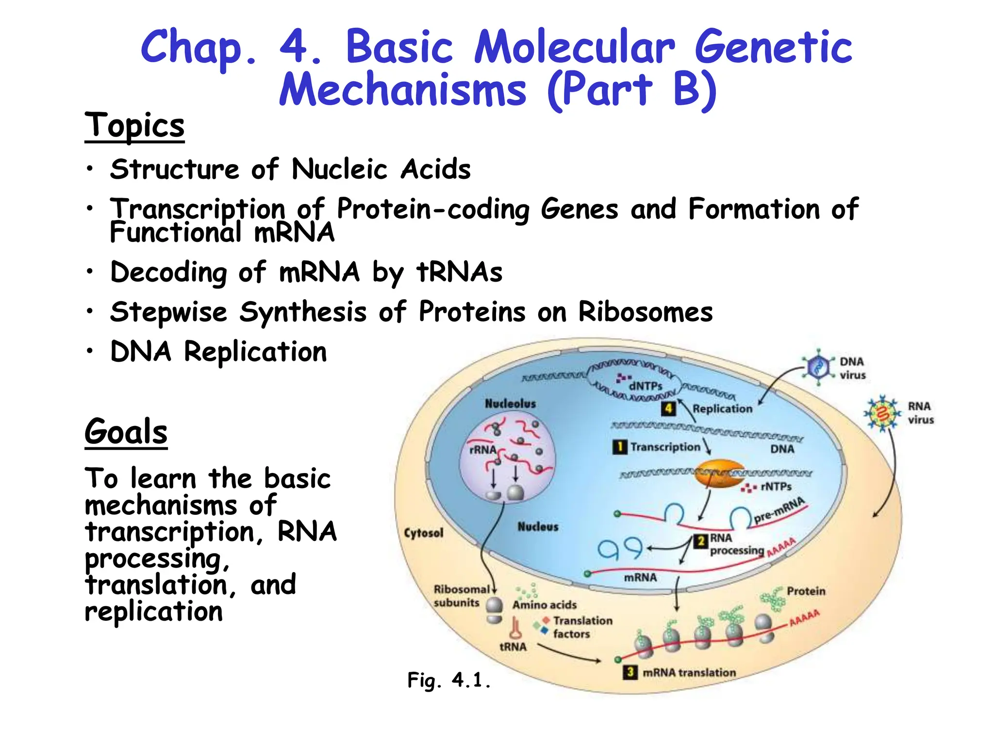
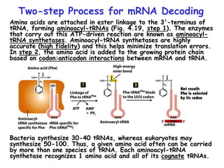
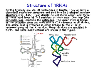
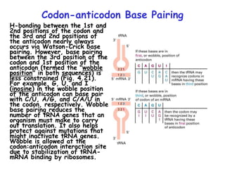
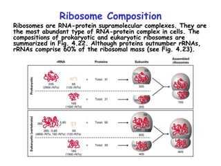
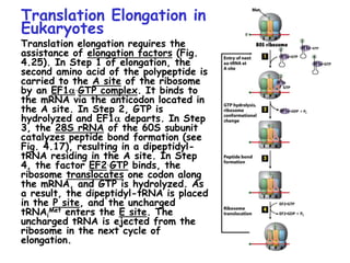
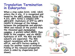
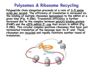
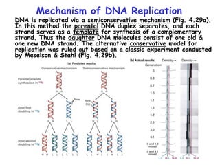
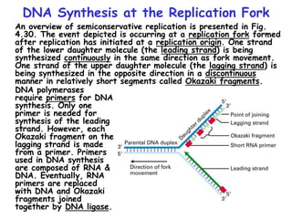
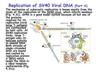
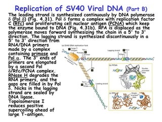
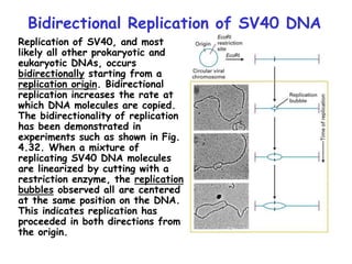
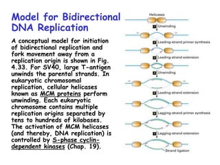
Chap. 4. Basic Molecular Genetic
Mechanisms (Part B)
Topics
• Structure of Nucleic Acids
• Transcription of Protein-coding Genes and Formation of
Functional mRNA
• Decoding of mRNA by tRNAs
• Stepwise Synthesis of Proteins on Ribosomes
• DNA Replication
Goals
To learn the basic
mechanisms of
transcription, RNA
processing,
translation, and
replication
Fig. 4.1.
The Three Roles of RNA in Translation
Protein translation by ribosomes requires three types of RNA
(Fig. 4.17). Messenger RNA (mRNA) specifies the amino acid
sequence of the protein. Each amino acid is selected based on
the order of triplet codons in mRNA. Transfer RNA (tRNA)
converts the
information in mRNA
codons into the
amino acid sequence
of the protein.
tRNAs carry amino
acids specified by
the codons and base
pair with the codons
via their anticodons.
Ribosomal RNA
(rRNA) makes up the
bulk of the mass of
the ribosome. One
rRNA species (28S
rRNA) is a ribozyme
that catalyzes the
reaction in which the
peptide bond is
formed.
The Genetic Code
The codons for the 20
standard amino acids are
specified by triplets of
bases known as the genetic
code (Table 4.1). Because
there are 43=64 possible
combinations of triplet
codons, most amino acids
are specified by more than
one codon (degeneracy).
61 codons specify amino
acids. Three do not (stop
or termination codons).
Termination codons tell
ribosomes where to end
translation of the mRNA.
Most commonly, the AUG
codon (specifying
methionine) serves as the
start codon, and tells the
ribosome where to begin
translation. Few deviations
from the standard genetic
code have been found,
providing strong evidence
that life on earth evolved
only once.
Reading of the Triplet Code
There are three potential reading frames in all mRNAs. However,
only one reading frame is used for translation, and is selected
based on the frame in which the AUG start codon appears.
Triplet codons are read in a non-overlapping, comma-less manner
(Fig. 4.18). Rarely are mRNAs read in more than one frame.
Likewise, frame-shifting is very uncommon.
Two-step Process for mRNA Decoding
Amino acids are attached in ester linkage to the 3'-terminus of
tRNA, forming aminoacyl-tRNAs (Fig. 4.19, step 1). The enzymes
that carry out this ATP-driven reaction are known as aminoacyl-
tRNA synthetases. Aminoacyl-tRNA synthetases are highly
accurate (high fidelity) and this helps minimize translation errors.
In step 2, the amino acid is added to the growing protein chain
based on codon:anticodon interactions between mRNA and tRNA.
Bacteria synthesize 30-40 tRNAs, whereas eukaryotes may
synthesize 50-100. Thus, a given amino acid often can be carried
by more than one species of tRNA. Each aminoacyl-tRNA
synthetase recognizes 1 amino acid and all of its cognate tRNAs.
Structure of tRNAs
tRNAs typically are 70-80 nucleotides in length. They all have a
cloverleaf secondary structure and fold into an L-shaped tertiary
structure (Fig. 4.20). Four double-helical stems occur, and three
of these have loops of 7-8 residues at their ends. One loop (the
anticodon loop) contains the anticodon. The upper stem is known
as the acceptor stem and ends with a CCA sequence in all tRNAs.
The amino acid is attached in ester linkage to the 2' or 3'
hydroxyl group of the A residue. Many residues are modified in
tRNA, and some modifications are shown in the figure.
aa
Codon-anticodon Base Pairing
H-bonding between the 1st and
2nd positions of the codon and
the 3rd and 2nd positions of
the anticodon nearly always
occurs via Watson-Crick base
pairing. However, base pairing
between the 3rd position of the
codon and 1st position of the
anticodon (termed the "wobble
position" in both sequences) is
less constrained (Fig. 4.21).
For example, G, U, and I
(inosine) in the wobble position
of the anticodon can base pair
with C/U, A/G, and C/A/U in
the codon, respectively. Wobble
base pairing reduces the
number of tRNA genes that an
organism must make to carry
out translation. It also helps
protect against mutations that
might inactivate tRNA genes.
Wobble is allowed at the
codon:anticodon interaction site
due to stabilization of tRNA-
mRNA binding by ribosomes.
Ribosome Composition
Ribosomes are RNA-protein supramolecular complexes. They are
the most abundant type of RNA-protein complex in cells. The
compositions of prokaryotic and eukaryotic ribosomes are
summarized in Fig. 4.22. Although proteins outnumber rRNAs,
rRNAs comprise 60% of the ribosomal mass (see Fig. 4.23).
Overview of Eukaryotic Translation Initiation
Like transcription, translation is mechanistically divided into
initiation, elongation, and termination stages. All stages require
translation factors in addition to ribosomes, mRNA, and aa-
tRNAs. Prior to initiation of translation, the 60S and 40S
subunits of the 80S eukaryotic ribosome occur in their
dissociated states. As described next, the assembly of the 80S
ribosome initiation complex at the start codon of the mRNA
proceeds via binding of the mRNA and a charged Met-tRNAi
Met
initiator tRNA to the 40S subunit, with subsequent addition of
the 60S subunit.
Translation Initiation
in Eukaryotes I
Translation initiation in
eukaryotes begins with three
components/complexes that
are shown near the top of
Fig. 4.24. These are 1) the
40S ribosomal subunit, to
which the eIF1, eIF1A, and
eIF3 initiation factors are
bound; 2) the eIF2.GTP +
Met-tRNAi
Met ternary
complex; and 3) a circular
mRNA formed by the binding
of the eIF4 cap-binding
complex at the 5’ end of the
mRNA to poly(A) binding
protein (PABP) associated
with the 3’ end of the
mRNA. These components
associate in Steps 2 and 4
of the diagram, placing
Met-tRNAi
Met in the P site
of the 40S subunit.
Translation Initiation
in Eukaryotes II
In the next stage of
initiation, the mRNA is
scanned in the 5’ to 3’
direction until the first AUG
start codon is brought into
the P site (Steps 5 & 6).
Then the hydrolysis of GTP
by eIF2 generates a stable
48S initiation complex in
which the initiator tRNA
(Met-tRNAi
Met) is H-bonded
to the AUG codon.
Translation Initiation
in Eukaryotes III
In the final stages of
initiation, all initiation
factors except eIF1A
dissociate from the 48S
initiation complex and the
80S subunit and eIF5B.GTP
complex add on (Step 7).
After eIF5B hydrolyzes
GTP, the last initiation
factors depart, and the
stable 80S initiation complex
is created (Step 8). This
complex contains the
complete E (exit), P
(peptidyl-tRNA), and A
(aminoacyl-tRNA) binding
sites, with Met-tRNAi
Met
bound to the P site.
Translation Elongation in
Eukaryotes
Translation elongation requires the
assistance of elongation factors (Fig.
4.25). In Step 1 of elongation, the
second amino acid of the polypeptide is
carried to the A site of the ribosome
by an EF1a.GTP complex. It binds to
the mRNA via the anticodon located in
the A site. In Step 2, GTP is
hydrolyzed and EF1a departs. In Step
3, the 28S rRNA of the 60S subunit
catalyzes peptide bond formation (see
Fig. 4.17), resulting in a dipeptidyl-
tRNA residing in the A site. In Step
4, the factor EF2.GTP binds, the
ribosome translocates one codon along
the mRNA, and GTP is hydrolyzed. As
a result, the dipeptidyl-tRNA is placed
in the P site, and the uncharged
tRNAi
Met enters the E site. The
uncharged tRNA is ejected from the
ribosome in the next cycle of
elongation.
Translation Termination
in Eukaryotes
When a stop codon (UAA, UAG, UGA)
enters the A site, it is recognized and
bound by the eRF1 release factor (Fig.
4.27). eRF1 forms a complex with
eRF3.GTP. Hydrolysis of GTP by eRF3
results in cleavage of the linkage
between the polypeptide and peptidyl-
tRNA and release of the protein from
the ribosomal post-termination
complex. A protein called ABCE1 then
binds to the complex, and via ABCE1
hydrolysis of ATP, the 40S and 60S
subunits are separated. The 40S
subunit recombines with the eIF1,
eIF1A, and eIF3 factors making it
ready for another round of initiation.
Folding of the released polypeptide
chain is aided by chaperones (not
shown).
Polysomes & Ribosome Recycling
Polypeptide chain elongation proceeds at a rate of 3-5 amino
acids per second. The efficiency of translation is increased via
the binding of multiple ribosomes (polysomes) to the mRNA at a
given time (Fig. 4.28b). Translation efficiency is further
increased due to the complex between poly(A)-binding protein
(PABP) and the eIF4-mRNA 5'-cap that occurs in mRNA (Fig.
4.28b). This circular complex positions ribosomes that have just
terminated translation of the message near its 5' end. These
ribosomes are recycled and rapidly reinitiate another round of
translation.
Mechanism of DNA Replication
DNA is replicated via a semiconservative mechanism (Fig. 4.29a).
In this method the parental DNA duplex separates, and each
strand serves as a template for synthesis of a complementary
strand. Thus the daughter DNA molecules consist of one old &
one new DNA strand. The alternative conservative model for
replication was ruled out based on a classic experiment conducted
by Meselson & Stahl (Fig. 4.29b).
DNA Synthesis at the Replication Fork
An overview of semiconservative replication is presented in Fig.
4.30. The event depicted is occurring at a replication fork formed
after replication has initiated at a replication origin. One strand
of the lower daughter molecule (the leading strand) is being
synthesized continuously in the same direction as fork movement.
One strand of the upper daughter molecule (the lagging strand) is
being synthesized in the opposite direction in a discontinuous
manner in relatively short segments called Okazaki fragments.
DNA polymerases
require primers for DNA
synthesis. Only one
primer is needed for
synthesis of the leading
strand. However, each
Okazaki fragment on the
lagging strand is made
from a primer. Primers
used in DNA synthesis
are composed of RNA &
DNA. Eventually, RNA
primers are replaced
with DNA and Okazaki
fragments joined
together by DNA ligase.
Replication of SV40 Viral DNA (Part A)
The mechanism of eukaryotic replication is known mostly from the
study of the replication of the SV40 virus, which infects monkeys
(Fig. 4.31). SV40 is a good model system because all but one of
the proteins
required for its
replication (viral
large T-antigen)
are synthesized
by host cells. At
SV40 replication
forks, large T-
antigen uses its
helicase activity
to unwind DNA.
Both strands of
single-stranded
DNA are bound
and coated by
replication protein
A (RPA) which
keeps the DNA in
a ideal template
conformation (Fig.
4.31c).
Replication of SV40 Viral DNA (Part B)
The leading strand is synthesized continuously by DNA polymerase
d (Pol d) (Fig. 4.31). Pol d forms a complex with replication factor
C (Rfc) and proliferating cell nuclear antigen (PCNA) which keep
the enzyme bound to DNA (Fig. 4.31b). RPA is displaced as the
polymerase moves forward synthesizing the chain in a 5' to 3'
direction. The lagging strand is synthesized discontinuously in a
5' to 3' direction from
RNA/DNA primers
made by a complex
containing primase and
Pol a. The 3' ends of
primers are elongated
by a second Pol
d/Rfc/PCNA complex.
RNase H degrades the
RNA primers, and the
gaps are filled in by Pol
d. Nicks in the lagging
strand are sealed by
DNA ligase.
Topoisomerase I
reduces positive
supercoiling ahead of
large T-antigen.
Bidirectional Replication of SV40 DNA
Replication of SV40, and most
likely all other prokaryotic and
eukaryotic DNAs, occurs
bidirectionally starting from a
replication origin. Bidirectional
replication increases the rate at
which DNA molecules are copied.
The bidirectionality of replication
has been demonstrated in
experiments such as shown in Fig.
4.32. When a mixture of
replicating SV40 DNA molecules
are linearized by cutting with a
restriction enzyme, the replication
bubbles observed all are centered
at the same position on the DNA.
This indicates replication has
proceeded in both directions from
the origin.
Model for Bidirectional
DNA Replication
A conceptual model for initiation
of bidirectional replication and
fork movement away from a
replication origin is shown in Fig.
4.33. For SV40, large T-antigen
unwinds the parental strands. In
eukaryotic chromosomal
replication, cellular helicases
known as MCM proteins perform
unwinding. Each eukaryotic
chromosome contains multiple
replication origins separated by
tens to hundreds of kilobases.
The activation of MCM helicases
(and thereby, DNA replication) is
controlled by S-phase cyclin-
dependent kinases (Chap. 19).

13-miller-chap-4b-lecture.urey miller experiment.

  • 1.
    Chap. 4. BasicMolecular Genetic Mechanisms (Part B) Topics • Structure of Nucleic Acids • Transcription of Protein-coding Genes and Formation of Functional mRNA • Decoding of mRNA by tRNAs • Stepwise Synthesis of Proteins on Ribosomes • DNA Replication Goals To learn the basic mechanisms of transcription, RNA processing, translation, and replication Fig. 4.1.
  • 2.
    The Three Rolesof RNA in Translation Protein translation by ribosomes requires three types of RNA (Fig. 4.17). Messenger RNA (mRNA) specifies the amino acid sequence of the protein. Each amino acid is selected based on the order of triplet codons in mRNA. Transfer RNA (tRNA) converts the information in mRNA codons into the amino acid sequence of the protein. tRNAs carry amino acids specified by the codons and base pair with the codons via their anticodons. Ribosomal RNA (rRNA) makes up the bulk of the mass of the ribosome. One rRNA species (28S rRNA) is a ribozyme that catalyzes the reaction in which the peptide bond is formed.
  • 3.
    The Genetic Code Thecodons for the 20 standard amino acids are specified by triplets of bases known as the genetic code (Table 4.1). Because there are 43=64 possible combinations of triplet codons, most amino acids are specified by more than one codon (degeneracy). 61 codons specify amino acids. Three do not (stop or termination codons). Termination codons tell ribosomes where to end translation of the mRNA. Most commonly, the AUG codon (specifying methionine) serves as the start codon, and tells the ribosome where to begin translation. Few deviations from the standard genetic code have been found, providing strong evidence that life on earth evolved only once.
  • 4.
    Reading of theTriplet Code There are three potential reading frames in all mRNAs. However, only one reading frame is used for translation, and is selected based on the frame in which the AUG start codon appears. Triplet codons are read in a non-overlapping, comma-less manner (Fig. 4.18). Rarely are mRNAs read in more than one frame. Likewise, frame-shifting is very uncommon.
  • 5.
    Two-step Process formRNA Decoding Amino acids are attached in ester linkage to the 3'-terminus of tRNA, forming aminoacyl-tRNAs (Fig. 4.19, step 1). The enzymes that carry out this ATP-driven reaction are known as aminoacyl- tRNA synthetases. Aminoacyl-tRNA synthetases are highly accurate (high fidelity) and this helps minimize translation errors. In step 2, the amino acid is added to the growing protein chain based on codon:anticodon interactions between mRNA and tRNA. Bacteria synthesize 30-40 tRNAs, whereas eukaryotes may synthesize 50-100. Thus, a given amino acid often can be carried by more than one species of tRNA. Each aminoacyl-tRNA synthetase recognizes 1 amino acid and all of its cognate tRNAs.
  • 6.
    Structure of tRNAs tRNAstypically are 70-80 nucleotides in length. They all have a cloverleaf secondary structure and fold into an L-shaped tertiary structure (Fig. 4.20). Four double-helical stems occur, and three of these have loops of 7-8 residues at their ends. One loop (the anticodon loop) contains the anticodon. The upper stem is known as the acceptor stem and ends with a CCA sequence in all tRNAs. The amino acid is attached in ester linkage to the 2' or 3' hydroxyl group of the A residue. Many residues are modified in tRNA, and some modifications are shown in the figure. aa
  • 7.
    Codon-anticodon Base Pairing H-bondingbetween the 1st and 2nd positions of the codon and the 3rd and 2nd positions of the anticodon nearly always occurs via Watson-Crick base pairing. However, base pairing between the 3rd position of the codon and 1st position of the anticodon (termed the "wobble position" in both sequences) is less constrained (Fig. 4.21). For example, G, U, and I (inosine) in the wobble position of the anticodon can base pair with C/U, A/G, and C/A/U in the codon, respectively. Wobble base pairing reduces the number of tRNA genes that an organism must make to carry out translation. It also helps protect against mutations that might inactivate tRNA genes. Wobble is allowed at the codon:anticodon interaction site due to stabilization of tRNA- mRNA binding by ribosomes.
  • 8.
    Ribosome Composition Ribosomes areRNA-protein supramolecular complexes. They are the most abundant type of RNA-protein complex in cells. The compositions of prokaryotic and eukaryotic ribosomes are summarized in Fig. 4.22. Although proteins outnumber rRNAs, rRNAs comprise 60% of the ribosomal mass (see Fig. 4.23).
  • 9.
    Overview of EukaryoticTranslation Initiation Like transcription, translation is mechanistically divided into initiation, elongation, and termination stages. All stages require translation factors in addition to ribosomes, mRNA, and aa- tRNAs. Prior to initiation of translation, the 60S and 40S subunits of the 80S eukaryotic ribosome occur in their dissociated states. As described next, the assembly of the 80S ribosome initiation complex at the start codon of the mRNA proceeds via binding of the mRNA and a charged Met-tRNAi Met initiator tRNA to the 40S subunit, with subsequent addition of the 60S subunit.
  • 10.
    Translation Initiation in EukaryotesI Translation initiation in eukaryotes begins with three components/complexes that are shown near the top of Fig. 4.24. These are 1) the 40S ribosomal subunit, to which the eIF1, eIF1A, and eIF3 initiation factors are bound; 2) the eIF2.GTP + Met-tRNAi Met ternary complex; and 3) a circular mRNA formed by the binding of the eIF4 cap-binding complex at the 5’ end of the mRNA to poly(A) binding protein (PABP) associated with the 3’ end of the mRNA. These components associate in Steps 2 and 4 of the diagram, placing Met-tRNAi Met in the P site of the 40S subunit.
  • 11.
    Translation Initiation in EukaryotesII In the next stage of initiation, the mRNA is scanned in the 5’ to 3’ direction until the first AUG start codon is brought into the P site (Steps 5 & 6). Then the hydrolysis of GTP by eIF2 generates a stable 48S initiation complex in which the initiator tRNA (Met-tRNAi Met) is H-bonded to the AUG codon.
  • 12.
    Translation Initiation in EukaryotesIII In the final stages of initiation, all initiation factors except eIF1A dissociate from the 48S initiation complex and the 80S subunit and eIF5B.GTP complex add on (Step 7). After eIF5B hydrolyzes GTP, the last initiation factors depart, and the stable 80S initiation complex is created (Step 8). This complex contains the complete E (exit), P (peptidyl-tRNA), and A (aminoacyl-tRNA) binding sites, with Met-tRNAi Met bound to the P site.
  • 13.
    Translation Elongation in Eukaryotes Translationelongation requires the assistance of elongation factors (Fig. 4.25). In Step 1 of elongation, the second amino acid of the polypeptide is carried to the A site of the ribosome by an EF1a.GTP complex. It binds to the mRNA via the anticodon located in the A site. In Step 2, GTP is hydrolyzed and EF1a departs. In Step 3, the 28S rRNA of the 60S subunit catalyzes peptide bond formation (see Fig. 4.17), resulting in a dipeptidyl- tRNA residing in the A site. In Step 4, the factor EF2.GTP binds, the ribosome translocates one codon along the mRNA, and GTP is hydrolyzed. As a result, the dipeptidyl-tRNA is placed in the P site, and the uncharged tRNAi Met enters the E site. The uncharged tRNA is ejected from the ribosome in the next cycle of elongation.
  • 14.
    Translation Termination in Eukaryotes Whena stop codon (UAA, UAG, UGA) enters the A site, it is recognized and bound by the eRF1 release factor (Fig. 4.27). eRF1 forms a complex with eRF3.GTP. Hydrolysis of GTP by eRF3 results in cleavage of the linkage between the polypeptide and peptidyl- tRNA and release of the protein from the ribosomal post-termination complex. A protein called ABCE1 then binds to the complex, and via ABCE1 hydrolysis of ATP, the 40S and 60S subunits are separated. The 40S subunit recombines with the eIF1, eIF1A, and eIF3 factors making it ready for another round of initiation. Folding of the released polypeptide chain is aided by chaperones (not shown).
  • 15.
    Polysomes & RibosomeRecycling Polypeptide chain elongation proceeds at a rate of 3-5 amino acids per second. The efficiency of translation is increased via the binding of multiple ribosomes (polysomes) to the mRNA at a given time (Fig. 4.28b). Translation efficiency is further increased due to the complex between poly(A)-binding protein (PABP) and the eIF4-mRNA 5'-cap that occurs in mRNA (Fig. 4.28b). This circular complex positions ribosomes that have just terminated translation of the message near its 5' end. These ribosomes are recycled and rapidly reinitiate another round of translation.
  • 16.
    Mechanism of DNAReplication DNA is replicated via a semiconservative mechanism (Fig. 4.29a). In this method the parental DNA duplex separates, and each strand serves as a template for synthesis of a complementary strand. Thus the daughter DNA molecules consist of one old & one new DNA strand. The alternative conservative model for replication was ruled out based on a classic experiment conducted by Meselson & Stahl (Fig. 4.29b).
  • 17.
    DNA Synthesis atthe Replication Fork An overview of semiconservative replication is presented in Fig. 4.30. The event depicted is occurring at a replication fork formed after replication has initiated at a replication origin. One strand of the lower daughter molecule (the leading strand) is being synthesized continuously in the same direction as fork movement. One strand of the upper daughter molecule (the lagging strand) is being synthesized in the opposite direction in a discontinuous manner in relatively short segments called Okazaki fragments. DNA polymerases require primers for DNA synthesis. Only one primer is needed for synthesis of the leading strand. However, each Okazaki fragment on the lagging strand is made from a primer. Primers used in DNA synthesis are composed of RNA & DNA. Eventually, RNA primers are replaced with DNA and Okazaki fragments joined together by DNA ligase.
  • 18.
    Replication of SV40Viral DNA (Part A) The mechanism of eukaryotic replication is known mostly from the study of the replication of the SV40 virus, which infects monkeys (Fig. 4.31). SV40 is a good model system because all but one of the proteins required for its replication (viral large T-antigen) are synthesized by host cells. At SV40 replication forks, large T- antigen uses its helicase activity to unwind DNA. Both strands of single-stranded DNA are bound and coated by replication protein A (RPA) which keeps the DNA in a ideal template conformation (Fig. 4.31c).
  • 19.
    Replication of SV40Viral DNA (Part B) The leading strand is synthesized continuously by DNA polymerase d (Pol d) (Fig. 4.31). Pol d forms a complex with replication factor C (Rfc) and proliferating cell nuclear antigen (PCNA) which keep the enzyme bound to DNA (Fig. 4.31b). RPA is displaced as the polymerase moves forward synthesizing the chain in a 5' to 3' direction. The lagging strand is synthesized discontinuously in a 5' to 3' direction from RNA/DNA primers made by a complex containing primase and Pol a. The 3' ends of primers are elongated by a second Pol d/Rfc/PCNA complex. RNase H degrades the RNA primers, and the gaps are filled in by Pol d. Nicks in the lagging strand are sealed by DNA ligase. Topoisomerase I reduces positive supercoiling ahead of large T-antigen.
  • 20.
    Bidirectional Replication ofSV40 DNA Replication of SV40, and most likely all other prokaryotic and eukaryotic DNAs, occurs bidirectionally starting from a replication origin. Bidirectional replication increases the rate at which DNA molecules are copied. The bidirectionality of replication has been demonstrated in experiments such as shown in Fig. 4.32. When a mixture of replicating SV40 DNA molecules are linearized by cutting with a restriction enzyme, the replication bubbles observed all are centered at the same position on the DNA. This indicates replication has proceeded in both directions from the origin.
  • 21.
    Model for Bidirectional DNAReplication A conceptual model for initiation of bidirectional replication and fork movement away from a replication origin is shown in Fig. 4.33. For SV40, large T-antigen unwinds the parental strands. In eukaryotic chromosomal replication, cellular helicases known as MCM proteins perform unwinding. Each eukaryotic chromosome contains multiple replication origins separated by tens to hundreds of kilobases. The activation of MCM helicases (and thereby, DNA replication) is controlled by S-phase cyclin- dependent kinases (Chap. 19).