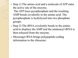
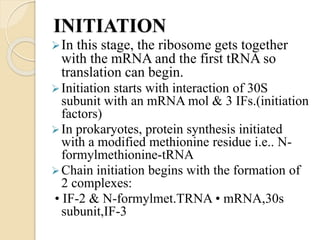
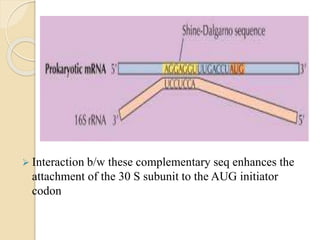
GROUP#7
TOPIC :
TRANSLATION IN
PROKARYOTES
Syeda Palwasha
BBTF16E96
INTRODUCTION
Prokaryotes:
Do not have true nucleus.
Have nucleoid region that contains cell’s DNA and RNA not
surrounded by nuclear envelope.
Prokaryotic cells are usually between 0.1 to 5
micrometers in length.
Prokaryotic cells have a higher surface-area-to-volume
ratio.
Translation in prokaryotes
 In molecularbiology and genetics, translation is the
process in which ribosomes in the cytoplasm
or ER synthesize proteins after the process
of transcription of DNA to RNA in the cell's nucleus.
The entire process is called gene expression.
 mRNA is decoded in a ribosome to produce
polypeptide.
 In prokaryotes (bacteria),translation occurs in cytosol.
 Tetracyclin,streptomycin,erthromycin,anisomycin and
puromycin are number of antibiodies that inhibits
translation.
 Rate of translation up to 17-21 amino acid residues per
second.
Tools of translation
 Mainly: Ribosomes, tRNA.
 mRNA, Initiation factors, elongation factors, termination
factors, amino acids etc.
Ribosomes
 Protein synthesising factories of the cell.
 70S & 80S
 Structure: A (aminoacyl) site, P (peptidyl) site & E (exit) site.
And also an mRNA binding site. The mRNA-binding site
binds a sequence near the 5 prime end of the mRNA, placing
the mRNA in the proper position for the translation of its first
codon . The binding sites are all located at or near the
interface between the large and small subunits
Transfer-RNA
tRNA molecules contain:
1) Three major loops.
2) Four base-paired regions,.
3) An anticodon triplet and
4) A 3 prime terminal sequence of CCA (where the
appropriate amino acid can be attached by an
ester bond).
 During maturation of the tRNA molecule a number
of nucleotides are modified in tRNA specific ways
The modified nucleotides in the tRNA structure
are :
 inosine (I),
 methylinosine (mI),
 dihydrouridine (D),
 ribothymidine (T),
 pseudouridine (¥) and
 methylguanosine (Gm).
Faysal Ghafoor
BBTF17E114
Translation
 Occurs in 3 stages:
initiation, elongation & termination
But first amino acid activated by enzymes.
PROCESS OF TRANSLATION
Amino acid activation:
 Amino acid activation (also known
as aminoacylation or tRNA charging) refers to the
attachment of an amino acid to its Transfer
RNA(tRNA).
 Twenty different aminoacyl-tRNA synthetases link
amino acids to the correct tRNAs.
 In two chemical steps, aminoacyl-tRNA catalyzes the
formation of an ester bond between the carboxyl
group of an amino acid and the 3 prime hydroxyl
(OH) group of the appropriate tRNA.
 Step 1) The amino acid and a molecule of ATP enter
the active site of the enzyme.
The ATP loses pyrophosphate and the resulting
AMP bonds covalently to the amino acid. The
pyrophosphate is hydrolyzed into two phosphate
groups.
 Step 2) The tRNA covalently bonds to the amino
acid to displace the AMP and the aminoacyl tRNA is
then released from the enzyme.
 Messenger RNA brings polypeptide-coding
information to the ribosome.
Nadia Sehrish
BBTF17E124
INITIATION
In this stage, the ribosome gets together
with the mRNA and the first tRNA so
translation can begin.
Initiation starts with interaction of 30S
subunit with an mRNA mol & 3 IFs.(initiation
factors)
In prokaryotes, protein synthesis initiated
with a modified methionine residue i.e.. N-
formylmethionine-tRNA
Chain initiation begins with the formation of
2 complexes:
• IF-2 & N-formylmet.TRNA • mRNA,30s
subunit,IF-3
Shine-Dalgarno sequence
 Bacterial genes are often transcribed in groups
(called operons), so one bacterial mRNA can
contain the coding sequences for several genes. A
Shine-Dalgarno sequence marks the start of each
coding sequence, letting the ribosome find the
right start codon for each gene.
 The mRNA-binding site is composed, at least in
part, of a portion of the 16S rRNA of the small
ribosomal subunit.
The 3 prime end of the 16S rRNA bears a
pyrimidine-rich stretch that base pairs with the
Shine-Dalgarno sequence of the mRNA.
 Interaction b/w these complementary seq enhances the
attachment of the 30 S subunit to the AUG initiator
codon
 Both the complexes combine with each other with IF-1
and 1 mol of GTP
 50 S subunit gets added to the complex structure. IF-3 is
released. The addition of 50S subunit utilizes GTP which
in turn triggers the release of IF-1 and IF-2
 Addition of 50S to the complex,positions N-formyl met
tRNA in the P site directly with the anticodon of the
tRNA aligned with AUG codon of mRNA
 With the AUG initiatior codon positioned in the P site, 2d
codon in the mRNA positions in such a way that it
corresponds to the incoming aminoacyl-tRNA in the A -
site
Ayesha
BBTF17E103
CHAIN ELONGATION
 In this stage, amino acids are brought to the
ribosome by tRNAs and linked together to form a
chain.
 Elongation begins with the binding of the second
aminoacyl tRNA at the ribosomal aminoacyl (A)
site.
 The tRNA is escorted to the A site by the
elongation factor EFTu, which also carries two
bound GTPs. As the tRNA binds, the GTPs are
hydrolyzed and EF-Tu is released.
 A peptide bond formed b/w the N-f-met-Trna at
the P site& 2nd amino acyl tRNA at the A site;
catalyzed by the peptidyl transferase
 Transfer of N-f-met. to aminoacyl-tRNA at A site
forming a peptidyl tRNA at that position and leaving
an uncharged tRNA at the P site
 Peptidyl trna translocated to the P site & uncharged
Trna is translocated to E domain.
 A site unoccupied ; new aminoacyl-TRNA bind to A
site &process continues
Tehreem Masood
BBTF17E76
CHAIN TERMINATION
 In the last stage, the finished polypeptide is released
to go and do its job in the cell.
 When a stop codon (UAG, UAA, or UGA) arrives at
the A site, it is recognized and bound by a protein
release factor. (RFs).2 classes of RFs: ClassI & Class
II. In E.coli 2 Class I RFs are seen- RF-1 ,RF-2
 RFs bind to the termination codon at A site &
stimulate hydrolysis of bond b/w tRNA&polypeptide
chain at P site , resulting in release of complete
polypeptide from ribosome.
 Termination is completed by release of mRNA mol
from ribosome & dissociation of ribosomes into its
subunits.
Recycling
 The post-termination complex formed by the end
of the termination step consists of mRNA with the
termination codon at the A-site, an uncharged
tRNA in the P site, and the intact 70S ribosome.
 Ribosome recycling step is responsible for the
disassembly of the post-termination ribosomal
complex. Once the nascent protein is released in
termination, Ribosome Recycling Factor and
Elongation Factor G (EF-G) function to release
mRNA and tRNAs from ribosomes and dissociate
the 70S ribosome into the 30S and 50S subunits.
 IF3 then replaces the deacylated tRNA releasing
the mRNA. All translational components are now
free for additional rounds of translation.
Polysomes
 The complex of one mRNA and a number of
ribosomes is called a polysome or polyribosome.
 Translation is carried out by more than one ribosome
simultaneously. Because of the relatively large size
of ribosomes, they can only attach to sites on mRNA
35 nucleotides apart.
M.Bilal
BBTF17E122
Regulation of Translation
 When bacterial cells run out of nutrients, they
enter stationary phase and down regulate/block
protein synthesis.
 In E.coli,70S ribosomes form 90S dimers upon
binding with a small 6.5 kDa protein, ribosome
modulation factor RMF.
 These intermediate ribosome dimers can
subsequently bind a hibernation promotion
factor molecule to form a mature 100S ribosomal
particle, in which the dimerization interface is made
by the two 30S subunits of the two participating
ribosomes. The ribosome dimers represent a
hibernation state and are translationally inactive.
 A third protein that can bind to ribosomes when E.
coli cells enter the stationary phase is YfiA .
 RMF blocks ribosome binding to mRNA by
preventing interaction of the messenger with 16S
rRNA.
 The nearly identical binding sites of HPF and
YfiA overlap with those of the mRNA, transfer
RNA, and initiation factors, which prevents
translation initiation.
 When bound to the ribosomes the C-terminal tail
of E.coli YfiA interferes with the binding of
RMF, thus preventing dimerization and resulting
in the formation of translationally inactive
monomeric 70S ribosomes.
 In addition to ribosome dimerization, the joining
of the two ribosomal subunits can be blocked
by RsfS (ribosome silencing factor). RsfS binds
to L14, a protein of the large ribosomal subunit,
and thereby blocks joining of the small subunit to
form a functional 70S ribosome, slowing down or
blocking translation entirely.
 Another ribosome-dissociation factor
in Escherichia coli is HflX, previously a GTPase
of unknown function.
 HflX is a heat shock–induced ribosome-splitting
factor capable of dissociating vacant as well as
mRNA-associated ribosomes.

DOC-20190508-WA0001.pptx

  • 1.
  • 2.
  • 3.
    INTRODUCTION Prokaryotes: Do not havetrue nucleus. Have nucleoid region that contains cell’s DNA and RNA not surrounded by nuclear envelope. Prokaryotic cells are usually between 0.1 to 5 micrometers in length. Prokaryotic cells have a higher surface-area-to-volume ratio.
  • 4.
    Translation in prokaryotes In molecularbiology and genetics, translation is the process in which ribosomes in the cytoplasm or ER synthesize proteins after the process of transcription of DNA to RNA in the cell's nucleus. The entire process is called gene expression.  mRNA is decoded in a ribosome to produce polypeptide.  In prokaryotes (bacteria),translation occurs in cytosol.  Tetracyclin,streptomycin,erthromycin,anisomycin and puromycin are number of antibiodies that inhibits translation.  Rate of translation up to 17-21 amino acid residues per second.
  • 5.
    Tools of translation Mainly: Ribosomes, tRNA.  mRNA, Initiation factors, elongation factors, termination factors, amino acids etc. Ribosomes  Protein synthesising factories of the cell.  70S & 80S  Structure: A (aminoacyl) site, P (peptidyl) site & E (exit) site. And also an mRNA binding site. The mRNA-binding site binds a sequence near the 5 prime end of the mRNA, placing the mRNA in the proper position for the translation of its first codon . The binding sites are all located at or near the interface between the large and small subunits
  • 7.
    Transfer-RNA tRNA molecules contain: 1)Three major loops. 2) Four base-paired regions,. 3) An anticodon triplet and 4) A 3 prime terminal sequence of CCA (where the appropriate amino acid can be attached by an ester bond).
  • 9.
     During maturationof the tRNA molecule a number of nucleotides are modified in tRNA specific ways The modified nucleotides in the tRNA structure are :  inosine (I),  methylinosine (mI),  dihydrouridine (D),  ribothymidine (T),  pseudouridine (¥) and  methylguanosine (Gm).
  • 10.
  • 11.
    Translation  Occurs in3 stages: initiation, elongation & termination But first amino acid activated by enzymes.
  • 12.
    PROCESS OF TRANSLATION Aminoacid activation:  Amino acid activation (also known as aminoacylation or tRNA charging) refers to the attachment of an amino acid to its Transfer RNA(tRNA).  Twenty different aminoacyl-tRNA synthetases link amino acids to the correct tRNAs.  In two chemical steps, aminoacyl-tRNA catalyzes the formation of an ester bond between the carboxyl group of an amino acid and the 3 prime hydroxyl (OH) group of the appropriate tRNA.
  • 13.
     Step 1)The amino acid and a molecule of ATP enter the active site of the enzyme. The ATP loses pyrophosphate and the resulting AMP bonds covalently to the amino acid. The pyrophosphate is hydrolyzed into two phosphate groups.  Step 2) The tRNA covalently bonds to the amino acid to displace the AMP and the aminoacyl tRNA is then released from the enzyme.  Messenger RNA brings polypeptide-coding information to the ribosome.
  • 15.
  • 16.
    INITIATION In this stage,the ribosome gets together with the mRNA and the first tRNA so translation can begin. Initiation starts with interaction of 30S subunit with an mRNA mol & 3 IFs.(initiation factors) In prokaryotes, protein synthesis initiated with a modified methionine residue i.e.. N- formylmethionine-tRNA Chain initiation begins with the formation of 2 complexes: • IF-2 & N-formylmet.TRNA • mRNA,30s subunit,IF-3
  • 17.
    Shine-Dalgarno sequence  Bacterialgenes are often transcribed in groups (called operons), so one bacterial mRNA can contain the coding sequences for several genes. A Shine-Dalgarno sequence marks the start of each coding sequence, letting the ribosome find the right start codon for each gene.  The mRNA-binding site is composed, at least in part, of a portion of the 16S rRNA of the small ribosomal subunit. The 3 prime end of the 16S rRNA bears a pyrimidine-rich stretch that base pairs with the Shine-Dalgarno sequence of the mRNA.
  • 18.
     Interaction b/wthese complementary seq enhances the attachment of the 30 S subunit to the AUG initiator codon
  • 19.
     Both thecomplexes combine with each other with IF-1 and 1 mol of GTP  50 S subunit gets added to the complex structure. IF-3 is released. The addition of 50S subunit utilizes GTP which in turn triggers the release of IF-1 and IF-2  Addition of 50S to the complex,positions N-formyl met tRNA in the P site directly with the anticodon of the tRNA aligned with AUG codon of mRNA  With the AUG initiatior codon positioned in the P site, 2d codon in the mRNA positions in such a way that it corresponds to the incoming aminoacyl-tRNA in the A - site
  • 22.
  • 23.
    CHAIN ELONGATION  Inthis stage, amino acids are brought to the ribosome by tRNAs and linked together to form a chain.  Elongation begins with the binding of the second aminoacyl tRNA at the ribosomal aminoacyl (A) site.  The tRNA is escorted to the A site by the elongation factor EFTu, which also carries two bound GTPs. As the tRNA binds, the GTPs are hydrolyzed and EF-Tu is released.  A peptide bond formed b/w the N-f-met-Trna at the P site& 2nd amino acyl tRNA at the A site; catalyzed by the peptidyl transferase
  • 24.
     Transfer ofN-f-met. to aminoacyl-tRNA at A site forming a peptidyl tRNA at that position and leaving an uncharged tRNA at the P site  Peptidyl trna translocated to the P site & uncharged Trna is translocated to E domain.  A site unoccupied ; new aminoacyl-TRNA bind to A site &process continues
  • 26.
  • 27.
    CHAIN TERMINATION  Inthe last stage, the finished polypeptide is released to go and do its job in the cell.  When a stop codon (UAG, UAA, or UGA) arrives at the A site, it is recognized and bound by a protein release factor. (RFs).2 classes of RFs: ClassI & Class II. In E.coli 2 Class I RFs are seen- RF-1 ,RF-2  RFs bind to the termination codon at A site & stimulate hydrolysis of bond b/w tRNA&polypeptide chain at P site , resulting in release of complete polypeptide from ribosome.  Termination is completed by release of mRNA mol from ribosome & dissociation of ribosomes into its subunits.
  • 29.
    Recycling  The post-terminationcomplex formed by the end of the termination step consists of mRNA with the termination codon at the A-site, an uncharged tRNA in the P site, and the intact 70S ribosome.  Ribosome recycling step is responsible for the disassembly of the post-termination ribosomal complex. Once the nascent protein is released in termination, Ribosome Recycling Factor and Elongation Factor G (EF-G) function to release mRNA and tRNAs from ribosomes and dissociate the 70S ribosome into the 30S and 50S subunits.  IF3 then replaces the deacylated tRNA releasing the mRNA. All translational components are now free for additional rounds of translation.
  • 30.
    Polysomes  The complexof one mRNA and a number of ribosomes is called a polysome or polyribosome.  Translation is carried out by more than one ribosome simultaneously. Because of the relatively large size of ribosomes, they can only attach to sites on mRNA 35 nucleotides apart.
  • 31.
  • 32.
    Regulation of Translation When bacterial cells run out of nutrients, they enter stationary phase and down regulate/block protein synthesis.  In E.coli,70S ribosomes form 90S dimers upon binding with a small 6.5 kDa protein, ribosome modulation factor RMF.  These intermediate ribosome dimers can subsequently bind a hibernation promotion factor molecule to form a mature 100S ribosomal particle, in which the dimerization interface is made by the two 30S subunits of the two participating ribosomes. The ribosome dimers represent a hibernation state and are translationally inactive.
  • 33.
     A thirdprotein that can bind to ribosomes when E. coli cells enter the stationary phase is YfiA .  RMF blocks ribosome binding to mRNA by preventing interaction of the messenger with 16S rRNA.  The nearly identical binding sites of HPF and YfiA overlap with those of the mRNA, transfer RNA, and initiation factors, which prevents translation initiation.  When bound to the ribosomes the C-terminal tail of E.coli YfiA interferes with the binding of RMF, thus preventing dimerization and resulting in the formation of translationally inactive monomeric 70S ribosomes.
  • 35.
     In additionto ribosome dimerization, the joining of the two ribosomal subunits can be blocked by RsfS (ribosome silencing factor). RsfS binds to L14, a protein of the large ribosomal subunit, and thereby blocks joining of the small subunit to form a functional 70S ribosome, slowing down or blocking translation entirely.  Another ribosome-dissociation factor in Escherichia coli is HflX, previously a GTPase of unknown function.  HflX is a heat shock–induced ribosome-splitting factor capable of dissociating vacant as well as mRNA-associated ribosomes.