3. TABLE OF CONTENT
What Is Chest Xray
Common Uses Of The Procedure
Preparation
Positioning Of Patient For Chest Xray
For Posterior Anterior View
For Lateral View
Radiation Protection Principle
Risks
Limitations Of Chest Xray
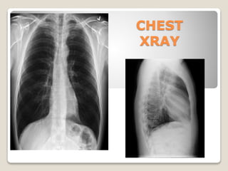
4. WHAT IS CHEST XRAY
The chest x-ray is the most commonly
performed diagnostic x-ray examination.
The chest x-ray produces images of the
heart, lungs, airways, blood vessels and the
bones of the spine and chest.
5. Common Uses Of The Procedure
Chest Xray Is Used To Check The Following;
The condition of your lungs: Chest X-rays can detect cancer,
infection or air collecting in the space around a lung, which can
cause the lung to collapse. They can also show chronic lung
conditions, such as emphysema or cystic fibrosis, as well as
complications related to these conditions.
Heart-related lung problems: Chest X-rays can show changes or
problems in your lungs that stem from heart problems
The size and outline of your heart: Changes in the size and shape
of your heart may indicate heart failure, fluid around the heart or
heart valve problems.
Blood vessels: Because the outlines of the large vessels near your
heart — the aorta and pulmonary arteries and veins — are visible
on X-rays, they may reveal aortic aneurysms, other blood vessel
problems or congenital heart disease.
6. Common Uses Of The Procedure
Calcium deposits: Chest X-rays can detect the presence of
calcium in your heart or blood vessels. Its presence may indicate
fats and other substances in your vessels, damage to your heart
valves, coronary arteries, heart muscle or the protective sac that
surrounds the heart.
Fractures: Rib or spine fractures or other problems with bone
may be seen on a chest X-ray.
Postoperative changes: Chest X-rays are useful for monitoring
your recovery after you've had surgery in your chest, such as on
your heart, lungs or esophagus.
A pacemaker, defibrillator or catheter: Pacemakers and
defibrillators have wires attached to your heart to help control
your heart rate and rhythm.
Diagnose many conditions and diseases such as asthma, pleurisy,
tumors e.t.c
7. PREPARATION FOR CHEST XRAY
A chest x-ray requires no special
preparation .
Patient may need to remove some
clothing and/or change into a gown for
the exam. Remove jewelry, removable
dental appliances, eyeglasses, and any
metal objects or clothing that might
interfere with the x-ray images.
8. POSITIONING OF PATIENT
FOR CHEST XRAY
Patient positioning
Normal PA radiograph of the
chest
FOR POSTERIOR-ANTERIOR VIEW
The patient is positioned facing the
receptor with the chin extended
and centered to the middle of the
top of the receptor.
The feet are paced slightly apart so
that the patient is able to remain
steady.
The median sagittal plane is
adjusted at right-angles to the
middle of the receptor; the
shoulders are rotated forward and
pressed downward in contact with
the receptor or vertical stand
9. POSITIONING OF PATIENT
FOR CHEST XRAY
Patient positioning
Lateral radiograph of the chest
FOR LATERAL VIEW
The erect patient is turned to bring
the side under investigation in
contact with the image receptor.
The median sagittal plane is
adjusted parallel to the image
receptor.
The arms are folded over the head
or raised above the head to rest on
a horizontal bar support.
The mid-axillary line is coincident
with the middle of the Bucky, and
the receptor is adjusted to include
the apices and the lower lobes to
the level of the 1st lumbar
vertebra.
11. Risks
There is always a slight chance of cancer from
excessive exposure to radiation. However, given
the small amount of radiation used in medical
imaging, the benefit of an accurate diagnosis far
outweighs the associated risk. However, doctors
don’t recommend X-rays if you are pregnant.
This is because radiation can harm your unborn
baby. If you believe you are pregnant, make sure
you tell your doctor.
12. LIMITATIONS OF CHEST
RADIOGRAPHY?
The chest x-ray is a very useful examination,
but it has limitations. Because some
conditions of the chest cannot be detected on
a conventional chest x-ray image, this
examination cannot necessarily rule out all
problems in the chest. For example, small
cancers may not show up on a chest x-ray. A
blood clot in the lungs, a condition called a
pulmonary embolism, cannot be seen on
chest x-rays.