SlideShare a Scribd company logo
1 of 57
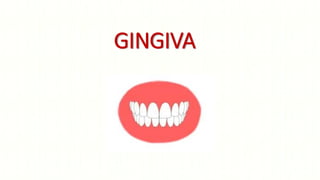
GINGIVA
Contents
• Introduction
• Definitions
• Development
• Macroscopic anatomy
• Marginal gingiva
• Attached gingiva
• Interdental gingiva
• Microscopic anatomy
• Gingival epithelium
• Gingival connective tissue
• Blood supply
• Nerve supply
• Lymphatic drainage
• Age changes
• Conclusion
• References
Introduction
Parts of oral mucosa
Definitions of Gingiva
• Carranza :
Is the part of oral mucosa that covers the alveolar process of jaw and
surrounds the neck of teeth in a collar like fashion.
• Shroeder :
It is the combination of epithelium and connective tissue and is defined
as that portion of oral mucous membrane, which in complete post-
eruptive dentition of a healthy young individual ,surrounds and is
attached to the teeth and the alveolar process.
• Grant :
Is the part of oral mucous membrane attached to the teeth and the
alveolar processes.
• Lindhe :
Is that part of masticatory mucosa covering the alveolar processes and
the cervical portions of teeth.
• GPT 2001:
The fibrous investing tissue covered by keratinized epithelium that
immediately surrounds a tooth and is contiguous with periodontal
ligament and with the mucosal tissues of the mouth .
Macroscopic features
Alveolar mucosa
Mucogingival junction
Marginal gingiva
Interdental gingiva
Free gingival groove
Attached gingiva
Marginal gingiva
Marginal or unattached gingiva is the terminal edge or
border of the gingiva surrounding the teeth in collar like
fashion.
(Carranza )
Free gingiva :That part of the gingiva that surrounds the
tooth and is not directly attached to the tooth surface.
(GPT2001)
Marginal gingiva :The most coronal portion of the
gingiva. Often used to refer to the free gingiva that
forms the wall of the gingival crevice in health .
(GPT 2001)
Free gingival groove
• In about 50% of cases, it is demarcated from
the adjacent attached gingiva by a shallow
linear depression, the Free gingival groove.
(Ainamo J,Loe H 1966)
• GPT 2001: A shallow, V-shaped groove or
indentation that is closely associated with the
apical extent of free gingiva and runs parallel
to the margin of the gingiva. The frequency of
its occurrence varies widely.
• Orban 1948 : A shallow V –shaped groove
which runs parallel to the margin of the
gingiva at a distance of 0.5-1.5mm
• Significance
Attached gingiva • GPT 2001 :The portion of the gingiva that is firm, dense,
stippled, and tightly bound to the underlying periosteum,
tooth, and bone.
• It is the distance between the mucogingival junction and
the projection on the external surface of the bottom of
the gingival sulcus or the periodontal pocket.
• Measurements
• A study done by Rajiv Subbaiah , Balaji Manohar on Indian
population – the average width of attached gingiva was
• Attached gingiva ranges 1mm- 9mm and appears to exhibit a
consistent pattern of variation throughout the dentition
(Bowers 1963)
• Attached gingiva on deciduous teeth < adult dentition, but the
pattern of variation is similar in two.
• The width of attached gingiva increases with age and in supra-
erupted teeth.
Inadequate zone of gingiva
• Dissipate the pull because of muscles of adjacent mucosa (Friedman 1957)
• Increase in subgingival plaque formation (Friedman 1962)
• Attachment loss and soft tissue recession due to apical spread of plaque
associated gingival lesions (Stern 1976)
• Along with decreased vestibular depth causes accumulation of food particles
during mastication and impedes oral hygiene measures (Gottsegen 1954)
Clinical significance
• Increases resistance to external injury and contribute in stabilization of gingival
margin.
• Against frictional forces
• Dissipating physiological forces exerted by the muscular fibres of the alveolar
mucosa on the gingival tissues.
How much zone of attached gingiva is necessary to
maintain the zone of attached gingiva?
Attached Gingiva around Implants
• Absence of keratinized mucosa increases the susceptibility of peri
implant lesions and plaque induced destructions
• Keratinized gingiva around implant has more hemidesmosomes.
Clinical significance
• Prevent spread of inflammation.
• Prevents recession of marginal tissue.
• Provides tight collar around implants.
• Enable patients to maintain good oral hygiene
Feature which are specific to attached gingiva
are:
• Deep rete pegs, Thick lamina propria.
• Abundant collagen with elastic fibers, Indistinct sub
mucosa.
Stippling
(Carranza):
It is a form of adaptive specialization or reinforcement for
function and feature of healthy gingiva .
(GPT 2001)
The pitted, orange-peel appearance frequently seen in
attached gingiva .
• Best viewed by drying Gingiva.
• Healthy Gingiva, More numerous in lower jaw (Cleaton
Jones 1978)
• Absence of stippling in Infancy, gingival diseases,
inflammation etc.
• The papillary layer of the connective tissue projects into
the elevation .
Interdental Papilla
• COHEN in 1959 first described interdental gingiva
• GPT 2001
Gingival Papilla : That portion of the gingiva that
occupies the interproximal spaces. The interdental
extension of the gingiva.
Anterior region- Pyramidal
Molar region-Papillae more flattened in buccolingual
direction (tent shaped)
• Shape of interdental gingiva depends on
• Contact relation between the two adjoining teeth
• Width of proximal tooth surfaces
• Course of CEJ
• Presence or absence of some degree of recession
• Interdental papilla has a shape in conformity with the outline of the
interdental contact surfaces which leads to formation of a concavity
called as ‘A col’
• GPT 2001
A valley-like depression of the interdental gingiva that connects facial
and lingual papillae and conforms to the shape of the interproximal
contact area.
• Seen in premolar and molar regions
• It is covered by a thin non-keratinised epithelium
Gingival sulcus
• (Carranza)
• Shallow crevice or space around the tooth bounded by
the surface of tooth on one side and the epithelium
lining the free margin of gingiva on other side
• (GPT 2001)
• The shallow fissure between the marginal gingiva and
the enamel or cementum.
• It is ‘V’ shaped.
• Clinically determined by introduction of the metallic
instrument, the periodontal probe and estimation of
distance it penetrates
• In periodontal health, the gingival crevice is known as
a sulcus, whereas in disease, it is called a pocket.
Gingival Sulcus
GPT 2001: Tissue fluid that seeps through the crevicular and junctional
epithelium. It is increased in the presence of inflammation.
Seeps through the thin sulcular epithelium
Cleanse material from the sulcus
Contains plasma proteins that improves adhesion of the epithelium to
the tooth
Antimicrobial properties
Antibody activity
Transduate /Exudate?
• Alfano (1974) and Pashley Hypothesis (1976)
• The initial fluid produced could simply represent interstitial fluid which
appears in the crevice as a result of an osmotic gradient.
• This initial, pre-inflammatory fluid was considered to be a transudate, and, on
stimulation, this changed to become an inflammatory exudate
• Brill in 1959 demonstrated that increased vascular permeability plays
an important role in the production of gingival fluid.
• GCF Flow
Microscopic anatomy
Overlying stratified squamous epithelium
Underlying core of connective tissue
Cells of gingival epithelium
Keratinocytes
• Have large round or oval nucleus with one or
more nuclei
• Cytoplasm is densely packed with organelles
• Golgi complex is prominent, lamellae of rough
endoplasmic reticulum and mitochondria present
• Most of ribosomes are present as free bodies
Clear cells /
Non Keratinocyte
• These cell types are often stellate and have
cytoplasmic extensions of various size and
appearance.
Melanocytes and spinous layer.
• They produce the pigment melanin.
• This cell contains melanin granules
• Tonofilaments or hemidesmosomes absent.
Langerhans cell
• Derived from the progenitor
cells present in the bone marrow.
• Present in suprabasal layers
• They have potential to present antigen to T-
lymphocytes and activate them
• They contain g-specific granules (Birbeck's granules)
and have marked adenosine triphosphatase activity
• They found in oral epithelium, smaller amount in
sulcular epithelium but not in junctional epithelium
Merkel cells
• Merkel cells are located in deeper layers of the epithelium.
• They harbor nerve endings and are connected to adjacent cells by
desmosomes.
• They have been identified as tactile perceptors.
Keratinized stratified
squamous epithelium.
4 classical epithelial strata
• Stratum Basale
• Stratum spinosum
• Stratum granulosum
• Stratum corneum
Stratum Basale
• Cells in the basal layer : Cylindrical or
Cuboidal
• In contact with the basement membrane
• Stratum germinativum : Progenitor cell
compartment of the epithelium
• The basal cells are found immediately adjacent to the connective
tissue and are separated from this tissue by the basement membrane
• This membrane appears as a structureless zone approximately 1–2
μm wide
• This reacts positively to a PAS stain (periodic acid-Schiff stain).
• Contains carbohydrate-glycoproteins
• Epithelium - Connective tissue junction :
• The boundary between the oral epithelium & underlying
connective tissue has a wavy course.
• Connective tissue portions project into the epithelium, called as
connective tissue papillae & are separated from each other by
epithelial ridges-so called rete pegs.
Stratum spinosum
• Consists of 10–20 layers of relatively large,
polyhedral cells.
• Presence of cytoplasmic processes resembling
spines.
• The cytoplasmic processes occur at regular
intervals and give the cells a prickly
appearance.
• Cohesion between the cells - “desmosomes”
• Acid phosphatase containing dense granules –
keratinosome or Odland bodies
Stratum granulosum
• Keratinocytes migrating from the
underlying stratum spinosum become
known as granular cells in this layer
• Electron dense keratohyalin bodies –
synthesis of keratin
• They are filled with histidine- and
cysteine-rich proteins that appear to
bind the keratin filaments together.
• Therefore, the main function of
keratohyalin granules is to bind
intermediate keratin filaments together
Stratum corneum
• There is an abrupt transition of the cells
from the stratum granulosum to the
stratum corneum because of a very
sudden keratinization.
• And it produces horny stratum corneum
• The cytoplasm of the cells in the stratum
corneum is filled with keratin.
Orthokeratinized
•No nuclei in the
stratum corneum
•Well-defined
stratum granulosum
Nonkeratinized
•Has neither
granulosum nor
corneum strata
•Superficial cells :
Viable nuclei
Parakeratinized
•Stratum corneum retains
pyknotic nuclei
•Keratohyaline granules :
Dispersed
•No stratum granulosum
Gingival epithelium
The epithelium covering the free gingiva
may be differentiated as follows:
• Oral epithelium (OE)
• Oral sulcular epithelium (OSE)
• Junctional epithelium (JE)
Oral Epithelium
Covers crest & outer surface of the
marginal gingiva & the surface of the
attached gingiva.
Thickness-0.2-0.3 mm
Orthokeratinized / Parakeratinized
Sulcular epithelium
• 1. Lines the gingival sulcus
• 2. Thin & Nonkeratinized
• 3. Stratified squamous epithelium
• 4. No rete pegs
• 5. Coronal limit of JE to the crest of to gingival
margin
• 6. Presence of cells with hydropic degeneration
• 7. Semipermeable membrane
Junctional epithelium
• GPT 2001 : A single or multiple layer of
nonkeratinizing cells adhering to the tooth
surface at the base of the gingival crevice.
Formerly called epithelial attachment
• Junctional epithelium contains mainly 3 zones:
• Coronal : more permeable
• Middle : adhesion
• Apical :germination
Features
• Collar like band of stratified squamous , non-keratinized epithelium.
• Thickness : 3-4 layers thick (early life) & 10-20 layers (as age increases)
• Thickness: Coronally 10-29 cells & apically 1-2 cells
• Length : 0.25 to 1.35mm.
• Cells are arranged into basal and supra basal layers
• Absence of granular or cornified layer
• Intercellular space is wider
• Highly permeable
• Attaches to tooth surface by means of an internal basal lamina and to
the connective tissue by an external basal lamina.
• The internal basal lamina consists of a lamina densa and lamina
lucida to which hemidesmosomes are attached
• Hemidesmosomes may act as specific sites of signal transduction & may
participate in gene expression, cell proliferation & cell differentiation
(Jones JC,Hopkinson SB et al)
The attachment of the junctional epithelium to the tooth is reinforced by the
gingival fibers, which brace the marginal gingiva against the tooth surface.
Therefore the junctional epithelium and gingival fibers are considered a
functional unit called as dentogingival unit.
(Listgarten et al, 1970)
Functions
• Provides attachment to the tooth.
• Act as a barrier against plaque bacteria & invading microbes.
• Rapid cell division & funneling of junctional epithelial cells towards the
sulcus hinders bacterial colonization & repair of damaged tissues
rapidly.
• Active antimicrobial substances are produced by junctional epithelial
cells which includes defensins, lysosomal enzymes, calprotein &
cathelicidin.
• Allows the access of GCF , inflammatory cells & components of the
immunological host defense to the gingival margin.
- two way movement:
1. from connective tissue to crevice
2. from crevice to connective tissue
• Epithelial cells activated by microbial substances secrete chemokines
examples: IL1, IL6, IL8 & TNF alpha that attract & activate professional
defense cells such as lymphocytes & PMNs.
Gingival connective tissue
• Lamina Propria
• Papillary layer subjacent to the
epithelium.
• Consists of papillary projections between
the epithelial rete pegs.
• Reticular layer contiguous with the
periosteum of the alveolar bone
Fibers in gingiva
• The connective tissue of gingiva is referred to as lamina propria
• Approximately 60-65% of the connective tissue compartment of
healthy gingiva is occupied by collagen, with the individual fibrils
highly organized into discrete and easily discernible fiber bundles
(Arnim and Hargerman 1953)
• Collagen fiber bundles have been referred to as the Gingival
Ligament, which is contiguous with the periodontal ligament.
•Functions of gingival fibers
• To brace the marginal gingiva firmly against the tooth
• To provide the rigidity necessary to withstand the forces of
mastication without being deflected away from the tooth surface.
• To unite the free marginal gingiva with the cementum of the root
and the adjacent attached gingiva.
Cells of connective tissue
• The periodontium is a dynamic structure and the cells normally
present in the soft connective tissues of the periodontium reflect
this dynamism.
• Individual cells comprise approximately 8% by volume of the
connective tissue compartment
(Schroeder H, Munzel-Pedrazzoli S, Page R 1973)
Fibroblast
• The preponderant cellular element in the
gingival connective tissue .
• Mesenchymal origin
• Role in the development, maintenance, and
repair of gingival connective tissue.
• Although the biologic and clinical significance of
fibroblast heterogeneity is not year clear, it
seems that this is necessary for the normal
functioning of tissues in health, disease, and
repair .
PLASMA CELL
PMN MAST
CELL
LYMPHOCYTE MACROPHAGE
Correlation of clinical and microscopic features
Color
Contour
Consistency
Surface texture
Shape
Position
Active eruption –
Movement of teeth
in direction of
occlusal plane.
Passive eruption-
Exposure of tooth
by apical migration
of gingiva.
Rate of Active
eruption is in pace
with tooth wear in
order to preserve
vertical dimension.
Clinical Considerations
• Biological width- is defined as the dimension
of soft tissue which is attached to the portion
of the tooth coronal to the crest of alveolar
bone.
• Gingival Biotype
• Thick / Thin
• Thick biotype- reacts to surgical and
restorative insults with pocket formation.
• Thin biotype- Bony dehiscence and
fenestration defects, reacts to surgical or
restorative interventions with soft tissue
recession
Blood supply
Lymphatic drainage
Lymphatics beneath the junctional epithelium extend into the
periodontal ligament and accompany the blood vessels.
Gingival innervation
• Nerves in the periodontal ligament
• Labial, buccal and palatal nerves
• Nerve structures are present in the
connective tissue.
• Meissner type tactile corpuscles – Touch
receptors
• Krause type of end bulbs-Temperature
receptors seen as coiled terminals.
• Fine fibers in the papilla pain receptors.
• All are found in the free and attached
gingiva.
Age changes in connective tissue
• Decreased keratinization
• Decreased connective tissue cellularity
• Decreased oxygen consumption
• Atrophy of connective tissue with loss of elasticity
• Greater amount of intercellular substance
• Reduced or no changes in stippling
• Increase in width of attached gingiva with constant location of the
mucogingival junction throughout the life
conclusion
• Gingiva is the part of oral mucosa that covers the alveolar process of
jaw and surrounds the neck of teeth.
• Anatomically divided into marginal, attached and interdental gingiva
• Microscopically consists of outer epithelium ,sulcular epithelium and
junctional epithelium
• Connective tissue consists of cellular and fibrous components
embedded in matrix.
References
• Carranza’s clinical periodontology, 10th edition
• Lindhe ‘s Clinical periodontogy and implant dentistry, 5th edition
• Angelo Mariotti . The extracellular matrix of the periodontium:
dynamic and interactive tissues. Periodotology 2000, Vol. 3, 1993, 39-
63
• Antonio Nanci & Dieter D. Bosshardt. Structure of periodontal tissues
in health and disease. Periodontology 2000, Vol. 40, 2006, 11–28
• Malathi K .Attached gingiva : A Review . IJSRR 2013, 3(2),188-198
• Marja T .Structure and function of the tooth–epithelial interface in
health and disease. Periodontology 2000, Vol. 31, 2003, 12–31
THANK YOU

More Related Content

Similar to GINGIVA.pptx

Gingiva in health and disease
Gingiva in health and diseaseGingiva in health and disease
Gingiva in health and diseaseDr. vasavi reddy
 
GINGIVA-DR JEEVANAND DESHMUKH.pptx
GINGIVA-DR JEEVANAND DESHMUKH.pptxGINGIVA-DR JEEVANAND DESHMUKH.pptx
GINGIVA-DR JEEVANAND DESHMUKH.pptxDentalYoutube
 
Ultrastructure of gingiva
Ultrastructure of gingiva Ultrastructure of gingiva
Ultrastructure of gingiva naseemashraf2
 
003.biology of periodontal tissues
003.biology of periodontal tissues003.biology of periodontal tissues
003.biology of periodontal tissuesDr.Jaffar Raza BDS
 
Width of attached gingiva and its significance
Width of attached gingiva and its significance Width of attached gingiva and its significance
Width of attached gingiva and its significance Hudson Jonathan
 
Structural anatomy and development of periodontium
Structural anatomy and development of periodontiumStructural anatomy and development of periodontium
Structural anatomy and development of periodontiumPeriSrivani
 
gingiva.pptx
gingiva.pptxgingiva.pptx
gingiva.pptxAvitaRath
 
GINGIVA Macroscopic features
GINGIVA Macroscopic featuresGINGIVA Macroscopic features
GINGIVA Macroscopic featuresSaiLakshmi128
 
Gingiva / rotary endodontic courses by indian dental academy
Gingiva / rotary endodontic courses by indian dental academyGingiva / rotary endodontic courses by indian dental academy
Gingiva / rotary endodontic courses by indian dental academyIndian dental academy
 

Similar to GINGIVA.pptx (20)

Gingiva......
Gingiva......Gingiva......
Gingiva......
 
Gingiva
GingivaGingiva
Gingiva
 
Gingiva in health and disease
Gingiva in health and diseaseGingiva in health and disease
Gingiva in health and disease
 
GINGIVA.pptx
GINGIVA.pptxGINGIVA.pptx
GINGIVA.pptx
 
Gingiva
Gingiva Gingiva
Gingiva
 
GINGIVA-DR JEEVANAND DESHMUKH.pptx
GINGIVA-DR JEEVANAND DESHMUKH.pptxGINGIVA-DR JEEVANAND DESHMUKH.pptx
GINGIVA-DR JEEVANAND DESHMUKH.pptx
 
Gingiva and its diseases
Gingiva and its diseasesGingiva and its diseases
Gingiva and its diseases
 
Ultrastructure of gingiva
Ultrastructure of gingiva Ultrastructure of gingiva
Ultrastructure of gingiva
 
Gingiva
GingivaGingiva
Gingiva
 
003.biology of periodontal tissues
003.biology of periodontal tissues003.biology of periodontal tissues
003.biology of periodontal tissues
 
Anatomy of periodontium
Anatomy of periodontiumAnatomy of periodontium
Anatomy of periodontium
 
Width of attached gingiva and its significance
Width of attached gingiva and its significance Width of attached gingiva and its significance
Width of attached gingiva and its significance
 
gigiva2ndseminar- 2.pdf
gigiva2ndseminar- 2.pdfgigiva2ndseminar- 2.pdf
gigiva2ndseminar- 2.pdf
 
Gingiva
Gingiva Gingiva
Gingiva
 
Gingiva
Gingiva Gingiva
Gingiva
 
Structural anatomy and development of periodontium
Structural anatomy and development of periodontiumStructural anatomy and development of periodontium
Structural anatomy and development of periodontium
 
gingiva.pptx
gingiva.pptxgingiva.pptx
gingiva.pptx
 
GINGIVA Macroscopic features
GINGIVA Macroscopic featuresGINGIVA Macroscopic features
GINGIVA Macroscopic features
 
Gingiva. Lilly & Bong.pptx
Gingiva. Lilly & Bong.pptxGingiva. Lilly & Bong.pptx
Gingiva. Lilly & Bong.pptx
 
Gingiva / rotary endodontic courses by indian dental academy
Gingiva / rotary endodontic courses by indian dental academyGingiva / rotary endodontic courses by indian dental academy
Gingiva / rotary endodontic courses by indian dental academy
 

Recently uploaded

Hot Call Girl In Chandigarh 👅🥵 9053'900678 Call Girls Service In Chandigarh
Hot  Call Girl In Chandigarh 👅🥵 9053'900678 Call Girls Service In ChandigarhHot  Call Girl In Chandigarh 👅🥵 9053'900678 Call Girls Service In Chandigarh
Hot Call Girl In Chandigarh 👅🥵 9053'900678 Call Girls Service In ChandigarhVip call girls In Chandigarh
 
Leading transformational change: inner and outer skills
Leading transformational change: inner and outer skillsLeading transformational change: inner and outer skills
Leading transformational change: inner and outer skillsHelenBevan4
 
pOOJA sexy Call Girls In Sector 49,9999965857 Young Female Escorts Service In...
pOOJA sexy Call Girls In Sector 49,9999965857 Young Female Escorts Service In...pOOJA sexy Call Girls In Sector 49,9999965857 Young Female Escorts Service In...
pOOJA sexy Call Girls In Sector 49,9999965857 Young Female Escorts Service In...Call Girls Noida
 
Call Girls Secunderabad 7001305949 all area service COD available Any Time
Call Girls Secunderabad 7001305949 all area service COD available Any TimeCall Girls Secunderabad 7001305949 all area service COD available Any Time
Call Girls Secunderabad 7001305949 all area service COD available Any Timedelhimodelshub1
 
Call Girls Service Chandigarh Grishma ❤️🍑 9907093804 👄🫦 Independent Escort Se...
Call Girls Service Chandigarh Grishma ❤️🍑 9907093804 👄🫦 Independent Escort Se...Call Girls Service Chandigarh Grishma ❤️🍑 9907093804 👄🫦 Independent Escort Se...
Call Girls Service Chandigarh Grishma ❤️🍑 9907093804 👄🫦 Independent Escort Se...High Profile Call Girls Chandigarh Aarushi
 
Call Girls Service Chandigarh Gori WhatsApp ❤9115573837 VIP Call Girls Chandi...
Call Girls Service Chandigarh Gori WhatsApp ❤9115573837 VIP Call Girls Chandi...Call Girls Service Chandigarh Gori WhatsApp ❤9115573837 VIP Call Girls Chandi...
Call Girls Service Chandigarh Gori WhatsApp ❤9115573837 VIP Call Girls Chandi...Niamh verma
 
Vip sexy Call Girls Service In Sector 137,9999965857 Young Female Escorts Ser...
Vip sexy Call Girls Service In Sector 137,9999965857 Young Female Escorts Ser...Vip sexy Call Girls Service In Sector 137,9999965857 Young Female Escorts Ser...
Vip sexy Call Girls Service In Sector 137,9999965857 Young Female Escorts Ser...Call Girls Noida
 
College Call Girls Hyderabad Sakshi 9907093804 Independent Escort Service Hyd...
College Call Girls Hyderabad Sakshi 9907093804 Independent Escort Service Hyd...College Call Girls Hyderabad Sakshi 9907093804 Independent Escort Service Hyd...
College Call Girls Hyderabad Sakshi 9907093804 Independent Escort Service Hyd...delhimodelshub1
 
Call Girls in Mohali Surbhi ❤️🍑 9907093804 👄🫦 Independent Escort Service Mohali
Call Girls in Mohali Surbhi ❤️🍑 9907093804 👄🫦 Independent Escort Service MohaliCall Girls in Mohali Surbhi ❤️🍑 9907093804 👄🫦 Independent Escort Service Mohali
Call Girls in Mohali Surbhi ❤️🍑 9907093804 👄🫦 Independent Escort Service MohaliHigh Profile Call Girls Chandigarh Aarushi
 
VIP Call Girls Hyderabad Megha 9907093804 Independent Escort Service Hyderabad
VIP Call Girls Hyderabad Megha 9907093804 Independent Escort Service HyderabadVIP Call Girls Hyderabad Megha 9907093804 Independent Escort Service Hyderabad
VIP Call Girls Hyderabad Megha 9907093804 Independent Escort Service Hyderabaddelhimodelshub1
 
Call Girls LB Nagar 7001305949 all area service COD available Any Time
Call Girls LB Nagar 7001305949 all area service COD available Any TimeCall Girls LB Nagar 7001305949 all area service COD available Any Time
Call Girls LB Nagar 7001305949 all area service COD available Any Timedelhimodelshub1
 
Call Girls Kukatpally 7001305949 all area service COD available Any Time
Call Girls Kukatpally 7001305949 all area service COD available Any TimeCall Girls Kukatpally 7001305949 all area service COD available Any Time
Call Girls Kukatpally 7001305949 all area service COD available Any Timedelhimodelshub1
 
Vip Kolkata Call Girls Cossipore 👉 8250192130 ❣️💯 Available With Room 24×7
Vip Kolkata Call Girls Cossipore 👉 8250192130 ❣️💯 Available With Room 24×7Vip Kolkata Call Girls Cossipore 👉 8250192130 ❣️💯 Available With Room 24×7
Vip Kolkata Call Girls Cossipore 👉 8250192130 ❣️💯 Available With Room 24×7Miss joya
 
VIP Call Girl Sector 88 Gurgaon Delhi Just Call Me 9899900591
VIP Call Girl Sector 88 Gurgaon Delhi Just Call Me 9899900591VIP Call Girl Sector 88 Gurgaon Delhi Just Call Me 9899900591
VIP Call Girl Sector 88 Gurgaon Delhi Just Call Me 9899900591adityaroy0215
 
Hot Call Girl In Ludhiana 👅🥵 9053'900678 Call Girls Service In Ludhiana
Hot  Call Girl In Ludhiana 👅🥵 9053'900678 Call Girls Service In LudhianaHot  Call Girl In Ludhiana 👅🥵 9053'900678 Call Girls Service In Ludhiana
Hot Call Girl In Ludhiana 👅🥵 9053'900678 Call Girls Service In LudhianaRussian Call Girls in Ludhiana
 
Dehradun Call Girls Service 7017441440 Real Russian Girls Looking Models
Dehradun Call Girls Service 7017441440 Real Russian Girls Looking ModelsDehradun Call Girls Service 7017441440 Real Russian Girls Looking Models
Dehradun Call Girls Service 7017441440 Real Russian Girls Looking Modelsindiancallgirl4rent
 
Call Girls in Hyderabad Lavanya 9907093804 Independent Escort Service Hyderabad
Call Girls in Hyderabad Lavanya 9907093804 Independent Escort Service HyderabadCall Girls in Hyderabad Lavanya 9907093804 Independent Escort Service Hyderabad
Call Girls in Hyderabad Lavanya 9907093804 Independent Escort Service Hyderabaddelhimodelshub1
 
Call Girls Hyderabad Krisha 9907093804 Independent Escort Service Hyderabad
Call Girls Hyderabad Krisha 9907093804 Independent Escort Service HyderabadCall Girls Hyderabad Krisha 9907093804 Independent Escort Service Hyderabad
Call Girls Hyderabad Krisha 9907093804 Independent Escort Service Hyderabaddelhimodelshub1
 
Local Housewife and effective ☎️ 8250192130 🍉🍓 Sexy Girls VIP Call Girls Chan...
Local Housewife and effective ☎️ 8250192130 🍉🍓 Sexy Girls VIP Call Girls Chan...Local Housewife and effective ☎️ 8250192130 🍉🍓 Sexy Girls VIP Call Girls Chan...
Local Housewife and effective ☎️ 8250192130 🍉🍓 Sexy Girls VIP Call Girls Chan...Russian Call Girls Amritsar
 

Recently uploaded (20)

Hot Call Girl In Chandigarh 👅🥵 9053'900678 Call Girls Service In Chandigarh
Hot  Call Girl In Chandigarh 👅🥵 9053'900678 Call Girls Service In ChandigarhHot  Call Girl In Chandigarh 👅🥵 9053'900678 Call Girls Service In Chandigarh
Hot Call Girl In Chandigarh 👅🥵 9053'900678 Call Girls Service In Chandigarh
 
Leading transformational change: inner and outer skills
Leading transformational change: inner and outer skillsLeading transformational change: inner and outer skills
Leading transformational change: inner and outer skills
 
pOOJA sexy Call Girls In Sector 49,9999965857 Young Female Escorts Service In...
pOOJA sexy Call Girls In Sector 49,9999965857 Young Female Escorts Service In...pOOJA sexy Call Girls In Sector 49,9999965857 Young Female Escorts Service In...
pOOJA sexy Call Girls In Sector 49,9999965857 Young Female Escorts Service In...
 
Call Girls in Lucknow Esha 🔝 8923113531 🔝 🎶 Independent Escort Service Lucknow
Call Girls in Lucknow Esha 🔝 8923113531  🔝 🎶 Independent Escort Service LucknowCall Girls in Lucknow Esha 🔝 8923113531  🔝 🎶 Independent Escort Service Lucknow
Call Girls in Lucknow Esha 🔝 8923113531 🔝 🎶 Independent Escort Service Lucknow
 
Call Girls Secunderabad 7001305949 all area service COD available Any Time
Call Girls Secunderabad 7001305949 all area service COD available Any TimeCall Girls Secunderabad 7001305949 all area service COD available Any Time
Call Girls Secunderabad 7001305949 all area service COD available Any Time
 
Call Girls Service Chandigarh Grishma ❤️🍑 9907093804 👄🫦 Independent Escort Se...
Call Girls Service Chandigarh Grishma ❤️🍑 9907093804 👄🫦 Independent Escort Se...Call Girls Service Chandigarh Grishma ❤️🍑 9907093804 👄🫦 Independent Escort Se...
Call Girls Service Chandigarh Grishma ❤️🍑 9907093804 👄🫦 Independent Escort Se...
 
Call Girls Service Chandigarh Gori WhatsApp ❤9115573837 VIP Call Girls Chandi...
Call Girls Service Chandigarh Gori WhatsApp ❤9115573837 VIP Call Girls Chandi...Call Girls Service Chandigarh Gori WhatsApp ❤9115573837 VIP Call Girls Chandi...
Call Girls Service Chandigarh Gori WhatsApp ❤9115573837 VIP Call Girls Chandi...
 
Vip sexy Call Girls Service In Sector 137,9999965857 Young Female Escorts Ser...
Vip sexy Call Girls Service In Sector 137,9999965857 Young Female Escorts Ser...Vip sexy Call Girls Service In Sector 137,9999965857 Young Female Escorts Ser...
Vip sexy Call Girls Service In Sector 137,9999965857 Young Female Escorts Ser...
 
College Call Girls Hyderabad Sakshi 9907093804 Independent Escort Service Hyd...
College Call Girls Hyderabad Sakshi 9907093804 Independent Escort Service Hyd...College Call Girls Hyderabad Sakshi 9907093804 Independent Escort Service Hyd...
College Call Girls Hyderabad Sakshi 9907093804 Independent Escort Service Hyd...
 
Call Girls in Mohali Surbhi ❤️🍑 9907093804 👄🫦 Independent Escort Service Mohali
Call Girls in Mohali Surbhi ❤️🍑 9907093804 👄🫦 Independent Escort Service MohaliCall Girls in Mohali Surbhi ❤️🍑 9907093804 👄🫦 Independent Escort Service Mohali
Call Girls in Mohali Surbhi ❤️🍑 9907093804 👄🫦 Independent Escort Service Mohali
 
VIP Call Girls Hyderabad Megha 9907093804 Independent Escort Service Hyderabad
VIP Call Girls Hyderabad Megha 9907093804 Independent Escort Service HyderabadVIP Call Girls Hyderabad Megha 9907093804 Independent Escort Service Hyderabad
VIP Call Girls Hyderabad Megha 9907093804 Independent Escort Service Hyderabad
 
Call Girls LB Nagar 7001305949 all area service COD available Any Time
Call Girls LB Nagar 7001305949 all area service COD available Any TimeCall Girls LB Nagar 7001305949 all area service COD available Any Time
Call Girls LB Nagar 7001305949 all area service COD available Any Time
 
Call Girls Kukatpally 7001305949 all area service COD available Any Time
Call Girls Kukatpally 7001305949 all area service COD available Any TimeCall Girls Kukatpally 7001305949 all area service COD available Any Time
Call Girls Kukatpally 7001305949 all area service COD available Any Time
 
Vip Kolkata Call Girls Cossipore 👉 8250192130 ❣️💯 Available With Room 24×7
Vip Kolkata Call Girls Cossipore 👉 8250192130 ❣️💯 Available With Room 24×7Vip Kolkata Call Girls Cossipore 👉 8250192130 ❣️💯 Available With Room 24×7
Vip Kolkata Call Girls Cossipore 👉 8250192130 ❣️💯 Available With Room 24×7
 
VIP Call Girl Sector 88 Gurgaon Delhi Just Call Me 9899900591
VIP Call Girl Sector 88 Gurgaon Delhi Just Call Me 9899900591VIP Call Girl Sector 88 Gurgaon Delhi Just Call Me 9899900591
VIP Call Girl Sector 88 Gurgaon Delhi Just Call Me 9899900591
 
Hot Call Girl In Ludhiana 👅🥵 9053'900678 Call Girls Service In Ludhiana
Hot  Call Girl In Ludhiana 👅🥵 9053'900678 Call Girls Service In LudhianaHot  Call Girl In Ludhiana 👅🥵 9053'900678 Call Girls Service In Ludhiana
Hot Call Girl In Ludhiana 👅🥵 9053'900678 Call Girls Service In Ludhiana
 
Dehradun Call Girls Service 7017441440 Real Russian Girls Looking Models
Dehradun Call Girls Service 7017441440 Real Russian Girls Looking ModelsDehradun Call Girls Service 7017441440 Real Russian Girls Looking Models
Dehradun Call Girls Service 7017441440 Real Russian Girls Looking Models
 
Call Girls in Hyderabad Lavanya 9907093804 Independent Escort Service Hyderabad
Call Girls in Hyderabad Lavanya 9907093804 Independent Escort Service HyderabadCall Girls in Hyderabad Lavanya 9907093804 Independent Escort Service Hyderabad
Call Girls in Hyderabad Lavanya 9907093804 Independent Escort Service Hyderabad
 
Call Girls Hyderabad Krisha 9907093804 Independent Escort Service Hyderabad
Call Girls Hyderabad Krisha 9907093804 Independent Escort Service HyderabadCall Girls Hyderabad Krisha 9907093804 Independent Escort Service Hyderabad
Call Girls Hyderabad Krisha 9907093804 Independent Escort Service Hyderabad
 
Local Housewife and effective ☎️ 8250192130 🍉🍓 Sexy Girls VIP Call Girls Chan...
Local Housewife and effective ☎️ 8250192130 🍉🍓 Sexy Girls VIP Call Girls Chan...Local Housewife and effective ☎️ 8250192130 🍉🍓 Sexy Girls VIP Call Girls Chan...
Local Housewife and effective ☎️ 8250192130 🍉🍓 Sexy Girls VIP Call Girls Chan...
 

GINGIVA.pptx

  • 2. Contents • Introduction • Definitions • Development • Macroscopic anatomy • Marginal gingiva • Attached gingiva • Interdental gingiva • Microscopic anatomy • Gingival epithelium • Gingival connective tissue • Blood supply • Nerve supply • Lymphatic drainage • Age changes • Conclusion • References
  • 4. Definitions of Gingiva • Carranza : Is the part of oral mucosa that covers the alveolar process of jaw and surrounds the neck of teeth in a collar like fashion. • Shroeder : It is the combination of epithelium and connective tissue and is defined as that portion of oral mucous membrane, which in complete post- eruptive dentition of a healthy young individual ,surrounds and is attached to the teeth and the alveolar process.
  • 5. • Grant : Is the part of oral mucous membrane attached to the teeth and the alveolar processes. • Lindhe : Is that part of masticatory mucosa covering the alveolar processes and the cervical portions of teeth. • GPT 2001: The fibrous investing tissue covered by keratinized epithelium that immediately surrounds a tooth and is contiguous with periodontal ligament and with the mucosal tissues of the mouth .
  • 6. Macroscopic features Alveolar mucosa Mucogingival junction Marginal gingiva Interdental gingiva Free gingival groove Attached gingiva
  • 7. Marginal gingiva Marginal or unattached gingiva is the terminal edge or border of the gingiva surrounding the teeth in collar like fashion. (Carranza ) Free gingiva :That part of the gingiva that surrounds the tooth and is not directly attached to the tooth surface. (GPT2001) Marginal gingiva :The most coronal portion of the gingiva. Often used to refer to the free gingiva that forms the wall of the gingival crevice in health . (GPT 2001)
  • 8. Free gingival groove • In about 50% of cases, it is demarcated from the adjacent attached gingiva by a shallow linear depression, the Free gingival groove. (Ainamo J,Loe H 1966) • GPT 2001: A shallow, V-shaped groove or indentation that is closely associated with the apical extent of free gingiva and runs parallel to the margin of the gingiva. The frequency of its occurrence varies widely. • Orban 1948 : A shallow V –shaped groove which runs parallel to the margin of the gingiva at a distance of 0.5-1.5mm • Significance
  • 9. Attached gingiva • GPT 2001 :The portion of the gingiva that is firm, dense, stippled, and tightly bound to the underlying periosteum, tooth, and bone. • It is the distance between the mucogingival junction and the projection on the external surface of the bottom of the gingival sulcus or the periodontal pocket. • Measurements • A study done by Rajiv Subbaiah , Balaji Manohar on Indian population – the average width of attached gingiva was
  • 10. • Attached gingiva ranges 1mm- 9mm and appears to exhibit a consistent pattern of variation throughout the dentition (Bowers 1963) • Attached gingiva on deciduous teeth < adult dentition, but the pattern of variation is similar in two. • The width of attached gingiva increases with age and in supra- erupted teeth.
  • 11. Inadequate zone of gingiva • Dissipate the pull because of muscles of adjacent mucosa (Friedman 1957) • Increase in subgingival plaque formation (Friedman 1962) • Attachment loss and soft tissue recession due to apical spread of plaque associated gingival lesions (Stern 1976) • Along with decreased vestibular depth causes accumulation of food particles during mastication and impedes oral hygiene measures (Gottsegen 1954) Clinical significance • Increases resistance to external injury and contribute in stabilization of gingival margin. • Against frictional forces • Dissipating physiological forces exerted by the muscular fibres of the alveolar mucosa on the gingival tissues.
  • 12. How much zone of attached gingiva is necessary to maintain the zone of attached gingiva?
  • 13. Attached Gingiva around Implants • Absence of keratinized mucosa increases the susceptibility of peri implant lesions and plaque induced destructions • Keratinized gingiva around implant has more hemidesmosomes. Clinical significance • Prevent spread of inflammation. • Prevents recession of marginal tissue. • Provides tight collar around implants. • Enable patients to maintain good oral hygiene
  • 14. Feature which are specific to attached gingiva are: • Deep rete pegs, Thick lamina propria. • Abundant collagen with elastic fibers, Indistinct sub mucosa. Stippling (Carranza): It is a form of adaptive specialization or reinforcement for function and feature of healthy gingiva . (GPT 2001) The pitted, orange-peel appearance frequently seen in attached gingiva . • Best viewed by drying Gingiva. • Healthy Gingiva, More numerous in lower jaw (Cleaton Jones 1978) • Absence of stippling in Infancy, gingival diseases, inflammation etc. • The papillary layer of the connective tissue projects into the elevation .
  • 15. Interdental Papilla • COHEN in 1959 first described interdental gingiva • GPT 2001 Gingival Papilla : That portion of the gingiva that occupies the interproximal spaces. The interdental extension of the gingiva. Anterior region- Pyramidal Molar region-Papillae more flattened in buccolingual direction (tent shaped) • Shape of interdental gingiva depends on • Contact relation between the two adjoining teeth • Width of proximal tooth surfaces • Course of CEJ • Presence or absence of some degree of recession
  • 16. • Interdental papilla has a shape in conformity with the outline of the interdental contact surfaces which leads to formation of a concavity called as ‘A col’ • GPT 2001 A valley-like depression of the interdental gingiva that connects facial and lingual papillae and conforms to the shape of the interproximal contact area. • Seen in premolar and molar regions • It is covered by a thin non-keratinised epithelium
  • 17. Gingival sulcus • (Carranza) • Shallow crevice or space around the tooth bounded by the surface of tooth on one side and the epithelium lining the free margin of gingiva on other side • (GPT 2001) • The shallow fissure between the marginal gingiva and the enamel or cementum. • It is ‘V’ shaped. • Clinically determined by introduction of the metallic instrument, the periodontal probe and estimation of distance it penetrates • In periodontal health, the gingival crevice is known as a sulcus, whereas in disease, it is called a pocket.
  • 18. Gingival Sulcus GPT 2001: Tissue fluid that seeps through the crevicular and junctional epithelium. It is increased in the presence of inflammation. Seeps through the thin sulcular epithelium Cleanse material from the sulcus Contains plasma proteins that improves adhesion of the epithelium to the tooth Antimicrobial properties Antibody activity
  • 19. Transduate /Exudate? • Alfano (1974) and Pashley Hypothesis (1976) • The initial fluid produced could simply represent interstitial fluid which appears in the crevice as a result of an osmotic gradient. • This initial, pre-inflammatory fluid was considered to be a transudate, and, on stimulation, this changed to become an inflammatory exudate • Brill in 1959 demonstrated that increased vascular permeability plays an important role in the production of gingival fluid. • GCF Flow
  • 20. Microscopic anatomy Overlying stratified squamous epithelium Underlying core of connective tissue Cells of gingival epithelium
  • 21. Keratinocytes • Have large round or oval nucleus with one or more nuclei • Cytoplasm is densely packed with organelles • Golgi complex is prominent, lamellae of rough endoplasmic reticulum and mitochondria present • Most of ribosomes are present as free bodies
  • 22. Clear cells / Non Keratinocyte • These cell types are often stellate and have cytoplasmic extensions of various size and appearance. Melanocytes and spinous layer. • They produce the pigment melanin. • This cell contains melanin granules • Tonofilaments or hemidesmosomes absent.
  • 23. Langerhans cell • Derived from the progenitor cells present in the bone marrow. • Present in suprabasal layers • They have potential to present antigen to T- lymphocytes and activate them • They contain g-specific granules (Birbeck's granules) and have marked adenosine triphosphatase activity • They found in oral epithelium, smaller amount in sulcular epithelium but not in junctional epithelium
  • 24. Merkel cells • Merkel cells are located in deeper layers of the epithelium. • They harbor nerve endings and are connected to adjacent cells by desmosomes. • They have been identified as tactile perceptors.
  • 25. Keratinized stratified squamous epithelium. 4 classical epithelial strata • Stratum Basale • Stratum spinosum • Stratum granulosum • Stratum corneum
  • 26. Stratum Basale • Cells in the basal layer : Cylindrical or Cuboidal • In contact with the basement membrane • Stratum germinativum : Progenitor cell compartment of the epithelium
  • 27. • The basal cells are found immediately adjacent to the connective tissue and are separated from this tissue by the basement membrane • This membrane appears as a structureless zone approximately 1–2 μm wide • This reacts positively to a PAS stain (periodic acid-Schiff stain). • Contains carbohydrate-glycoproteins • Epithelium - Connective tissue junction : • The boundary between the oral epithelium & underlying connective tissue has a wavy course. • Connective tissue portions project into the epithelium, called as connective tissue papillae & are separated from each other by epithelial ridges-so called rete pegs.
  • 28. Stratum spinosum • Consists of 10–20 layers of relatively large, polyhedral cells. • Presence of cytoplasmic processes resembling spines. • The cytoplasmic processes occur at regular intervals and give the cells a prickly appearance. • Cohesion between the cells - “desmosomes” • Acid phosphatase containing dense granules – keratinosome or Odland bodies
  • 29. Stratum granulosum • Keratinocytes migrating from the underlying stratum spinosum become known as granular cells in this layer • Electron dense keratohyalin bodies – synthesis of keratin • They are filled with histidine- and cysteine-rich proteins that appear to bind the keratin filaments together. • Therefore, the main function of keratohyalin granules is to bind intermediate keratin filaments together
  • 30. Stratum corneum • There is an abrupt transition of the cells from the stratum granulosum to the stratum corneum because of a very sudden keratinization. • And it produces horny stratum corneum • The cytoplasm of the cells in the stratum corneum is filled with keratin.
  • 31. Orthokeratinized •No nuclei in the stratum corneum •Well-defined stratum granulosum Nonkeratinized •Has neither granulosum nor corneum strata •Superficial cells : Viable nuclei Parakeratinized •Stratum corneum retains pyknotic nuclei •Keratohyaline granules : Dispersed •No stratum granulosum
  • 32. Gingival epithelium The epithelium covering the free gingiva may be differentiated as follows: • Oral epithelium (OE) • Oral sulcular epithelium (OSE) • Junctional epithelium (JE)
  • 33. Oral Epithelium Covers crest & outer surface of the marginal gingiva & the surface of the attached gingiva. Thickness-0.2-0.3 mm Orthokeratinized / Parakeratinized
  • 34. Sulcular epithelium • 1. Lines the gingival sulcus • 2. Thin & Nonkeratinized • 3. Stratified squamous epithelium • 4. No rete pegs • 5. Coronal limit of JE to the crest of to gingival margin • 6. Presence of cells with hydropic degeneration • 7. Semipermeable membrane
  • 35. Junctional epithelium • GPT 2001 : A single or multiple layer of nonkeratinizing cells adhering to the tooth surface at the base of the gingival crevice. Formerly called epithelial attachment • Junctional epithelium contains mainly 3 zones: • Coronal : more permeable • Middle : adhesion • Apical :germination
  • 36. Features • Collar like band of stratified squamous , non-keratinized epithelium. • Thickness : 3-4 layers thick (early life) & 10-20 layers (as age increases) • Thickness: Coronally 10-29 cells & apically 1-2 cells • Length : 0.25 to 1.35mm. • Cells are arranged into basal and supra basal layers • Absence of granular or cornified layer • Intercellular space is wider • Highly permeable
  • 37. • Attaches to tooth surface by means of an internal basal lamina and to the connective tissue by an external basal lamina. • The internal basal lamina consists of a lamina densa and lamina lucida to which hemidesmosomes are attached • Hemidesmosomes may act as specific sites of signal transduction & may participate in gene expression, cell proliferation & cell differentiation (Jones JC,Hopkinson SB et al)
  • 38. The attachment of the junctional epithelium to the tooth is reinforced by the gingival fibers, which brace the marginal gingiva against the tooth surface. Therefore the junctional epithelium and gingival fibers are considered a functional unit called as dentogingival unit. (Listgarten et al, 1970) Functions • Provides attachment to the tooth. • Act as a barrier against plaque bacteria & invading microbes. • Rapid cell division & funneling of junctional epithelial cells towards the sulcus hinders bacterial colonization & repair of damaged tissues rapidly. • Active antimicrobial substances are produced by junctional epithelial cells which includes defensins, lysosomal enzymes, calprotein & cathelicidin.
  • 39. • Allows the access of GCF , inflammatory cells & components of the immunological host defense to the gingival margin. - two way movement: 1. from connective tissue to crevice 2. from crevice to connective tissue • Epithelial cells activated by microbial substances secrete chemokines examples: IL1, IL6, IL8 & TNF alpha that attract & activate professional defense cells such as lymphocytes & PMNs.
  • 40. Gingival connective tissue • Lamina Propria • Papillary layer subjacent to the epithelium. • Consists of papillary projections between the epithelial rete pegs. • Reticular layer contiguous with the periosteum of the alveolar bone
  • 41.
  • 42. Fibers in gingiva • The connective tissue of gingiva is referred to as lamina propria • Approximately 60-65% of the connective tissue compartment of healthy gingiva is occupied by collagen, with the individual fibrils highly organized into discrete and easily discernible fiber bundles (Arnim and Hargerman 1953) • Collagen fiber bundles have been referred to as the Gingival Ligament, which is contiguous with the periodontal ligament.
  • 43. •Functions of gingival fibers • To brace the marginal gingiva firmly against the tooth • To provide the rigidity necessary to withstand the forces of mastication without being deflected away from the tooth surface. • To unite the free marginal gingiva with the cementum of the root and the adjacent attached gingiva.
  • 44.
  • 45. Cells of connective tissue • The periodontium is a dynamic structure and the cells normally present in the soft connective tissues of the periodontium reflect this dynamism. • Individual cells comprise approximately 8% by volume of the connective tissue compartment (Schroeder H, Munzel-Pedrazzoli S, Page R 1973)
  • 46. Fibroblast • The preponderant cellular element in the gingival connective tissue . • Mesenchymal origin • Role in the development, maintenance, and repair of gingival connective tissue. • Although the biologic and clinical significance of fibroblast heterogeneity is not year clear, it seems that this is necessary for the normal functioning of tissues in health, disease, and repair .
  • 48. Correlation of clinical and microscopic features Color Contour Consistency Surface texture Shape Position
  • 49. Active eruption – Movement of teeth in direction of occlusal plane. Passive eruption- Exposure of tooth by apical migration of gingiva. Rate of Active eruption is in pace with tooth wear in order to preserve vertical dimension.
  • 50. Clinical Considerations • Biological width- is defined as the dimension of soft tissue which is attached to the portion of the tooth coronal to the crest of alveolar bone. • Gingival Biotype • Thick / Thin • Thick biotype- reacts to surgical and restorative insults with pocket formation. • Thin biotype- Bony dehiscence and fenestration defects, reacts to surgical or restorative interventions with soft tissue recession
  • 52. Lymphatic drainage Lymphatics beneath the junctional epithelium extend into the periodontal ligament and accompany the blood vessels.
  • 53. Gingival innervation • Nerves in the periodontal ligament • Labial, buccal and palatal nerves • Nerve structures are present in the connective tissue. • Meissner type tactile corpuscles – Touch receptors • Krause type of end bulbs-Temperature receptors seen as coiled terminals. • Fine fibers in the papilla pain receptors. • All are found in the free and attached gingiva.
  • 54. Age changes in connective tissue • Decreased keratinization • Decreased connective tissue cellularity • Decreased oxygen consumption • Atrophy of connective tissue with loss of elasticity • Greater amount of intercellular substance • Reduced or no changes in stippling • Increase in width of attached gingiva with constant location of the mucogingival junction throughout the life
  • 55. conclusion • Gingiva is the part of oral mucosa that covers the alveolar process of jaw and surrounds the neck of teeth. • Anatomically divided into marginal, attached and interdental gingiva • Microscopically consists of outer epithelium ,sulcular epithelium and junctional epithelium • Connective tissue consists of cellular and fibrous components embedded in matrix.
  • 56. References • Carranza’s clinical periodontology, 10th edition • Lindhe ‘s Clinical periodontogy and implant dentistry, 5th edition • Angelo Mariotti . The extracellular matrix of the periodontium: dynamic and interactive tissues. Periodotology 2000, Vol. 3, 1993, 39- 63 • Antonio Nanci & Dieter D. Bosshardt. Structure of periodontal tissues in health and disease. Periodontology 2000, Vol. 40, 2006, 11–28 • Malathi K .Attached gingiva : A Review . IJSRR 2013, 3(2),188-198 • Marja T .Structure and function of the tooth–epithelial interface in health and disease. Periodontology 2000, Vol. 31, 2003, 12–31