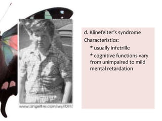
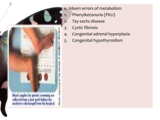
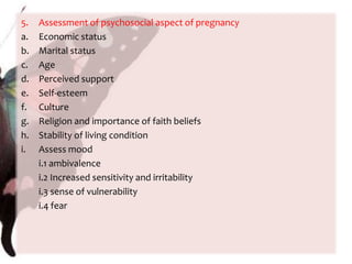
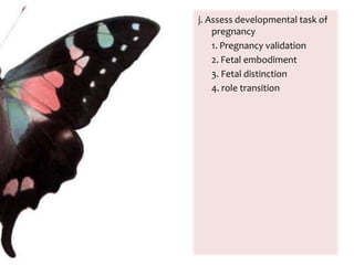
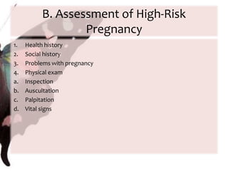
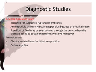
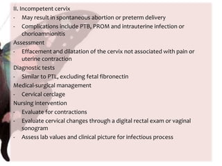
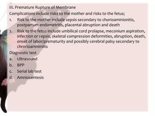
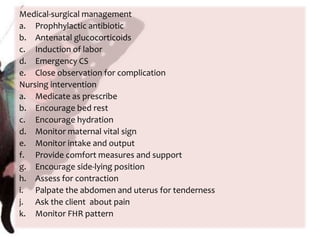
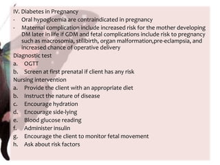
The document provides information on anatomy and physiology changes during pregnancy and assessment of high-risk conditions. It discusses the uterus, cervix, vagina and other organs. It also covers fetal development stages and complications that can arise. Common high-risk conditions addressed include preterm labor, incompetent cervix, premature rupture of membranes, diabetes, and abruptio placenta. Nursing interventions are outlined for monitoring and managing clients with various complications.


![Prenatal[3]](https://cdn.slidesharecdn.com/ss_thumbnails/prenatal3-120201220429-phpapp02-thumbnail.jpg?width=640&height=640&fit=bounds)


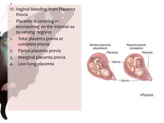
![Prenatal[2]](https://cdn.slidesharecdn.com/ss_thumbnails/prenatal2-120201220630-phpapp01-thumbnail.jpg?width=640&height=640&fit=bounds)
![Prenatal[2]](https://cdn.slidesharecdn.com/ss_thumbnails/prenatal2-120201201254-phpapp02-thumbnail.jpg?width=640&height=640&fit=bounds)
![Prenatal[3]](https://cdn.slidesharecdn.com/ss_thumbnails/prenatal3-120201200622-phpapp01-thumbnail.jpg?width=640&height=640&fit=bounds)


