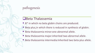
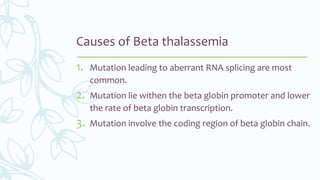
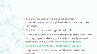
2. INTRODUCTION
Early also called anemia splenica infantum.
Autosomal dominant disorder caused by mutation
that decrease the synthesis of alpha and beta
globin chain.
Chain structurally normal but imbalance in
production of two diffrant chains.
On basis of alpha and beta globin chain on
chromosome no 11 thalassemia divided into Alpha
and Beta thalassemia
3. pathogenesis
Beta Thalassemia
B* in which no beta globin chains are produced.
Beta plus,in which there is reduced in synthesis of globin.
Beta thalassemia minor:one abnormal allele.
Beta thalassemia major:inherited two abnormal allele.
Beta thalassemia intermedia:Inherited two beta plus allele.
4. Causes of Beta thalassemia
1. Mutation leading to aberrant RNA splicing are most
common.
2. Mutation lie withen the beta globin promoter and lower
the rate of beta globin transcription.
3. Mutation involve the coding region of beta globin chain.
5. Two mechanisms contribute to the anemia:
reduced synthesis of beta globin leads to inadequate HbA
formation.
Result in microcytic and hypochromic cells.
Reduce beta chain and excess of unpaired alpha chain which
form aggregate and damage the red cell membrane and
erythroid precursor called infective erythropoiesis.
Extravasculae hemolysis:reduced red cell life span.
Inappropiate increase iron absorption and causes iron
7. MORPHOLOGY
• Red cells are small(microcytic) and pale(hypochromic) but
regular shape.
• “Target cells’’ dark red central puddle shaped.
• Blood smear show
microcytosis,hypochromia,poikilocytosis(variation in cell
size),anisocytosis(variation in cell shape).
• Expanded marrow compeletly fill the intramedullary space
of skeleton,invade bone cortex,impair bone
growth,skeletal deformatities.
8. Clinical features:
Mental retardation,microcytic and hypochromic
anemia,cardiac dysfunction from secondary
hemochromatosis.
Diagonosis:
Hb electrophoresis……..reduce HbA and increase level of
HbF
Prenatal diagnosis:by DNA analysis