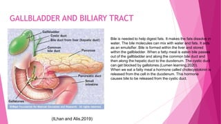
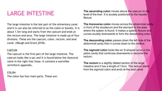
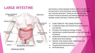
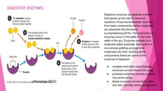
This slide is about the physiology of the human body. It talks about
The gross structure of the digestive system
It explains the process of chemical and mechanical digestion
It talks about the specialised cells of the digestive system and their function and their
2. DIGESTION AND PROCESS OF DIGESTION
Digestion is the process where complex food molecules are broken down into simple, soluble molecules
which can be absorbed and used by the cells of our body to maintain all life processes such as growth and
other life processes.(Hurst,2013).
Ingestion is the process of taking food through the mouth into the alimentary canal through the mouth.
Propulsion is the movement of food mixed with saliva through the gastrointestinal tract.it involves
swallowing and peristalsis. Swallowing also called deglutition is the process through which food moves
down the alimentary canal while the epiglottis is closed to prevent food from entering the larynx,
Peristalsis is the series of involuntary contraction and the relaxation of the smooth muscles in order to
allow food to move through the gastrointestinal tract and other tubular organs(Hurst,2013)
3. DIGESTIVE SYSTEM: OVERVIEW
According to Marieb (2004), The digestive system is made up of the alimentary canal or the gastrointestinal tract
and its accessory organs.The length of the gastrointestinal tract is about 8-10 meters long in an adult.
The alimentary canal helps to digest food we eat and absorb the useful bits or nutrients needed for the body for
growth and repairing worn out tissues.
The alimentary canal consist of the mouth,pharynx, oesophagus,stomach,small intestine(illium), large
intestine(colon),rectum and the anus.
The accessory organs are also composed of liver, gallbladder ,pancreas and the salivary gland.They are called the
accessory organs because they they are responsible for the secretion of chemical substances like enzymes
necessary for chemical digestion.(Marieb,2004)
The alimentary canal together with the various accessory organs helps to complete the process of digestion.
5. DIGESTION AND PROCESS OF DIGESTION
Physical digestion refers to the breaking of food into tiny pieces in
the mouth using the teeth. It involves chewing and swallowing.
Chewing helps to increase the surface area and the churning of the
food in the stomach.
Churning is the process where the smooth muscles of the digestive
system squeeze the food and mix it with digestive juices. The food
turns into a creamy paste called chyme.
Chemical digestion is the breaking down of food furtherly into
smaller molecules using enzymes. Enzymes are globular proteins
which act as proteins act as biological catalysts. They speed up the
rate of a chemical reaction. The various glands and accessory
organs are responsible for the secretion of enzymes .
Absorption is a way by which the food nutrients or useful bits of
substances diffuses through the digestive tract and enter the
bloodstream to be transported to the various parts of the body.
Elimination refers to the process where food that was not able to
be digested and absorbed is removed through the anus as feces.
This process can also be called defecation(Waugh and Grant,2018).
(Shutterstock,2019)
6. STRUCTURE OF THE ALIMENTARY CANAL
The alimentary canal is a long tube that starts from the
mouth through to the anus. It is about 5m in length for an
adult. (Waugh and Grant,2018). The basic covering of the
digestive tract follows the same arrangement from the
oesophagus downwards with the exception of the mouth
and the pharynx. The alimentary canal consist of four
different layers of tissues. These are submucosa, muscle
layer, and mucosa,adventitia or serosa.
Adventitia is the outer covering. It consists of loose
fibrous tissues in the thorax(upper section of the body )
and (the lower part) abdomen is covered with serous
membrane or serosa known as peritoneum.
The largest serous membrane of the body is made of the
peritoneum. It is a membrane single It is like a closed sac that
contains serous fluid. It acts as a barrier against the spread of
infection.
There are two kinds of the peritoneum. Visceral and parietal
peritoneum and are in close contact. Their friction is reduced
by peritoneal fluid secreted by peritoneal cells.
Muscle layers of the alimentary canal are made up of two
layers of smooth muscle.These are the multi smooth muscle
and visceral smooth muscle. The multi smooth muscle is
located within the walls of the blood vessels and contracts or
relaxes based on the release of hormones (Waugh and
Grant,2018).
7. STRUCTURE OF THE ALIMENTARY CANAL
The visceral smooth muscles are spindle-shaped
cells in close contact with one another. They are
mostly located within the walls of organs like the
stomach, intestines, and uterus.Peristalsis is the
series of involuntary contraction and the relaxation
of the smooth muscles in order to allow food to move
through the gastrointestinal tract and other tubular
organs. Peristalsis transports food through the
alimentary canal, and bile transported from the
gallbladder into the duodenum.
Mucosa. The lining of the mucosa is made of layers of
mucous membrane which is also made of columnar
epithelium.It has the functions of protection, absorption,
and secretion. The thin layers of the mucosa have
connective tissues that give support to the blood vessels
and lymphatic tissues.(Waugh and Grant ,2018),
The mucus membrane is responsible for the secretion of
saliva in the mouth through saliva and the gastric juices in the
walls of the stomach.
The submucosa is composed of areolar connective tissue
that contains collagen and some elastic fibers. It connects the
mucosa to the muscle layer. It also has nerves, blood
vessels, lymph vessels, and different kinds of lymphoid
tissues. The blood vessels are the venules arterioles and
capillaries (Waugh and Grant,2018),
The nerve plexus of the submucosa is made of
sympathetic and parasympathetic nerves which regulate the
increase or decrease in glandular secretion
(Shutter stock,2019)
9. MOUTH ,TEETH AND TONGUE
The mouth is made up of teeth, tongue saliva, and other
organs that facilitate digestion.
Mastication of food -Mechanical breakdown of food by
chewing the food using The various kind of teeth and
mixing with saliva.(lumen learning,2020)
Saliva from the salivary gland contains the enzyme amylase
which initiates the process of chemical digestion. It
lubricates food.
There is the digestion of carbohydrate and lipids
The tongue rolls the food and turns it into a bolus for easy
swallowing.
(Lumen learning, 2020)
10. SALIVARY GLAND
The salivary glands found in the mouth secrete saliva that moistens and softens the food particles. Saliva contains the
enzyme, salivary amylase. The job of this enzyme breaks the bonds between the monosaccharide sugar units of
disaccharides, oligosaccharides, and starches which can be used by the body. (Waugh and Grant,2018).The salivary
amylase converts amylose and amylopectin into smaller chains of glucose, called dextrins and maltose. The
concentration increase of maltose within the mouth that results from the mechanical and chemical breakdown of
starches in whole grains are what causes them to be sweet.
Only about five percent of starches are broken
down in the mouth.
The required pH of the salivary amylase is 5.8 to 7.4.
The parotid glands are located on all sides of the face ;
below the external acoustic meatus.
(Adapted from Shutterstock,2019)
11. SALIVARY GLAND
COMPONENTS OF SALIVA
The saliva is a combination of different substances
from the salivary gland and the mucous gland of the oral
mucosa. The walls of the salivary gland secrete almost
one and half liters of saliva every day . It is made up of
water, mineral salts salivary amylase-a digestive
enzyme, mucus antimicrobial
substances(immunoglobulins and the enzyme lysozyme.
FUNCTIONS OF THE SALIVA
The salivary amylase breaks complex carbohydrates into
simple sugars which can be absorbed into the
bloodstream.(Waugh and Grant,2018).
Maltose➝ glucose+glucose
Lactose➝ glucose and galactose
Sucrose➝glucose and fructose
● The saliva moistens and lubricates the food before it
can be transformed into a bolus to be swallowed
which is then swallowed.
● The saliva also lubricates and cleans the mouth to
prevent wear and tear of the mucus membrane.
● The saliva also contains lysozyme and
immunoglobulins which protect the mouth against
invading harmful bacteria.
● The salivary gland stimulate the sense of taste when
it mixes with the food
12. Epithelial cell
There are three main types of epithelial cells
(squamous, cuboidal, and columnar), and these
epithelial cells form the thin tissues/membranes
that covers many structures in the alimentary
canal.Most of the epithelial cells have microvilli
which increases surface areas for absorption
(Bandoim, 2019). The lining of the epithelial cells in
the small intestine is only one cell thick this allows
rapid diffusion molecules into and out of the small
intestine. Goblet cells which are found on the
epithelial cells secrete mucus which helps to
lubricate food (Waugh and Grant ,2018).
(Shutterstock ,2019)
microvilli
Peroxisome
13. UVULA, PHARYNX, LARYNX AND EPIGLOTTIS
The uvula is a a soft tissue structure which is made up of muscles and
connective and is lined with a mucus membrane.(Yazaki and
Sifrim,2012) It is commonly referred to as a piece of flesh that
hangs at the back of the throat.The uvula prevents food from
entering the trachea.When food touches the uvula, a signal is
carried to the brain which causes the epiglottis to close.
The pharynx, or throat, is the passageway leading from the mouth
and nose to the esophagus and larynx. The pharynx gives way to
the the passage of swallowed solids and liquids into the
oesophagus,The larynx is the organ that connects the lower part of
the pharynx with the trachea. It serves as a valve to guard the air
passages, especially during swallowing(Yazaki and Sifrim,2012)
The epiglottis is a cartilaginous structure which is found in the
throat behind the tongue and at the entrance of the larynx. It is
also found sideways at the rear of the tongue and hyoid bone.
Normally the epiglottis points upwards allowing free passage of air
into the trachea and larynx. Its main function is to prevent food
and liquid from entering the larynx area during swallowing.
(Admin,2016).
14. OESOPHAGUS
The esophagus is a long tubed structure of 8 inches in length between the
pharynx and the stomach. It is covered by moist pink tissue called the
mucosa. (Yazaki and Sifrim,2012)It runs behind the trachea and heart,
and ahead of the spine. Just before entering the stomach, and passes
through the diaphragm. The upper esophageal sphincter (UES) could also
be a bundle of muscles at the very best of the esophagus. The muscles of
the upper esophageal sphincter are used when breathing, eating,
belching, and vomiting. The lower esophageal sphincter (LES) contains a
bundle of muscles at the low end of the esophagus where it is connected
to the stomach. The Lower esophageal sphincter closed to block acid and
stomach contents from traveling backward from the stomach. The LES
muscles are not controlled by voluntary control. The main function of the
esophagus is to propel swallowed food or fluid into the stomach and to
stop or clear esophageal reflux . This function is achieved through an
organization of pattern that involves a sensory pathway, neural reflexes,
and a motor response that includes esophageal tone, peristalsis, and
shortening. The esophagus is controlled by highly complex voluntary and
involuntary mechanisms (Yazaki and Sifrim,2012),
(Yazaki and Sifrim,2012)
15. SMOOTH MUSCLE CELLS The smooth muscle of the alimentary canal
(the digestive tract) controls the movement
of substance involuntarily example is the
the peristaltic waves that move swallowed
food and nutrients.
(Visiblebody,2018).
(Visiblebody,2018).
nucleus
16. STOMACH
The stomach is a muscular organ in the form of a “J” shape situated in the hypochondriac regions of the abdominal
cavity. The stomach receives food from the esophagus(Belieza,2017) As food reaches the top of the esophagus, it
enters the stomach through a muscular valve called the lower esophageal sphincter. The walls of the stomach secrete
acid and enzymes that digest food. Ridges of muscle a tissue called rugae line which provides a large area for expansion.
The stomach muscles contract periodically, churning food to reinforce digestion. The pyloric valve may be a muscular
valve that opens to permit food to pass from the stomach to the tiny intestine. The stomach is made of
Four basic regions; cardia, fundus, body, and pylorus.
The presence of an inner oblique smooth muscle layer gives
the muscular the power to vigorously churn and blend food.
(Belieza,2017)
17. STOMACH
The wall of the stomach similar layer as most of the organs
of the alimentary tract, but with adaptations to the
mucosa and muscularis for the unique functions of this
organ. (Harkhani,2018)
In addition to the standard circular and longitudinal
smooth muscle layers, the muscular has an inner oblique
smooth muscle layer.
The gastric pits of the stomach are composed of gastric
glands that contain a variety of cells. One gland has been
taken out and enlarged at the right.
Harkhasni,2018)
18. STOMACH
The gastric gland secrete secretes gastric juice.
The juice contains hydrochloric acid(HCL), Potassium
chloride(KCL),
and Sodium chloride
The HCL pays an important role in digestion by activating
digesting
enzymes which break down the long chain of amino acid.
(Harkhani,2018)
GASTRIC SECRETING CELLS
Chief cells secrete pepsinogen.
Pepsinogen is an inactive enzyme also called zymogen.
Parietal Cells secrete HCL and intrinsic factor.The HCL is
needed to activate the protein-digesting enzyme called
pepsin and it is the cause of the high acidity content of
the stomach and also the intrinsic factor; glycoprotein
helps in the absorption of vitamin B12 in the small
intestines.Mucus cells secrete mucus and alkaline.
(Harkhani,2018) The alkaline helps to neutralize the
HCL.(Lindenmeyer,2019)
Lindenmeyer ,C.(2019)
(Shutterstock,2019)
19. STOMACH
FUNCTIONS The stomach release protease which are
protein-digesting enzymes called pepsin and
hydrochloric acid which kills bacteria. Proteases work
suitably in acidic condition with a PH of 2
The stomach temporarily stores food, which passes from
the esophagus to the stomach where it is held for 2
hours or longer. (Harkhani,2018)
mixing and breakdown of food by contraction and
relaxation of the muscle layers in the stomach.This is
also known as churning.
Absorption of useful bits into the blood.
(Instant Anatomy,2018)
20. CHIEF CELLS
Chief cells are responsible for the secretion of
pepsinogen. They are composed of basally located
nuclei and a basophilic cytoplasm which contains a
rough endoplasmic reticulum and many secretory
granules that contain pepsinogen. Pepsinogen are
secreted into the lumen of the gastric gland. The chief
cells have a lot of rough endoplasmic reticulum to for
for protein synthesis. It has microvilli which increases
the surface area for secretion. It has golgi apparatus
for the package and transport of protein
(Waugh and Grant
,2018)
(Shutterstock,2019)
21. PARIETAL CELL
The parietal cell also known as the oxyntic cell is
found in the fundus and cardia region of the
stomach.It is responsible for the secretion
hydrochloric acid (HCL) which is used for the
activation of enzymes.It has intracellular canaliculi
which increases the surface area for secretion of
HCL.The microvilli also increases the surface area.The
cytoplasm of the parietal cells contains a lot parietal
cells mitochondria which converts the energy
obtained from food into energy in the form of
ATP.(Waugh and Grant,2018)
The cell uses this ATP to pump hydrogen ions. This makes the cytoplasm and the mitochondria acidophilic
in nature.
*(Shutterstock,2019)
22. SMALL INTESTINE
The small intestine is the longest tube of muscle found
within the digestive tract between the stomach and the
small intestine. (Harkhani,2018)
It is about 3-5m long with a diameter of 2.5cm to 3cm.
Functions
Digestion
Most of the chemical digestion occurs in the small intestine
Most of the enzymes used for digestion in the small
intestine are secreted by the liver and pancreas.
Digestion of carbohydrates ,proteins and lipids occur here.
Harkhani,,2018)
23. SMALL INTESTINE
Useful bits of nutrients from digested food pass through
the walls of the small intestines and are absorbed
either diffusion or active transport into the bloodstream
to be transported to the vital parts of the body
Villi Microvilli helps to increase the surface area of the
villi i for absorption of nutrients parts of the body.
(Harkhani,2018).
Lacteals helps the absorption of lipids from the intestines.
The epithelial layer is single in order to reduce the
diffusion distance between the capillaries and the
intestinal walls.
Intestinal crypts are found between villi and release juices
that act as a fluid transporter of nutrients .
Membrane proteins and mitochondria enable active
transport of nutrients into cells (contents then passively
diffuse into bloodstream)
Adapted from (Harkhani,2018).
24. PANCREAS
The pancreas is a creamy pink accessory organ that is
located in the epigastric and the hypochondriac part of
the abdominal cavity. It weighs about 60g and has 12-15
cm of length. It has a large head, a body, and a small
tail. The head is found in the head of the duodenum,
the body is found at the back of the stomach and the
tail is located at the front of the left kidney and close to
the spleen, The pancreas is both an exocrine and an
endocrine gland.
The pancreas produces natural juices called pancreatic
enzymes that break down foods molecules . These juices are
transported through the pancreas via ducts. They are emptied
into the upper part of the small intestine called the
duodenum.The pancreas produces about 8 ounces of
digestive juice filled with enzymes every day. These are some
of the different enzymes:
Lipase. These enzyme works together with bile, which your
liver produces, to break down fat in your diet. Insufficient lipase
in the body can cause the poor absorption of vitamin A, D, E,
K). Diarrhea and fatty bowel movements. are symptoms of poor
fat absorption include
● Protease. These enzymes break down proteins into
amino acid molecules which the cells can use. Allergic
reactions can be caused by undigested proteins in some
people.
● Amylase. This enzyme helps break down starches into
sugar, which the cells can use for energy(Waugh and
Grant ,2018).
(Bandoim,2012)
25. PANCREAS The pancreatic acinar cells are made of highly
specialized structures developed for synthesis, storage,
and secretion of digestive enzymes.(Instant,2012). The
acinar cell arises from the same pancreatic progenitor as
duct and islet cells and is tightly polarized. Its apical pole
is densely packed with zymogen granules that secrete
digestive enzymes by exocytosis.
The acinar cells have more mitochondria which supply
the cell with energy.They also have microvilli to increase
surface area for absorption.
The acinar cells of the pancreas . (Pressbooks,2017)
26. LIVER
The liver is the biggest gland in the body and is found in
the upper part of the abdominal cavity. It is pink and
weighs about 1 to 2.3kg. It is located in the upper part of
the abdominal cavity and at the right hypochondriac
region. The liver is linked to the two large blood vessels:
the hepatic artery and the portal vein. It is located in the
right upper quadrant, resting just below the diaphragm.
The liver close to the right side of the stomach.
Basically, the liver is divided into four lobes: left, right,
caudate, and quadrate. The lobes are also divided into
lobules, the functional units of the liver. Each lobule is
composed of millions of hepatic cells that are the basic
metabolic cells of the liver. (Waugh and Grant,2018).
FUNCTIONS OF THE LIVER
The liver is responsible for the regulation of all the chemical
levels in the blood and the secretion of bile. Production of
bile, which helps carry away waste and break down fats in the
small intestine during digestion. It helps to produce certain
proteins for blood plasma.(Waugh and Grant,2018).
Production of cholesterol and special proteins to assist carry
fats through the body. It helps within the conversion of
glucose into glycogen for storage (glycogen can later be
converted back to glucose for energy) and to balance and
make glucose as required
Regulation of blood levels of amino acids, that form the
building blocks of proteins
Processing of hemoglobin to be used of its iron content (the
liver stores iron)
Conversion of poisonous ammonia to urea (urea is an
outcome of protein metabolism and is excreted within the
urine)
It also helps in clearing the blood of drugs and other
poisonous substances.
28. GALLBLADDER AND BILIARY TRACT
The gallbladder is a pear-shaped, hollow structure
found under the liver and at the right side of the
abdomen. Its primary function is to store and
concentrate bile, a yellow-brown digestive enzyme
produced by the liver. The gallbladder is part of the
biliary tract.
BILE
Bile is a greenish yellow, thick, sticky fluid which consists
of bile salts, dissolved charged particles, such as sodium
and bicarbonate),cholesterol, bile pigments, and other
fats (lipids). The bile has two main roles:
● The removal of excess amount of hemoglobin and
excess amount of cholesterol from the body.
● Helps in digestion.
BILE SALT helps in the digestion process by making
cholesterol, fats, and fat-soluble vitamins easier to absorb
from the intestine.
BILIRUBIN is the main bile pigment of the bile. Thisis a
waste product that is produced from hemoglobin which is the
protein that carries oxygen in the blood) and is excreted in
bile. Hemoglobin is released through the destruction of red
blood cells(Hopkins,2020).
29. GALLBLADDER AND BILIARY TRACT
(ILhan and Alis,2019)
Bile is needed to help digest fats. It makes the fats dissolve in
water. The bile molecules can mix with water and fats. It acts
as an emulsifier. Bile is formed within the liver and stored
within the gallbladder. When a fatty meal is eaten bile passes
out of the gallbladder and along the common bile duct and
then along the hepatic duct to the duodenum. The cystic duct
can get blocked by gallstones.(Lumen learning,2020).
When we eat a fatty meal a hormone called cholecystokinin is
released from the cell in the duodenum. This hormone
causes bile to be released from the cystic duct.
30. LARGE INTESTINE
The large intestine is the last part of the alimentary canal
and it is can also be referred to as the colon or bowels. It is
about 1.5m long and starts from the caecum and ends at
the rectum and anus. The large intestine is made up of four
divisions. These are the caecum, colon, rectum, and anal
canal. (Waugh and Grant,2018).
CAECUM
The caecum is the first part of the large intestine. The
caecum looks like a sac and it is found below the ileocecal
valve in the right iliac fossa. It contains a wormlike
vermiform appendix,
COLON
The colon has four main parts. These are:
The ascending colon moves above the caecum to the
level of the liver. It is acutely positioned to the hepatic
flexure
The transverse colon moves across the abdominal cavity
in front of the duodenum and the stomach to the area
where the spleen is found. it makes a splenic flexure and
curves acutely downwards to form the descending colon.
The descending colon passes down the left side of the
abdominal cavity then it curves down to the midline.
The sigmoid colon looks like an S-shaped curve in the
pelvic cavity that continues downwards to become the
rectum,
The rectum is a slightly dilated section of the large
intestine and it has a length of 13cm. The rectum starts
from the sigmoid colon and ends at the anal canal
31. LARGE INTESTINE anal canal is a short passage of about 3.8cm in length. It is
the last section of the alimentary canal and it is controlled by
two muscular sphincters. These are internal sphincter -
smooth muscle (involuntary) and external sphincter The-
skeletal muscle (voluntary). Harkhani,(2018).
● FUNCTIONS OF THE LARGE INTESTINE
● It reabsorbs water and mineral ions such as sodium
and chloride
● formation and temporary storage of feces
● maintaining a resident population of over 500 species
of bacteria
● bacterial fermentation of indigestible materials.Further
breaks down food residues
● Absorbs most residual water, electrolytes, and
vitamins produced by bacteria
● Propels feces toward the rectum
● Eliminates feces
(Harkhani,2018)
32. DIGESTIVE ENZYMES
Digestive enzymes are globular proteins
that speed up the rate of chemical
reactions. Enzymes breakdown food into
their basic components before molecules
are absorbed into the blood.
(Lumenlearning,2018). The production of
enzymes occur in the cells of the inner
walls of the gut. Enzymes operate on a
molecule called substrate their action is
one among splitting up large food
molecules into their ‘building block’
components.these are some of the
enzymes of digestion.
● amylase and other carbohydrase
enzymes convert starch into sugar
● protease enzymes convert proteins
into amino acids
● lipase enzymes convert lipids (fats
and oils) into fatty acids and glycerol
(Akoranga,2012)
33. DIGESTIVE ENZYMES
Each digestive enzyme works on specific substrate and at an optimal temperature.Any
temperature above these on the table might cause the enzymes to be denatured or not to be able
to work at their optimum best.(Barik,2019)
( Barik,2019).
34. SUMMARY OF THE DIGESTIVE SYSTEM
From (American cancer society,2044)
The digestive system
play a very major role
in the survival of all
living organism
especially human
beings.
The digestive system
is made of two
groups of organs.
The alimentary canal
which is composed of
the organs through
which food passes
and the accessory
organs which
secretes digestive
enzymes for the
chemical breakdown
of food(Nursing
times,2019).
The alimentary canal
Accessory organs
35. REFERENCES
Waugh,A. and Grant,A. (2018). Anatomy and Physiology of the digestive system.13th edition,Poland:
Elsevier publication.
Hurst,2013 The digestive system retrieved from https://onlinelibrary.wiley.com/doi/abs/10.1002/smi.2465.
Accessed on 12/11/2020.
Harkhani,D.(2018).Digestive System.retrieved from:https://www.slideshare.net/Dipaliharkhani21/anatomy-
and-physiology-of-the-digestive-system.accessed on 20/10/2020.
American Cancer Society. (2014). Stomach Cancer. Atlanta, GA: American Cancer Society. Retrieved from:
http://www.cancer.org/acs/groups/cid/documents/webcontent.retrieved on 11/11/20.
Pressbooks (2017) Digestion in humans. Retrieved from.http://pressbooks-
dev.oer.hawaii.edu/humannutrition/chapter/digestion-an/. Accessed on 29/10/20.:
Lumen learning (2020) The structure of the digestive system rerrieved from https://courses.lumenlearning.com/boundless-
ap/chapter/overview-of-the-digestive-system/. Accessed on 23/10/2020.
Belieza,A.(2017).Anatomy and Physiology.Structure of the stomach.laudis publication.pp 23-25.
Marieb,P.(2004).The digestive system.retrieved fromhttps://slideplayer.com/slide/7581792/accessed on 11/12/2020.
36. REFERENCES
Ilhan,M.and Alil, H.(2012)Acute Biliary pancreatitis https:https//www.intechopen.com/books/acute-pancreatitis/acute-
biliary-pancreatitis.accesed on 13/11/20.
Lumen learning(2020). The alimentary canal. Retrieved from:https://courses.lumenlearning.com/boundless-
ap/chapter/overview-of-the-digestive-system/.accessed on 10/11/2020.
Hopkins,J.(2020).Biliary Anatomy and functions retrieved
from:https://www.hopkinsmedicine.org/health/conditions.accessed on 09/11/20
Lindenmeyer ,C.(2019). Liver and the gallbladder.retrieved from :https://www.msdmanuals.com/home/liver-and-
gallbladder-disorders/.accessed on 15/11/2020.
Akoranga,P.(2012).Digestive Enzymes. retrieved from :https://www.sciencelearn.org.nz/resources/1840-digestive-
enzymes#.accessed on 17/11/2020.
Bandoim,L (2019). "Epithelial Cells: Definition, Function, Types & Examples" sciencing.com, reteieved from
https://sciencing.com/epithelial-cells-definition-function accessed on. 21 December 2020.
Admin,2016 The pharynx,retreived from https://www.therespiratorysystem.com/pharynx/ accessed on 29/11/2020.
37. REFERENCES
Yazaki and Sifrim,(2012).The oesophagus.retrieved from https://onlinelibrary.wiley.com/doi/abs/10.1111/j.1442-
2050.2011.01180.x.accessed on 21/11/2020.
Shutterstock,(2019). Parietal cells images.retrieved from https://shutterstock.com/images.accessed on 20/12/2020/
Barik,P. (2019).retrieved from https://www.learnpick.in/prime/documents/ppts/details/4879/enzyme accessed on
21/11/2020.
Nursing Times.(2015)The alimentary canal.retrieved from:https://www.nursingtimes.net/clinical-
archive/gastroenterology/gastrointestin.accessed on 10/11/20.
Instant, A. (2012)Systems physiology.retrieved from
https://www.instantanatomy.net/abdomen/areas/bowel/musclecoatsmucosalcells.html.accessed on 17/11/2020.
Lumen learning(2018). Retrieved from:https://courses.lumenlearning.com/boundless-ap/chapter/overview-of-the-
digestive-system/.accessed on 10/11/2020.
Visiblebody(2018 ) Smooth muscle cells.retreived from https://www.visiblebody.com/learn/muscular/muscle-types
Accessed on 18/12/2020.