
Recommended
More Related Content
What's hot
What's hot (20)
Similar to Human digestive system presentation
Similar to Human digestive system presentation (20)
Recently uploaded
Recently uploaded (20)
Human digestive system presentation
- 2. Objectives At the end of the lesson learners should be able to: Define the terminology associated with digestive system processes Label the alimentary canal. Draw the alimentary canal. Relate structures of digestive organs with their functions. Discuss the process of peristalsis. Group major organs of digestive tract Compare structures and functions (Mangino, 2015)
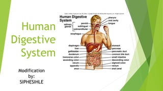
- 3. Structure of human Digestive tract Humans have a complete digestive tract starting from the mouth and ends with the anus. The major structures of human digestive tracts are: a) Mouth b) Pharynx c) Esophagus d) Stomach e) Small intestine f) Large intestine g) Rectum and h) Anus. (Saputra, 2017)
- 4. Digestion and Alimentary Canal The digestive system consist of all the organs which take part in the digestive of food. While alimentary canal are the pathways involved during the digestion of food starting from the mouth to the and ends in the anus. (Ling et.al, 2013)
- 5. The accessory organs of digestion are: Liver Salivary glands Gall Bladder Pancreas (Ghosh, 2017)
- 6. Why is digestion important? It is important for breaking down food into nutrients, which the body uses for energy, growth, and cell repair. (Sehgal, 2013)
- 7. Digestive Processes Ingestion Digestion – Mechanical and Chemical Absorption Assimilation Excretion (Bayoneta, 2016)
- 8. Mouth Ingestion – Food is placed in the mouth when eating. Teeth: a) Incisors they are used to cut and bite food. b) Canines they are used to tear of solid food c) Pre-molars and molars used to grind food finer. This is all called mechanical digestion (Ghosh, 2017)
- 9. Structure of the teeth. (Ghosh, 2017)
- 10. Saliva Secreted by the salivary (goo.gl/124gXQ) Michael mangino) glands(parotid gland, sublingual gland and sub- maxillary gland) It mixes with food to form bolus. Saliva contains the enzyme called amlyse and it function is that it breaks down cooked starch into maltose. This is called Chemical Digestion. (Mangino, 2015)
- 11. Tongue Mix food with saliva and help to push food between teeth. It also makes swallowing easier. (Ling et.al, 2013)
- 12. Esophagus The bolus is forced down into the esophagus, when the muscular pharynx contract and this is called swallowing. Peristalsis is the contraction and relaxation of circular and longitudinal muscles of the esophagus, pushes food downwards into stomach, through cardiac valve. No absorption takes place The epiglottis covers the trachea preventing food from going to the trachea when swallowing. (Ghosh, 2017)
- 13. Stomach Food enters the fundus region of the stomach through the cardiac valve. Remains for about 30-35 minutes before the muscular, longitudinal and oblique muscles starts to contracting and relaxing (peristalsis). Food move with circular movements in the stomach (corpus and pyloric regions) and mixes with gastric juices. This is called mechanical digestion (Saputra, 2017) and (Bayoneta, 2016)
- 14. Gastric juices – secreted after the hormone gastrin stimulates the parietal cells in the fundus region of stomach Gastric Juices consist of: a) HCL(Acidify stomach and neutralizes bolus, antiseptic, solution, emulsifies fats. b) Digestive enzymes (pepsin, rennin and lipase.) c) Mucus protect the inner lining of the stomach during enzyme activity. d) Water. (Saputra, 2017) and (Bayoneta, 2016)
- 15. Gastric juices with bolus and now called chym Some substances are absorbed in stomach. Water, glucose, salt and certain drugs and alcohol pass into blood capillaries of stomach wall (Saputra, 2017) and (Bayoneta, 2016)
- 16. Small Intestine Chym enters duodenum( 1st part of small intestine) through pyloric valve. Mix with bile (secreted from liver/gall bladder and pancreatic juices. (Ling et.al, 2013)
- 17. Chemical Digestion Secretin – hormone that stimulates pancreas to secrete pancreatic juice into duodenum. Pancreatic juice contain: Sodium bicarbonate (neutralizes the chym, antiseptic) and Digestive enzymes (Trypsin, amylase and lipase) Bile produced in liver and stored by the gall bladder. (Ling et.al, 2013)
- 18. Chym moves through jejunum( 2nd part of small intestines) Mixes with intestinal juice (succus entericus) – contains digestive enzymes for final digestion of food. (Bayoneta, 2016)
- 19. Large Intestine No digestion takes place in the colon. Undigested food particles from small intestine enter the caecum through ileocaecal valve. In colon – water is absorbed – chym becomes semi-solid. (Saputra, 2017)
- 20. Large Intestine Symbiotic bacteria present in colon act upon food rests – decomposing them & into faeces. Bacteria synthesize vitamins B groups and K – essential for blood clotting process. Peristalsis in colon – facilitated by mucus produced by numerous mucous glands. Mucus assists the movement of feces & protects the wall of the colon. (Saputra, 2017)
- 21. Rectum The food residue (mainly cellulose) stays on the rectum until it is excreted as feces in the anus. (Sehgal, 2013)
- 22. Anus Defecation (excretion) takes places in the anus. (Ghosh, 2017)
- 23. References Bayoneta, R, J. (2016). The Digestive System. Available from slideshare at http://goo.gl/fK6HJY (Accessed 17 September 2017). Ghosh, A,.(2017). The Digestive System. Available from slideshare at http://goo.gl/BGkE2q (Accessed 17 September 2017). Ling, W, Z,. Yee, L,C,. Heng, K,C,. Ji, C, K,. Tong, T, J,. (2013). Human Digestive System. Available from slideshare at http://goo.gl/rKzFHx (Accessed 17 September 2017). Mangino, M, J. (2015). Stomp e-Portfolio. Available from slideshare at http://goo.gl/124gXQ (Accessed 17 September 2017). Saputra M, R. (2017). Human Digestive System. Available from slideshare at http://goo.gl/T2PgT9 (Accessed 17 September 2017). Sehgal, K. (2013). Human Digestive System. Available from slideshare at http://goo.gl/hAXq9i (Accessed 17 September 2017).