
Recommended
More Related Content
What's hot
What's hot (20)
Similar to Anatomy and Physiology of Digestive System
Similar to Anatomy and Physiology of Digestive System (20)
More from iffat aisha
More from iffat aisha (14)
Recently uploaded
Recently uploaded (20)
Anatomy and Physiology of Digestive System
- 2. Process by which organisms obtain and utilize their food. There are two parts to Nutrition: 1. Ingestion- process of taking food into the digestive system so that it may be hydrolized or digested. 2. Digestion- the breakdown of food (either chemically or mechanically) in order to utilize nutrients
- 3. Micronutrients- vitamins, minerals, & water Macronutrients- proteins, lipids, carbohydrates, etc…
- 5. GI (gastrointestinal) tract = alimentary canal
- 6. Mouth mechanical digestion teeth breaking up food chemical digestion saliva Amylase: enzyme digests starch mucin slippery protein (mucus) protects soft lining of digestive system lubricates food for easier swallowing buffers neutralizes acid to prevent tooth decay anti-bacterial chemicals kill bacteria that enter mouth with food
- 7. Epiglottis flap of cartilage closes trachea (windpipe) when swallowing food travels down esophagus Peristalsis involuntary muscle contractions to move food along
- 8. Salivary glands release Serous and mucous fluid Digestion begins Amylase – breaks down carbohydrates Lipase for lipid digestion Chewing (mastication) and mixing of food with tongue Stomach muscles contract, acid and enzymes released Pancreas and gall bladder secrete 8
- 9. 1. Chewing a saltine? - 2. Saliva breaking the saltine down into molecules of glucose? - 3. Your tongue breaking pieces of a hamburger apart? 4. Pepsin (an enzyme) in your stomach breaking the hamburger into amino acids?
- 10. The back of the throat. Larynx- passage for air, closes when we swallow. Is approximately 15cm long.
- 11. Groups of specialized secretory cells. Found in the lining of the alimentary canal or accessory organs.
- 12. Food is temporarily stored here. Gastric juices are secreted. Has layers of muscle that line the inside. Mechanically and chemically breaks down food.
- 13. Functions food storage can stretch to fit ~2L food disinfect food HCl = pH 2 kills bacteria chemical digestion pepsin enzyme breaks down proteins But the stomach is made out of protein! What stops the stomach from digesting itself? mucus secreted by stomach cells protects stomach lining
- 14. Stomach functions: Storage of food Mixing via muscle contractions Release of H+ & Cl- and pH lowers Acidic (pH 1.5-2.5) (HCl). kills bacteria Degrades foods = chyme Cells release pepsinogen – a zymogen Pepsinogen converted to pepsin in low pH – cleaves proteins Digestion continues via Acid and pepsin Amylase, lipase 14
- 16. Salivary glands Moistening/lubricating fluid with enzymes Amylase helps break down starch; lipase - lipids Pancreas Release of digestive enzymes Release of bicarbonate (HCO3 - ) solution Endocrine functions = insulin & glucagon Liver Makes bile -- helps dissolve fats Receives and stores building blocks (aa, CHO, etc.) from intestine Makes blood proteins Detoxifies drugs Gallbladder Stores and concentrates bile (from liver) 16
- 17. Pouch structure located near the liver which concentrates and stores bile Bile duct – a long tube that carries BILE. The top half of the common bile duct is associated with the liver, while the bottom half of the common bile duct is associated with the pancreas, through which it passes on its way to the intestine.
- 18. Bile emulsifies lipids (physically breaks apart FATS) Bile is a bitter, greenish-yellow alkaline fluid, stored in the gallbladder between meals and upon eating is discharged into the duodenum where it aids the process of digestion.
- 19. An organ which secretes both digestive enzymes (exocrine) and hormones (endocrine) ** Pancreatic juice digests all major nutrient types. Nearly all digestion occurs in the small intestine & all digestion is completed in the SI.
- 20. Digestive enzymes digest proteins trypsin, chymotrypsin digest starch amylase Buffers neutralizes acid from stomach
- 22. Function produces bile bile stored in gallbladder until needed breaks up fats act like detergents to breakup fats Bile contains colors from old red blood cells collected in liver = Iron in RBC rusts & makes feces brown
- 23. Mouth break up food digest starch kill germs moisten food Liver produces bile - stored in gall bladder break up fats Pancreas produces enzymes to digest proteins & starch Stomach kills germs break up food digest proteins store food
- 24. Most chemical digestion takes place here. Simple sugars and proteins are absorbed into the inner lining. Fatty acids and glycerol go to lymphatic system. Lined with villi, which increase surface area for absorption, one cell thick.
- 25. Function chemical digestion major organ of digestion & absorption absorption through lining over 6 meters! small intestine has huge surface area = 300m2 (~size of tennis court) Structure 3 sections duodenum = most digestion jejunum = absorption of nutrients & water ileum = absorption of nutrients & water
- 26. 1st section of small intestines acid food from stomach mixes with digestive juices from: pancreas liver gall bladder
- 27. Much absorption is thought to occur directly through the wall without the need for special adaptations Almost 90% of our daily fluid intake is absorbed in the small intestine. Villi - increase the surface area of the small intestines, thus providing better absorption of materials
- 28. Absorption through villi & microvilli finger-like projections increase surface area for absorption
- 29. Function re-absorb water use ~9 liters of water every day in digestive juices > 90% of water reabsorbed not enough water absorbed diarrhea too much water absorbed constipation
- 30. Solid materials pass through the large intestine. These are undigestible solids (fibers). Water is absorbed. Vitamins K and B are reabsorbed with the water. Rectum- solid wastes exit the body.
- 31. Living in the large intestine is a community of helpful bacteria Escherichia coli (E. coli) produce vitamins vitamin K; B vitamins generate gases by-product of bacterial metabolism methane, hydrogen sulfide
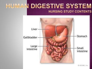
- 32. Copyright © The McGraw-Hill Companies, Inc. Permission required for reproduction or display. Accessory Digestive Organs Gastrointestinal (GI) Tract Teeth Tongue: mechanical processing, moistening, and mixing with salivary secretions Salivary glands: secretion of lubricating fluids with enzymes to breakdown carbohydrates and lipids Liver: synthesis of bile, storage of nutrients, many other functions Gallbladder: Storage, concentration and secretion of bile Pancreas: exocrine portion secrete buffers and digestive enzymes and endocrine portion secretes hormones Large intestine: dehydration and compaction of materials in preparation for elimination (3’) Small intestine: enzymatic digestion and absorption of nutrients (20’) Stomach: chemical breakdown of materials by acidic and enzymatic processing and mechanical mixing via muscular contractions ( 12”) Esophagus: conduit to the stomach (15”) Oral cavity Pharynx: muscular propulsion of materials into the esophagus Anus
- 33. ULCERS – erosion of the surface of the alimentary canal generally associated with some kind of irritant. APPENDICITIS – an inflammation of the appendix due to infection Common treatment is removal of the appendix via surgery
- 34. GALLSTONES – an accumulation of hardened cholesterol and/or calcium deposits in the gallbladder Can either be “passed” (OUCH!!) or surgically removed
- 35. Like and Share Nursing Study Contents