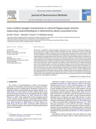
This document describes a study that used laser stimulation of neurons expressing channelrhodopsin-2 (ChR2) to study synaptic transmission in cultured hippocampal neurons. Recombinant adeno-associated virus (rAAV) was used to deliver the ChR2 gene to the neurons. Laser stimulation was able to activate action potentials in ChR2-expressing neurons. By voltage-clamping a neuron and scanning a laser, synaptic responses were observed at some locations, indicating spatial localization of stimulation. Pharmacological tests identified responses that were synaptic. While monosynaptic responses could not be entirely distinguished from polysynaptic ones, smaller amplitudes, simpler shapes, and latencies around 8 ms suggested monosynaptic interactions.
UTF-8''Final Assessing post-synaptic partners of Dentate Granule Cells in a M...Grant Pizzo
1) The document describes an experiment assessing how dentate granule cell (DGC) axon terminals and their post-synaptic partners change in a rat model of temporal lobe epilepsy (TLE).
2) The researcher induced seizures in rats using pilocarpine and later injected retroviruses to label dividing DGCs. Tissue was then analyzed to identify post-synaptic partners of DGCs using antibodies targeting interneurons and mossy cells.
3) Preliminary results showed that antibodies for GAD67 and GluR2 successfully labeled the correct cell types, but need further refinement, while an mGluR1/5 antibody did not label interneurons in the dentate gyrus
This document summarizes a study that investigated the role of presynaptic and postsynaptic TrkB signaling in the establishment of visual connectivity in Xenopus tadpoles. The researchers disrupted TrkB signaling in individual retinal ganglion cells (RGCs) or tectal neurons by overexpressing a dominant-negative TrkB receptor. They found that disrupting presynaptic TrkB signaling in RGCs decreased axon branching and stability, reduced synaptic maturation, and decreased the number of mature synaptic profiles. In contrast, disrupting postsynaptic TrkB signaling in tectal neurons did not alter synaptic number or dendritic morphology. This demonstrates that presynaptic TrkB signaling in RGCs is necessary for normal development of visual connectivity.
CRISPR/Cas9 is a powerful new technique for genome editing that allows DNA to be easily cut and modified. It involves using the Cas9 enzyme, guided by RNA, to create targeted double-stranded breaks in DNA which are then repaired, allowing the DNA sequence to be altered. This system was adapted from a bacterial immune system. CRISPR/Cas9 represents a major breakthrough as it is simpler, cheaper and more accurate than previous genome editing methods. It has already been used to edit genes in numerous organisms and holds promise for applications like correcting genetic diseases. However, off-target effects and ethical concerns surrounding its use in humans remain limitations that need to be addressed.
The document summarizes a presentation given by Evan Y. Snyder on strategies for enhancing neural stem cell efficacy in the central nervous system. Some key points:
1) Neural stem cells can rescue injured cells through direct cell-cell contact via gap junctions, as well as secreting diffusible factors. This contact may improve homing of stem cells to injured sites and influence host cell differentiation.
2) In disease models, neural stem cells appear to rescue endangered cells by modulating intracellular signaling pathways related to trophic factors and mitochondrial function. Cell-cell contact through gap junctions plays a role in this rescue.
3) Neural stem cells provide therapeutic benefits not just through cell replacement but also through parac
CRISPR/Cas9 is a genome editing technique that allows for highly specific modification of DNA. It involves a Cas9 protein guiding a customized RNA to a target location in the genome to cut DNA. Scientists adapted the natural CRISPR immune system found in bacteria for genome editing. CRISPR/Cas9 holds promise for treating genetic diseases and developing crops but also raises ethical concerns when applied to human germline cells due to heritable effects. Researchers continue improving targeting specificity and developing new Cas proteins for additional applications in genome editing and gene regulation.
This document summarizes the development of genome editing technologies leading up to the advent of CRISPR-Cas9. Early approaches relied on site-specific DNA recognition by oligonucleotides or proteins, but were limited. The discovery of CRISPR immune systems in bacteria revealed Cas9, an RNA-guided endonuclease that introduces targeted DNA double-strand breaks. Engineering the dual-RNA structure into a single guide RNA created a simple two-component system for programming Cas9 to cleave any DNA sequence. CRISPR-Cas9's simplicity, efficiency, and versatility has enabled widespread applications in biology research across many cell and organism types.
This document discusses several experiments involving gene delivery to the spinal cord using viral vectors. It first outlines different types of viral vectors like retroviruses, adenoviruses, and adeno-associated viruses. It then summarizes results from experiments delivering progranulin or other genes to the spinal cord using lentiviral vectors. The experiments found lentiviral vectors allowed long-term gene expression but did not find statistically significant effects on neuronal numbers or stress markers. Challenges included low transduction levels and inability to detect viral presence or upregulation of the delivered genes. Other experiments delivered genes to the spinal cord using pseudotyped lentiviruses and found transduction of motor neurons originating from intramuscular injections.
This document discusses genome editing using the CRISPR-Cas9 system. It begins by introducing three main genome editing technologies - zinc-finger nucleases, TALENs, and the CRISPR-Cas9 system. It then describes the key events in the discovery of CRISPR-Cas9, including its origins as a bacterial defense system. The document outlines the main components of the CRISPR-Cas9 system, including crRNA, tracrRNA, sgRNA, and Cas9. It also summarizes the two main steps in genome editing using CRISPR-Cas9 - knocking out genes and DNA repair. The document concludes by discussing opportunities for applying CRISPR-Cas9 technology across various
CRISPR: Opportunities and Challenges WebinarPreScouter
CRISPR is a genome-editing technique that provides a simple yet versatile method for making targeted changes to the genome of living cells. Researchers have already applied CRISPR to reverse disease-causing mutations and to engineer pest-resistant crops.
In this CRISPR webinar, PreScouter will be joined by experts and researchers to review CRISPR's opportunities and challenges. We will touch upon key challenges, such as reducing off-target effects, as well as new advances set to overcome some of the current limitations, such as novel CRISPR systems. Additionally, we'll highlight potential first applications of the technology within medicine and agriculture.
This is a great opportunity to not only learn from experts about how this disruptive technology is changing gene editing but also ask your personal questions about CRISPR.
Moderator:
This CRISPR discussion will be moderated by Charles Wright, the Medical Project Architect at PreScouter.
Panelists:
John G. Doench, Ph.D., is Associate Director of the Genetic Perturbation Platform at the Broad Institute.
C. B. Gurumurthy (Guru), BVSC, MVSC, Ph.D. Exec. MBA, is an Assistant Professor at the Department of Developmental Neuroscience, Munroe Meyer Institute for Genetics and Rehabilitation, and he serves as the Director of UNMC Mouse Genome Engineering Core Facility at the University of Nebraska Medical Center.
Shuibing Chen is an Assistant Professor in the Department of Surgery and Biochemistry at Weill Cornell Medical College, New York.
Rab5 Mediates an Amyloid Precursor Protein Signaling Pathway That Leads to Ap...Lucas Patzek
This document summarizes a study examining the role of Rab5, a protein involved in endocytosis, in neuronal apoptosis induced by familial Alzheimer's disease (FAD) mutations in amyloid precursor protein (APP). The study found that expression of a FAD mutant form of APP or the APP-binding protein APP-BP1 in neurons led to enlarged early endosomes, increased endocytosis, and apoptosis. Both APP-BP1 and Rab5 levels were elevated in early endosomes. Inhibition of Rab5 or dynamin activity, but not another endocytosis protein Eps15, prevented neuronal apoptosis induced by mutant APP or APP-BP1 without affecting amyloid-β levels. Expression of an activated Rab5 mutant further increased neuronal
This study examines synapse formation between transplanted GABAergic interneurons and newborn dentate granule cells (DGCs) in the hippocampus of mice with temporal lobe epilepsy. The results show that epileptic mice receiving transplants of GABAergic interneurons from the medial ganglionic eminence had fewer seizures than control mice. Newborn DGCs were labeled using retroviruses and showed a range of normal and hyperexcitable morphologies. Confocal microscopy revealed appositions and clusters of synaptic proteins between the dendrites of newborn DGCs and axons of transplanted interneurons, suggesting synapse formation between the cells. Future studies aim to further characterize this synaptic connectivity.
This document provides an overview of biotechnology principles and applications. It defines biotechnology as the application of technology to modify biological organisms by adding genes from other organisms. The document discusses how genes are identified, isolated, and manipulated to introduce desired traits. It describes techniques such as homology cloning, complementary genetics, and map-based cloning used to isolate genes. The document explains how genes are introduced into plants using transformation methods like Agrobacterium and biolistics. It provides examples of transgenic crops and their applications in agriculture.
This document describes an experiment to test the effectiveness of different bacterial transformation mixes in CRISPR-Cas9 systems. The standard transformation mix contains calcium chloride while the experimental mix adds polyethylene glycol and dimethyl sulfoxide. Bacteria are made competent using each mix and transformed with CRISPR components before plating. Preliminary results showed more growth with the standard mix, but the experimental mix supported slightly more antibiotic-resistant growth. Multiple errors prevented conclusions, requiring further replication and controls.
The document discusses biotechnology principles including gene manipulation techniques used to modify genes and introduce them into transgenic organisms. It defines biotechnology as applying technology to modify the biological function of an organism by adding genes from another. Gene manipulation starts at the DNA level by identifying genes that control traits of interest or modifying existing genes. Genes are then introduced into organisms using techniques like transformation to form transgenic organisms that express new traits.
CRISPR-Cas9 is a genome editing technique that allows DNA to be precisely cut and modified. It involves using the Cas9 enzyme, guided by RNA, to cut DNA at a specific target location. The cell's DNA repair machinery can then introduce changes by adding, removing, or replacing DNA segments. CRISPR-Cas9 was adapted from a natural defense system in bacteria against viruses. It holds promise for treating genetic diseases but also raises ethical concerns about editing human embryos or germline cells.
Have you considered that protein over-expression or inefficient mRNA knockdown may be masking physiological effects in your assays? Increasingly scientists are moving to endogenous gene-editing to characterise the function of their genes of interest.
Dr Chris Thorne from Cambridge Biotech Horizon Discovery discusses the ground breaking gene-editing technology CRISPR. The simplicity of experimental design has led to rapid adoption of the technology across the scientific community. However, challenges remain.
This Slidedeck focuses specifically on implementing CRISPR experiments, and explore a number of key considerations crucial to maximising chances of targeting success, whether your goal is to generate a knock-out or a knock-in. Chris also takes a look at some of the alternative uses of CRISPR, including sgRNA genome wide synthetic lethality screens.
The slides aim to support those researchers either planning to or already using CRISPR gene-editing in their lab. Horizon Discovery have also recently launched a program aimed specifically at academic cell biologists to promote the adoption of CRISPR by offering FREE CRISPR Reagents for knock-out cell line generation - more information available here. http://www.horizondiscovery.com/what-we-do/discovery-toolbox/genassist-crispr--raav-genome-editing-tools
CRISPR system is very simple, consisted of a Cas9 protein and a single guided RNA. With the guidance of sgRNA, Cas9 could cause a double stranded breaks in the target site.
1) The author worked in Dr. Jack Parent's neurodevelopment lab studying adult neurogenesis and regeneration using mouse, zebrafish, and iPSC models.
2) Specifically, the author worked with Dr. Palsamy Kanagaraj to study the inflammatory response and regeneration in the zebrafish telencephalon following induced brain injury.
3) The author assisted with experiments analyzing pro-inflammatory and anti-inflammatory cytokine expression over time after injury in zebrafish to understand differences in the inflammatory response and regeneration between fish and mice.
This study investigated the use of quantum dots (qdots) to label and visualize two endogenous synaptic proteins, GABAA-1 receptors and glutamate transporters (VGLUT1), in the rat cerebellum. Qdots allowed for the clear visualization of these proteins in very small presynaptic structures like parallel fiber varicosities that are below the diffraction limit of conventional microscopy. Specifically, qdots formed clusters around interneurons and isolated clusters on interneuron dendrites, revealing the presence of GABAA receptors. They also labeled sub-micrometer parallel fibers and 1-2 micrometer presynaptic varicosities containing VGLUT1. While double labeling with two qdot colors was attempted, some receptor sites remained unlabeled
This document summarizes research using a zebrafish model to study paclitaxel-induced peripheral neuropathy (PIPN). The research shows that paclitaxel treatment in zebrafish larvae induces neurodegeneration and impairs axon regeneration after injury. However, providing exogenous hydrogen peroxide or a regulatory pharmaceutical rescues axon regeneration in the presence of paclitaxel. This suggests these compounds may help prevent or treat PIPN.
This document provides an overview of CRISPR/Cas9 genome editing. It discusses the history and limitations of prior genome engineering techniques like recombinant DNA and zinc finger nucleases. It then explains how CRISPR/Cas9 works as a RNA-guided DNA endonuclease and how this allows it to efficiently and specifically edit genomes. The document outlines several applications of CRISPR/Cas9 like generating knockout animals and cell lines. It also notes some concerns about using the technique for human genome editing.
This document discusses recombinant DNA technology, which involves introducing foreign DNA into a host organism. It describes the basic steps of isolating DNA, cutting it with restriction enzymes, amplifying the gene with PCR, and ligating it into a vector for insertion into the host. The key tools used are restriction enzymes, DNA polymerase for PCR, ligase, and vectors to introduce the recombinant DNA into hosts such as bacteria. The goal is to generate organisms that produce desired proteins or have new traits.
The document discusses an experiment testing the effect of knocking down the C. elegans homolog of the human SynGAP gene, called gap-2, on short-term memory in worms. Worms that had gap-2 knocked down using RNAi showed impaired ability to retain short-term memory compared to control worms. This provides a model for how mutations in the human SynGAP gene could cause memory impairment and suggests gap-2 is important for short-term memory in worms.
Next generation sequencing (NGS) refers to modern DNA sequencing technologies that allow for high-speed, low-cost sequencing of entire genomes. NGS works by massively parallel sequencing of millions of DNA fragments. The Illumina sequencing by synthesis method is the most commonly used NGS approach. It involves library preparation, cluster generation on a flow cell, sequencing via reversible dye-terminator chemistry, and computational analysis of sequenced reads. Key advantages of NGS include its scalability, unlimited dynamic range, tunable coverage levels, and ability to multiplex many samples simultaneously in a single run.
Bioo Scientific - Simplify and Reduce Cost of mtDNA Isolation and Library PrepBioo Scientific
Mutations in mitochondrial DNA (mtDNA) have been implicated in various human disorders and in aging, making NGS analysis of mtDNA a priority for a number of labs. However, accurately determining the diversity of mtDNA has been difficult for a number of reasons. The standard methods for mitochondrial DNA extraction have a number of limitations making them inferior solutions for NGS library preparation. Bioo Scientific has commercialized a kit which overcomes these limitations of mtDNA isolation by selectively digesting linear nuclear DNA (nDNA) while leaving circular mtDNA intact. This technology has been incorporated into the NEXTflex mtDNA-Seq Kit which includes optimized reagents for the isolation of mtDNA and for the construction of Illumina mtDNA libraries. Libraries constructed using the NEXTflex mtDNA-Seq Kit are ideal for many NGS applications including heteroplasmy analysis.
Bioo Scientific - Absolute Quantitation for RNA-SeqBioo Scientific
PCR bias during RNA-seq library preparation can incorrectly amplify some transcripts over others, making accurate quantification of transcript numbers difficult. Molecular indexing uses unique molecular identifier adapters to label each transcript, correcting for PCR bias and improving RNA quantification. Published research shows molecular indexing returns over 15% of reads to libraries and improves accuracy, especially for highly expressed genes, by accounting for distinct fragments with identical start/stop sites eliminated by typical unique molecular identifier methods.
The long awaited hour was finally at hand as the UPES SPE Fest 2014 got under way on 6th Feb 2014. This fest saw the presence of a panel of estimated & valued dignitaries chief guest Mr. Rich Paces, Director Operations, Cairn India Limited along with Mr. Mohd Fiyazudeen - Reservoir Engineer, Mr. Sumit Malik - Principal Drilling Engineer and Mr. Pradeep Singh - Principal Operations Geologist, Cairn India Limited.
The Auditorium was jampacked with participants whose enthusiasm lent a great atmosphere to the fest. UPES hosted participants from 14 national universities & 6 international universities for the SPE Fest 2014.
The event was kicked off with the unveiling of the mascot and the launch of the 2013-2014 Annual magazine Energy Era. This ceremony heralded the gush of events that took place from 6th - 9th February 2014.
Here is our overview of what went down at the amazing 3 day SPE Fest!
The document outlines a business success program that aims to:
1) Help businesses in Vietnam prepare for upcoming international standards by developing their businesses and employees now according to these standards.
2) Advises that acquiring international standard training programs is the next best option if international trainers are unavailable internally.
3) Provides examples of how some Vietnamese businesses have already adopted best international practices and standards.
World War I began in 1914 due to rising nationalism, imperialism, militarism, and systems of alliances between European powers. The immediate cause was the assassination of Archduke Franz Ferdinand by Serbian nationalists. The US initially remained neutral but was drawn into the war in 1917 after German submarine warfare threatened American merchant shipping and the Zimmerman Telegram proposed an alliance between Germany and Mexico against the US. The addition of American troops and resources helped the Allies defeat the Central Powers by 1918. Over 20 million people died making it one of the deadliest conflicts in history.
Este documento presenta actualizaciones al procedimiento para calcular la evapotranspiración de referencia y del cultivo a partir de datos meteorológicos y coeficientes de cultivo. Incorpora avances en investigación y un procedimiento más preciso para determinar el uso de agua de los cultivos según recomendaciones de expertos de la FAO. Explica métodos para determinar la evapotranspiración del cultivo de referencia usando el método Penman-Monteith y provee procedimientos actualizados para estimar la evapotranspiración de diversos cultivos en diferentes etap
A Training Workshop on SELLING SOLAR LIGHTS: TECHNOLOGY AND FINANCE is going to be held on the 15th February 2014 at Center for Management Development, UPES, Village- Bidholi, Dehradun.
Find all the events and the timings here.
Crando Plugins en ImageJ [de http://www.imagingbook.com/fileadmin/goodies/ijt...drk28
This document provides a tutorial on writing plugins for the ImageJ image analysis program. It covers getting started, the plugin concept and architecture in ImageJ, image representation and processing APIs, user interface tools like windows and dialogs, advanced topics like movie import/export and I/O plugins, and troubleshooting. The goal is to explain everything needed to develop custom plugins that extend the functionality of ImageJ. Example plugins are provided throughout to demonstrate key concepts.
Mobile marketing is an emerging opportunity, especially in emerging markets like India where mobile penetration is high. Some key points:
1. Mobile marketing allows companies to reach a large customer base through targeted, personalized messages on wireless networks.
2. Emerging markets like India, China, and Brazil have seen strong growth in mobile users, with India having over 900 million subscribers.
3. Mobile marketing offers advantages over other media in reach, frequency of exposure, effectiveness of message delivery, convenience, localization, integration, and building relationships.
4. Emerging mobile marketing technologies that may be used include mobile websites, SMS alerts/reminders, mobile apps, coupons, QR codes, and mobile search advertising.
The University of Petroleum and Energy Studies (UPES), Dehradun, campus buzzed with excitement on the eighth day, i.e. June 28th, 2016, of Phase I of its 4th consecutive Orientation Program - ‘Monsoon Management Magic - M3’.
Poster - determining the effects of tau on synaptic density in a mouse model ...Shaun Croft, MScR
1. The document proposes that tau exacerbates amyloid-beta oligomer (AβO) toxicity at synapses, resulting in synapse loss and cognitive impairment in Alzheimer's disease.
2. To test this, the authors will use array tomography to analyze synaptic density in mouse models with and without human tau genes near amyloid plaques and determine if tau and AβOs colocalize at degenerating synapses.
3. Preliminary results show synapse loss around plaques and some colocalization of synapses and AβOs, supporting the hypothesis. Completing the data could further understanding of Alzheimer's pathology and identify new treatment targets.
Abstract In the mammalian neocortex, excitatory neurons provide excitation in both columnar and laminar dimensions, which is modulated further by inhibitory neurons. However, our understanding of intracortical excitatory and inhibitory synaptic inputs in relation to principal excitatory neurons remains incomplete, and it is unclear how local excitatory and inhibitory synaptic connections to excitatory neurons are spatially organized on a layer-by-layer basis. In the present study, we combined whole cell recordings with laser scanning photostimulation via glutamate uncaging to map excitatory and inhibitory synaptic inputs to single excitatory neurons throughout cortical layers 2/3–6 in the mouse primary visual cortex (V1). We find that synaptic input sources of excitatory neurons span the radial columns of laminar microcircuits, and excitatory neurons in different V1 laminae exhibit distinct patterns of layer-specific organizationofexcitatoryinputs.Remarkably,thespatialextentofinhibitoryinputsofexcitatory neurons for a given layer closely mirrors that of their excitatory input sources, indicating that excitatory and inhibitory synaptic connectivity is spatially balanced across excitatory neuronal networks. Strong interlaminar inhibitory inputs are found, particularly for excitatory neurons in layers 2/3 and 5. This differs from earlier studies reporting that inhibitory cortical connections to excitatory neurons are generally localized within the same cortical layer. On the basis of the functional mapping assays, we conducted a quantitative assessment of both excitatory and inhibitory synaptic laminar connections to excitatory cells at single cell resolution, establishing precise layer-by-layer synaptic wiring diagrams of excitatory neurons in the visual cortex.
This document describes an experiment that used near-infrared spectroscopy (NIRS) to noninvasively measure the optical properties of a songbird's brain. Researchers placed optical fibers on the head of anesthetized zebra finches to transmit laser light and collect the light after it passed through the brain tissue. They were able to measure the absorption and scattering coefficients of the caudal nidopallium region of the brain in vivo. This technique could help monitor brain activity and oxygenation levels in songbirds.
This document reviews various techniques that have been used to study neural crest cell migration, including:
1. Classic ablation experiments, which remove neural folds to observe structure development but have interpretive issues.
2. Explantation experiments, which culture neural crest cells but their potential varies depending on location.
3. Cell marking techniques like radioactive labeling but the label is diluted over generations.
4. The quail-chick chimera technique, which grafts quail neural tissue into chicks to track migration based on nuclear differences.
5. Cell lineage studies using fluorescent dyes to label and track single cells and their descendants.
6. Cell lineage studies using retroviruses to incorporate genetic markers into mouse
Neuroscience: Transforming Visual Percepts into Memoriesmustafa sarac
The document discusses a new study that recorded neural activity in the human medial temporal lobe (MTL) during a visual perception task. The study found that when participants consciously perceived visual stimuli, there was increased oscillatory activity in the theta (4-8 Hz) and gamma (70-200 Hz) frequency bands within local field potentials in the MTL. Specifically, theta band activity increased globally across MTL regions, while gamma band activity increased locally near neurons that responded selectively to the stimuli. This suggests that theta oscillations may coordinate neural activity across the MTL when a visual stimulus is consciously perceived, which could facilitate synaptic plasticity and memory formation for perceived stimuli.
This study examines the expression of nicotinic acetylcholine receptors (nAChRs) in neuroepithelial bodies (NEBs) of neonatal hamster lung. The key findings are:
1) NEB cells express mRNA for the β2 subunit of nAChRs and contain α4, α7, and β2 nAChR subunits, as shown by in situ hybridization and immunofluorescence.
2) Patch clamp recordings of NEB cells show that nicotine activates inward currents in a concentration-dependent manner, mediated by nAChRs.
3) The nicotine-induced currents have properties consistent with α3β2, α4β2, and α7
This document discusses using neuronal networks derived from human induced pluripotent stem cells to study neurological disorders like schizophrenia. It proposes culturing the neurons under different conditions: alone, with astrocytes, or with astrocyte-conditioned media. Multielectrode arrays would record the electrical activity of the networks. Specifically, networks overexpressing the schizophrenia gene NOS1AP could be studied. The document also compares two neuronal culturing media, NbActive4 and NDM, finding that NbActive4 increases spontaneous spike rates and bursting, suggesting it improves neuronal network growth and activity.
Brainbow - Combinatorial Fluorescent Protein Techniques to Map The Human Conn...Corbett Hall
Brainbow is a technique that uses fluorescent proteins to map neural connections in the brain. It involves genetically engineering mice so that individual neurons express different combinations of colors. This allows their connections to be traced visually. The technique was improved over time by incorporating additional genetic tools. Brainbow has been applied to map connectomes in mice, fish, flies and more. It provides a way to visualize neural circuits and how they change over time or with conditions like disease.
This document discusses various materials and therapies for peripheral nerve regeneration. It covers guidance therapies using nerve conduits made from natural and synthetic biomaterials. Biomolecular therapies involve delivering growth factors to promote regeneration. Cellular therapies utilize Schwann cells, stem cells, and genetically modified cells. Advanced techniques include nerve conduits fabricated using 3D printing, injection molding and aligned polymer fibers. Future areas of focus are multi-chamber conduits and stem cell therapies to further enhance regeneration.
1) The study investigates how localized protein translation in axons regulates presynaptic development at synapses.
2) The researchers developed a method to selectively repress cap-dependent translation in axons using a targeted translational repressor.
3) They found that repressing axonal translation enlarged synaptic vesicle recycling pools, and this effect was partly due to decreased levels of p35, a protein involved in regulating vesicle recycling pools. Local translation of p35 mRNA in axons normally helps regulate vesicle recycling.
This study analyzed dendritic spine morphology in the CA1 region of the hippocampus in a mouse model of Alzheimer's disease (AD). Three key findings were observed:
1) Dendritic spine necks in the stratum oriens layer were significantly shorter in AD mice compared to controls.
2) The frequency of dendritic spines with small head volumes increased in the stratum radiatum layer of AD mice.
3) These layer-specific changes to spine morphology in an AD mouse model may underlie the synaptic dysfunction and cognitive impairments seen in the disease. The changes reflect the effects of amyloid-beta overexpression on excitatory synapses in the hippocampus.
The role of machine learning in modelling the cellbutest
The document discusses the role of machine learning in modelling the cell. It provides an overview of cell biology and challenges in modelling the cell. It then discusses machine learning techniques like neural networks that can be used to model biological sequences and patterns. Specifically, it discusses how recurrent neural networks have been applied to predict subcellular localization of proteins to organelles like the endoplasmic reticulum and peroxisomes.
WIP is a negative regulator of neuronal maturation and synaptic activity. Mice lacking WIP (WIP-/-) have enlarged neuronal somas and overgrown neuritic and dendritic branches, especially during early development. WIP-/- neurons also have increased amplitude and frequency of miniature excitatory postsynaptic currents, indicating more mature synapses than in wild-type neurons. This reveals WIP as a previously unknown regulator of neuronal maturation and synaptic activity.
1) The study assessed changes in hippocampal dendritic spines of APP/PS1 transgenic mice, a model of Alzheimer's disease.
2) It found a substantial decrease in the frequency of large dendritic spines in plaque-free regions of the dentate gyrus in these mice compared to controls.
3) Dendrites passing through amyloid plaques also showed alterations in spine density and morphology, with lower spine density within plaques and higher density on dendrites contacting plaques.
This study examines the maturation of synaptic connections in the retinotectal system of Xenopus laevis tadpoles using electron microscopy. The results show that early in development, retinal ganglion cell axons form synapses onto filopodial processes, but later synapses form directly onto dendritic shafts and spines. Treatment with brain-derived neurotrophic factor increases the number of spine synapses and docked vesicles within 24 hours. These structural changes correlate with BDNF's role in functional synaptic maturation.
Activity-dependent transcriptional dynamics in mouse primary cortical and hum...Darya Vanichkina
Poster I presented at Lorne Genome 2012. Subsequently formed part of the paper
Barry G, Briggs JA, Vanichkina DP, Poth EM, Beveridge NJ, Ratnu VS, Nayler SP, Nones K, Hu J, Bredy TW, Nakagawa S, Rigo F, Taft RJ, Cairns MJ, Blackshaw S, Wolvetang EJ, Mattick JS (2013). The long non-coding RNA Gomafu is acutely regulated in response to neuronal activation and involved in schizophrenia-associated alternative splicing. Molecular psychiatry doi: 10.1038/mp.2013.45
Duplicate of http://figshare.com/articles/Activity_dependent_transcriptional_dynamics_in_mouse_primary_cortical_and_human_iPS_derived_neurons/978468
An Evolutionarily Conserved Long Noncoding RNA TUNA Controls Pluripotency and...Zach Rana
Here are 3 sentences summarizing the key points of the document:
1) An unbiased genome-scale RNAi screen in mouse embryonic stem cells identified 20 long noncoding RNAs (lincRNAs) required for maintenance of pluripotency, including linc86023 (also known as TUNA or megamind).
2) TUNA was found to form a complex with RNA-binding proteins at the promoters of pluripotency genes Nanog, Sox2, and Fgf4, and its depletion inhibited neural differentiation and impaired proliferation of mouse ESCs.
3) TUNA showed evolutionary conservation and central nervous system-restricted expression in vertebrates, and its knockdown
The basics for symbiosis of Optics and Genetics have been explained in this presentation. " How light can change the very way of life?" .This question has been addressed using relevant web content, consultations from book and through nature videos. This presentation was awarded the highest score in PHM805 at Dayalbagh Educational Institute, Agra.
DNA nanoball sequencing is a high-throughput sequencing method that determines the entire genomic sequence of an organism. It uses rolling circle replication to amplify small DNA fragments into DNA nanoballs. Fluorescent probes then bind to complementary DNA sequences on the nanoballs, and the probes' colors are recorded to identify the sequence. The method allows for high DNA concentration and adding new probes without disrupting the reaction. It has been used to identify mutations in cancer samples and estimate inter-generational mutation rates.
Mobile apps for scientists were discussed, including types like dedicated apps, web apps, and where to find them. Apps are available for tasks like molecular design, interacting with databases, and calculations. Features of tablets versus phones were compared. Future developments may include more powerful platforms, easier data entry, and cross-platform apps. An example problem of tracking calf embryo biopsy samples was presented, and how a mobile app could provide an integrated solution by simplifying data entry and syncing information across devices.
This document provides instructions for using an iPad app to manage freezer samples in a laboratory. The app allows users to check samples into and out of freezers by selecting the freezer, box location, and sample type. Notes can be added to samples, like expiration dates, and samples can be linked to experiment records. The app also enables searching for samples and automatically syncs all information with a PC or Mac, keeping the electronic records consistent across devices.
Managing samples in an electronic lab notebookAxiope Limited
The document discusses how an electronic lab notebook can help manage samples by allowing researchers to check samples in and out, configure containers and sample records, link samples to experiments, and share sample information securely between collaborators. Key benefits include fewer lost samples, clearer relationships between samples and experiments, reduced errors, improved searchability, and gains in productivity and efficiency.
Electronic Lab Notebooks in Biomedical ResearchAxiope Limited
The document discusses electronic lab notebooks (ELNs) and what problems they aim to solve for biomedical research labs. It explains that ELNs allow scientists to structure, share, and manage the large amounts of diverse data generated by research groups more effectively than paper notebooks. ELNs provide searchable metadata, controlled sharing capabilities, and remote access that make it easier to coordinate data across team members. They help biomedical research labs address the challenge of organizing and integrating the wide variety of data sets produced by multiple scientists over time.
The document discusses electronic lab notebooks (ELNs) and their benefits over traditional paper notebooks for biomedical research. An ELN is a software program that allows researchers to record lab notes online in a collaborative manner, enabling sharing of data and resources between lab members. ELNs help solve the challenges of coordinating different types of data generated by many researchers, and allow easy access to records from any location. The examples provided show how ELNs add structure to record keeping, enable customized sharing of information, and provide a familiar interface for streamlined communication and organization of research data.
2. 166 J. Wang et al. / Journal of Neuroscience Methods 183 (2009) 165–175
expression was reported to be stable for a year (Bi et al., 2006). neous postsynaptic currents (“mini”s) are tens of picoamperes or
We chose rAAV for introducing ChR2 in cultured hippocampal less (Wilcox et al., 1994). A low calcium solution combined with
neurons for several reasons. First and foremost, genetic modules microperfusion of a high calcium, hypertonic solution (Bekkers
introduced into rAAV are less prone to epigenetic gene silencing. and Stevens, 1995) has been used to reduce the amplitude of
Second, long-term expression, from months to years, is achievable. evoked postsynaptic currents by permitting activation of only a
Due to high rate of infectivity, rAAV can be used to introduce mul- small subset of synapses between two neurons. But without this
tiple genes into the same neurons in pre-selected brain regions kind of manipulation, postsynaptic currents evoked in microisland
(Shevtsova et al., 2005) without epigenetic silencing (Zhu et al., cultures are much larger than those recorded in brain slice experi-
2007). This broadens the experimental possibilities so that other ments.
genes whose products act as biosensors for different signaling sys- For these reasons, we have been interested in using mass cul-
tems, such as for calcium (Miyawaki, 2003; Palmer and Tsien, 2006; tures for synaptic plasticity experiments. These types of cultures
Wallace et al., 2008) and neurotransmitter release (Miesenbock et are relatively easy to grow and keep healthy, because neurons can
al., 1998), could also be introduced into the same neuron using be cultured at higher densities. But in our experience, it is difficult to
rAAV as the delivery method. This would make it possible to opti- find connected pairs by intracellular recording of randomly chosen
cally record functional neuronal connectivity without the need neurons, because the probability of connection is low. ChR2 could
to use patch pipettes. Moreover, by gene selective knockdown of potentially solve this problem, by allowing the screening of many
endogenous protein levels using small interfering RNA (siRNA) candidate neurons to find presynaptic partners of a single postsy-
(Fountaine et al., 2005), especially under control of the tetracycline- naptic neuron. We could do this by expressing ChR2 in the culture,
controlled systems (Hasan et al., 2004; Sprengel and Hasan, 2007), and then stimulating presynaptic neurons with a laser while patch
it should be possible to correlate how changes in gene activity affect recording from a single neuron. If a stimulated neuron is monosy-
neuronal circuits and animal behavior (Grillner, 2006; Kandel, naptically or polysynaptically connected to the recorded neuron, a
2001). synaptic response should be observed.
Additionally, the rAAV gene delivery method allows targeting For this purpose, we needed a delivery method for ChR2 that
of selective brain regions, which makes it especially powerful for in reliably resulted in viable cells, adequate expression levels, and
vivo applications. ChR2 has also been targeted to genetically defined expression during the right time window. We also needed a method
populations of neurons through cell-type specific promoters and of optical stimulation that was spatially precise and reliably evoked
Cre-dependent constructs to examine neural circuits based on cell action potentials.
types (Atasoy et al., 2008; Liewald et al., 2008). Availability of differ- In dissociated cultured neurons, experiments on synaptic phys-
ent AAV serotypes provides additional means to selectively target iology are typically done between one and three weeks in vitro
different neuron types (Burger et al., 2004; Shevtsova et al., 2005; (Arancio et al., 1995; Bekkers et al., 1990; Bekkers and Stevens, 1995;
Tenenbaum et al., 2004). Bi and Poo, 1998; Goda and Stevens, 1996; Gomperts et al., 2000;
Another feature of rAAV is that it shows low immunogenicity Kaplan et al., 2003). Earlier than one week, there is little or no synap-
over a long time span (Sun et al., 2002), a key safety criterium tic transmission (Gomperts et al., 2000). After three weeks, there is
that has made rAAV gene delivery the method of choice for thera- typically spontaneous synchronous bursting (Pasquale et al., 2008;
peutic treatment of animal diseases, including humans. Therefore, van Pelt et al., 2004a,b), which could interfere with plasticity exper-
rAAV-mediated delivery of ChR2 should not only help to investigate iments. For this reason, we were interested in characterizing the
functional brain circuits in living animals but it may also provide expression of ChR2 during this time window. Based on the fluo-
a plausible approach to treat neurological diseases which require rescence of a ChR2-YFP fusion protein, expression starts at about
deep brain stimulation (Gradinaru et al., 2007; Mehrkens et al., one week, and is strong after about two weeks in vitro. Infected
2008; Obeso et al., 1997). cells looked as healthy as uninfected cells, as they could not be
The preceding considerations are general reasons for using rAAV distinguished from each other in phase contrast images.
to introduce the ChR2 gene into neurons. Our goal in this paper To characterize the effectiveness of optical stimulation, we per-
was to develop the ChR2 method specifically for studying evoked formed patch clamp recordings of neurons while simultaneously
synaptic transmission in mass cultures of dissociated hippocampal illuminating them transiently. After two weeks in vitro, about 80%
neurons. of infected cells could be stimulated to generate action potentials
Dual intracellular recording from pairs of dissociated hippocam- using wide-field illumination. Stimulation by laser was more dif-
pal or neocortical neurons is a widely accepted method of studying ficult, and depended on the size of the illuminated spot. A 40 m
synaptic plasticity (Arancio et al., 1995; Bi and Poo, 1998; Goda and diameter spot stimulated approximately one third of infected neu-
Stevens, 1996; Kaplan et al., 2003). Such experiments are often done rons, whereas a 10 m diameter spot stimulated only one quarter.
with low density cultures. In one culture method, the substrate that This demonstrated a trade-off between the fraction of cells that
the neurons grow on is sprayed in a mist onto the coverslips, making can be stimulated, and the accuracy of spatial localization of stimu-
dots that are less than 1 mm in diameter. Then neurons are plated at lation. The luminance of the spot had little effect, if it was above
low density. The microdots physically limit the neurons’ growth so a threshold value. If a cell could be stimulated to fire an action
that some microislands end up with just a few neurons or even just potential, then this response was highly reliable every time it was
two (Bekkers and Stevens, 1991, 1995; Segal and Furshpan, 1990). illuminated.
Within such a microisland, the probability of connection is high, so Our next goal was to investigate the best means of using ChR2
that it is straightforward to find connected pairs of neurons (Kaplan to study synaptic transmission. We performed patch clamp record-
et al., 2003). In another culture method, neurons and glia are plated ings of neurons that were not expressing ChR2, or only weakly,
at the same time at low density, and this also leads to growth of in order to reduce the possibility or magnitude of direct stimula-
isolated pairs of neurons (Wilcox et al., 1994). tion by light. Then we scanned the laser across multiple locations
While the microisland technique makes it easier to record from arranged in a grid. Many types of responses were recorded in the
pairs, culturing healthy neurons becomes more challenging at patch clamped neuron, which appeared to be direct, monosynap-
lower densities. Furthermore, pairs of neurons on microislands tic, or polysynaptic. Based on classification of these responses, we
tend to be strongly coupled, probably by multiple synaptic con- propose a criterion for identifying possible monosynaptic pairs of
tacts (Segal and Furshpan, 1990). Evoked postsynaptic currents neurons using amplitude, shape, and latency of the recorded cur-
are typically hundreds of picoamperes or more, while sponta- rents.
3. J. Wang et al. / Journal of Neuroscience Methods 183 (2009) 165–175 167
2. Materials and methods 17.5, NaCl 9, MgCl2 1, HEPES 10, EGTA 0.2, pH 7.20 as described pre-
viously (Bi and Poo, 1998) with 300 g/ml Amphotericin-B (Sigma).
2.1. Preparation of rAAV Data were sampled at 10 kHz and collected using the Matlab XPC
target toolbox. Sutter micromanipulators were used to position the
The viral expression construct rAAV-PhSYN –ChR2-YFP was con- patch pipettes.
structed by subcloning the ChR2-YFP fragment (Boyden et al., 2005) The holding current for current clamp recordings was set to the
into an adeno-associated (serotype-2) viral expression cassette leak current, which was defined to be the current measured when
with the human synapsin promoter (PhSYN ), a woodchuck post- the neuron was voltage clamped at −70 mV.
transcriptionalregulatory element (WPRE), and a bovine growth
hormone (BGH) polyadenylation sequence (Shevtsova et al., 2005).
2.4. Optical stimulation
rAAV was prepared by transfecting HEK293 cells with the plasmid
rAAV-PhSYN –ChR2-YFP together with helper plasmids (Grimm et
Wide-field stimulation of patch clamped neurons was per-
al., 2003), pDp1 (serotype 1) and pDp2 (serotype 2)in a ratio of
formed with an unfiltered XCite lamp, attached to the epi-
3:1 harboring expression cassettes for replicase and capsid pro-
fluorescence illumination port on the microscope.
teins. Seventy-two hours after transfection, HEK293 cells were
Timing and synchronization with the electrophysiology for both
washed once with phosphate-buffered saline (PBS) and collected
wide-field and laser stimulation were achieved by triggering a
into 50 ml falcon tubes, pelleted by centrifugation and resuspended
mechanical shutter (Uniblitz) placed between the light source and
in lysis and digestion buffer supplemented with benzonase (Sigma)
the microscope. However, the opening time of the shutter did not
at 37 ◦ C for 30 min and with NaCl at 50 ◦ C for 30 min. Cell debris was
occur synchronously with the trigger. We measured the time for
removed by centrifugation at 6000 rpm and clear supernatant was
the shutters to open using a silicon photodiode (Thorlabs). The
frozen at −70 ◦ C. For harvesting pure virus, the supernatant was
delay in opening of the wide-field shutter (Model VS35) from the
thawed on ice and layered on an idoxanol (Progen, Germany) gra-
trigger time was measured to be 3.4 ± 0.1 ms (mean ± standard
dient (Auricchio et al., 2001). Centrifugation was done for 60 min,
deviation for five measurements), and the delay for the laser
and the virus located on top of the 40% idoxanol layer was removed
shutter (Model VS25) was 3.3 ± 0.04 ms (mean ± standard devi-
and placed into a 15 ml falcon tube. To remove salt and further
ation for five measurements). The reported latencies for the
concentrate the virus, the idoxanol gradient fraction was washed
electrophysiological signals were calculated by measuring the time
three times with PBS and concentrated to a volume of 200–300 l
between the point of interest and the shutter trigger signal and
using Amicon filters (Amersham). Infectious virus titers were deter-
then subtracting the mean shutter delay time from this differ-
mined in primary neuron cultures as described previously (Zhu et
ence.
al., 2007).
A guided laser stimulation system was designed using a 30 mW
488 nm solid state laser (Coherent). The laser output was coupled
2.2. Primary dissociated cell culture
to a fiber optic (Point Source), beam diameter expanded to 3 mm,
passed through neutral density filters, directed into galvos (Cam-
Dissociated primary cultures of rat hippocampal neurons were
bridge Technologies, model 6210H) to move the beam, and sent
prepared in 24-well plates as described previously (Hagler and
through a scan lens so that the spot was focused in the image
Goda, 2001). Hippocampi were extracted from P0 rat pups, rinsed
plane of the microscope (Olympus IX-70). A schematic of the optical
three times in HBSS, incubated with an enzyme solution contain-
system is shown in Fig. 3A.
ing 1 mM l-cysteine, 0.5 mM EDTA, 1.5 mM CaCl2 , 200 units Papain
The intensity of the laser measured at the sample ranged from
(Worthington), and 0.1 g/ml DNAse in a modified HBSS (con-
10 W to 1 mW. We measured the diameter of the laser spot size
tains an additional 25 mM HEPES, pH 7.3) for 30–40 min, and then
by sandwiching a drop of fluorescein between two coverslips and
mechanically triturated with a fire-polished pipette. The cells were
taking an image of the fluorescent excitation produced by the laser
counted on a hemocytometer and diluted in culture medium con-
spot. We plotted the intensity values across a line centered over
taining 6 mg/ml glucose, 1 mM Na–Pyruvate (Invitrogen), 10% fetal
the spot. From the resulting profile, we calculated the diameter of
bovine serum (Hyclone), 0.1% Mito serum extender (Invitrogen),
the spot as the full width at half maximum (FWHM) intensity. The
2% B27 (Invitrogen), and 1 mM HEPES (pH 7.3) in Basal Medium
luminance was calculated using the intensity measurement at the
Eagle (Invitrogen), so that the plating density was 50,000 cells/mL.
sample, divided by the area of the laser spot, calculated from the
The cells were plated on 12 mm German glass coverslips (Elec-
FWHM.
tron Microscopy Sciences), coated with a mixture of 5.5 mM acetic
To accurately stimulate specific locations in the culture, a map
acid and 0.9 mg/ml rat tail collagen (BD Biosciences). After 2 days,
of control voltages for the galvos and the corresponding laser spot
20 M Ara-C (Sigma) was added to prevent further growth of
location in the image was created at intervals of 100 pixels. Values
glia.
were interpolated between these points. Custom software was writ-
The virus (rAAV-PhSYN –ChR2) was added either to the cell sus-
ten in Matlab to guide the laser spot to particular spatial locations
pension just before plating or 1 day after plating by adding 1–3 l
and control the mechanical shutter.
of solution containing the virus for each well. The culture medium
was not changed after adding the virus.
3. Results
2.3. Electrophysiology
3.1. rAAV mediated delivery of ChR2-YFP into neurons
The culture was visualized using an inverted microscope (Olym-
pus IX-70) and a QICam CCD camera (Qimaging). Whole cell To monitor the expression levels of ChR2 in neurons, the ChR2
recordings of membrane potential and currents were obtained gene was fused to a yellow fluorescent indicator protein (YFP)
using a patch clamp amplifier (Axon Instruments). The bath solu- (Boyden et al., 2005) and cloned into a rAAV expression plas-
tion contained (in mM): NaCl 145, KCl 3, HEPES 10, CaCl2 3, glucose mid with the human synapsin promoter (PhSYN ) (Shevtsova et
8, MgCl2 2, pH 7.30 as described previously (Bi and Poo, 1998). To al., 2005) driving ChR2-YFP expression (rAAV-PhSYN –ChR2-YFP)
prevent washout of the intracellular milieu, we used a perforated (Fig. 1A). Virus with capsid proteins of the serotype 1 and 2 was
patch solution containing (in mM): potassium gluconate 136.5, KCl prepared as described previously (Zhu et al., 2007). The culture
4. 168 J. Wang et al. / Journal of Neuroscience Methods 183 (2009) 165–175
Fig. 1. (A) Expression cassette for rAAV-PhSYN –ChR2. The human synapsin promoter (PhSYN) drives expression of ChR2-EYFP. Labels: synthetic transcription blocker (TB), a
woodchuck hepatitis virus posttranscriptional control element (WPRE), polyadenylation signal (pA), inverted terminal repeats of AAV2 (ITR). (B) Left column shows phase
contrast images of primary hippocampal culture at DIV 7, DIV 14, and DIV 21, magnified 20×. Right column has the same field of view as the left. The 50 m scalebar applies
to all images. Images were taken using an Excite lamp and YFP filter so that YFP fluorescence could be observed. Exposure time was fixed for all the fluorescence images so
that visual comparison could be possible.
was infected with the virus, rAAV-PhSYN –ChR2-YFP, 1–2 days after neurons were done between DIV 14 and DIV 28 and made no dis-
plating.1 We observed YFP expression in the culture at 7, 14, and tinction between different time points in ChR2 expression (Boyden
21 days in vitro (DIV). Phase contrast and fluorescence images at et al., 2005; Schoenenberger et al., 2008).
20× magnification are compared in Fig. 1B for these different time We performed current clamp recordings of neurons that were
points. The exposure time is the same for all fluorescence images visibly expressing YFP (see Fig. 2A for diagram of experiment). We
(2 s), so that image intensity should be directly proportional to YFP applied the synaptic blockers CNQX, APV, and bicuculline to ensure
expression. Expression appears to begin around DIV 7, but does not that responses were due to direct stimulation by light, rather than
reach high levels until DIV 14. indirect stimulation through synaptic transmission. The neurons
were stimulated with 5 pulses of broadband visible light for 10 ms
3.2. Wide-field stimulation of neurons expressing ChR2 per pulse. Sample traces from 3 neurons at DIV 7, 14, and 21 are
shown in Fig. 2B.
We then assessed the effectiveness of wide-field stimulation as a Stimulation was defined as “reliable” if the neuron spiked in
function of days in vitro, spanning the time period from DIV 7 to DIV response to all 5 pulses of light. We quantified reliability as a func-
27. Previous experiments performed on dissociated hippocampal tion of days in vitro (Fig. 2D). The data were grouped so that week
1 included DIV 7 to DIV 13, week 2 included DIV 14 to DIV 20, and
week 3 included DIV 21 to DIV 26. In week 1, only 29% of neurons
1
Previous experiments in hippocampal culture have utilized lentiviral vectors for showed reliable stimulation (n = 7), consistent with the weak YFP
delivery of ChR2, applied at DIV7 (Boyden et al., 2005; Schoenenberger et al., 2008). signal reported above. This increased to 71% in week 2 (n = 14) and
5. J. Wang et al. / Journal of Neuroscience Methods 183 (2009) 165–175 169
Fig. 2. (A) Schematic of experimental setup showing patched neuron stimulated with widefield illumination. (B) Example recordings from neurons in current clamp from
five light stimulations at three different time points in the development of the cultures. The stimulations are indicated with a blue dot and are 10 ms each with frequency
6.25 Hz. Dashed line indicates threshold set at −10 mV, used to determine if neuron fired an action potential or not. (C) Zoomed in example of an action potential that was
stimulated by light. The blue line indicates the 10 ms duration of light stimulation. The latency to depolarization, td , is the time from the onset of light to the beginning of
the photopotential. The latency to the peak of the spike, denoted tsp , is the time from the onset of light to the peak of the action potential. These measurements are shown
in the bottom panel of D for different time points in development. (D) Upper panel shows the fraction of neurons which spike for every photo stimulus for three different age
groups (week 1, n = 7; week 2, n = 14; week 3, n = 4). A neuron was considered to spike if for each of five stimuli (shown in B), the neuron fired an action potential. Lower panel
shows the average latencies for photostimulation. Open squares indicate mean latency to depolarization within and across neurons; errorbars indicate standard deviation
within and across neurons (week 1, n = 35 photostimulations; week 2, n = 70 photostimulations; week 3, n = 20 photostimulations). Solid squares indicate mean latency to the
peak of the action potential for the same age groups. The mean latency was calculated from the average spike latency for individual neurons (week 1, n = 2 neurons; week
2, n = 11 neurons; week 3, n = 4 neurons). Error bars denote the standard deviation for the population of neurons. (For interpretation of the references to color in this figure
legend, the reader is referred to the web version of the article.)
100% in week 3 (n = 4), again consistent with stronger expression We also calculated the mean and standard deviation of the
levels as observed using fluorescence. latency from light onset to the peak of the action potential ( tsp , see
We calculated the mean and standard deviation of the Fig. 2C) for neurons which could be reliably stimulated and aver-
latency from the onset of light to the start of depolariza- aged the mean and standard deviation for the neurons by week. As
tion ( td , see Fig. 2C). As shown in Fig. 2D, for week 1, shown in Fig. 2D, for week 1, tsp = 8.8 ± 2.3 ms (n = 2 neurons);
td = 2.2 ± 0.7 ms (n = 7 neurons, 5 photostimulations/neuron); for week 2, tsp = 9.2 ± 2.1 ms (n = 11 neurons); and for week 3,
for week 2, td = 2.3 ± 0.8 ms (n = 14 neurons, 5 photostimula- tsp = 5.0 ± 0.8 ms (n = 4 neurons). Judging from the mean latency,
tions/neuron); and for week 3, td = 1.8 ± 0.2 ms (n = 4 neurons, 5 which decreased over time, and the standard deviation, the latency
photostimulations/neuron). The mean values show that the laten- to spiking is more variable than the latency to start of depolar-
cies were about the same for all weeks. The standard deviations are ization. This is not surprising, given that the latency to spiking is
rather small, indicating that the latencies were about the same for determined by the time to integrate to threshold, and therefore
all neurons.2 depends on many factors like the amplitude of the ChR2 current and
intrinsic properties of the neuron. Previous measurements found
that the latency to spiking was 8.0 ± 1.9 ms for DIV 14 hippocampal
2
Note that all confidence intervals in this paper are standard deviations, rather culture (Boyden et al., 2005), which is comparable to our measure-
than standard errors. This is to give some idea of the variability across a population. ments.
6. 170 J. Wang et al. / Journal of Neuroscience Methods 183 (2009) 165–175
3.3. Laser stimulation each spot size. The standard deviation of the radius was also calcu-
lated, to characterize the variability across neurons. For the 10 m
The preceding experiments were performed using wide-field spot, the radius of subthreshold responses was 102.4 ± 15.4 m
stimulation, which lacks the spatial resolution necessary for finding (n = 2), and for the 40 m spot, the radius of subthreshold responses
connected pairs of neurons. In our laser stimulation experiments, was 69.5 ± 2.3 m (n = 3). Likewise, the radii of superthreshold
we chose to focus on the time period between DIV 14 and DIV responses were as follows: 42.2 m (n = 1) for the 10 m spot and
21. This is before spontaneous bursting emerges in mass cultures 33.7 ± 11.6 m (n = 2) for the 40 m spot. The resolution had little
(Pasquale et al., 2008; van Pelt et al., 2004a,b), and our wide-field dependence on the size of the laser spot, indicating that the mor-
experiments suggested that it should be possible to stimulate neu- phology of the neuron could be more important than the spot size
rons during this time window. in determining the spatial extent of responses. This is perhaps due
Again we performed current clamp recordings of neurons, this to activation of ChR2 expressed in the proximal dendrites.
time stimulating them with a laser. Fig. 3B shows an example of a We could probably have obtained better spatial resolution by
patched neuron, and the blue dot indicates the location of a typical lowering luminance to the minimum value necessary for pro-
laser stimulation. For these measurements, we targeted the optical ducing spikes by somatic illumination, as was shown previously
stimulation to the soma. The experiments were performed in the (Schoenenberger et al., 2008). However, it is also important to know
presence of CNQX, APV, and bicuculline to ensure that responses the spatial resolution that is possible at luminances above the min-
were direct rather than synaptically mediated. imum value.
Initially, we tuned the laser spot to be approximately 1 m We might also have obtained better spatial resolution by lower-
in diameter. Since the soma of the neurons varied between 10 ing the spot diameter below 10 m. However, our experience with
and 20 m in diameter, this spot size would keep the stimulation the 1 m spot suggests that better spatial resolution would come
confined to the soma, resulting in high spatial resolution. While only at the cost of drastically reducing the fraction of neurons that
depolarization responses were observed, no spikes were stimulated can be stimulated reliably.
(data not shown). Turning up the intensity caused saturation of the
amplitude of the subthreshold depolarization, but still no spikes 3.5. Response latencies for laser stimulation
were observed.
Then we tried 10 and 40 m spot diameters, indicated by white We also measured the latencies from light onset to the peak of
circles in Fig. 3B, with various intensity values. For both spot diame- the action potentials evoked by laser stimulation targeted to the
ters we used a 10 ms laser pulse. If a neuron did not spike, increasing soma, similar to the measurements reported above for wide-field
the duration beyond this value did not make any difference. This is stimulation. Data were pooled from both 10 and 40 m laser spots,
consistent with previous experiments showing that the photocur- and various luminances greater than 500 mW/mm2 . For each of
rent decays after about 10 ms of photostimulation (Wang et al., three neurons with action potential responses, we plotted the mean
2007). and the standard deviation for the latency to the peak response
The threshold luminance for stimulation was about (Fig. 3C, lower panel). The average of the mean latencies across
500 mW/mm2,3 . Below this value, none of the neurons would neurons was 7.8 ± 2.6 ms (n = 3 neurons). The standard deviation
fire action potentials when stimulated with the 10 m spot (n = 4), of the mean latency across neurons was greater than the standard
and only one response was observed for the 40 m spot (n = 5). deviation of the latency for a single neuron.
For luminance values above this threshold, stimulation with the We also measured the latency to the start of the depolarization,
10 m spot was reliable for 20% of neurons (n = 5), and with the including both super- and subthreshold responses of 11 neurons.
40 m spot was reliable for 33% (n = 6) (Fig. 3C, upper panel). For a given spot size, there was little variability between neurons,
These percentages are lower than for wide-field stimulation. The so all of the data was lumped together. The mean and standard
40 m spot illuminated the entire soma and proximal dendrites. deviation of the latency were 0.9 ± 0.8 ms (n = 5) for the 10 m spot
Wide-field stimulation also includes more distant sites in the axon and 0 ± 0.4 ms (n = 6) for the 40 m spot.
and/or dendrites, which could account for its higher success rate. It is not surprising that the latency to spiking is quite variable
across neurons since it depends on the time required for the neuron
3.4. Spatial resolution of laser stimulation to integrate the ChR2 current to threshold. The latency to response
onset is less variable because it does not include this integration
We next characterized the spatial resolution of laser stim- time.
ulation by recording responses in a neuron while illuminating The latencies for laser illumination were similar to those for
multiple locations arranged in a grid. The grid locations are indi- wide-field stimulation, but slightly less. The latencies for wide-
cated by the blue circles in Fig. 4A and were spaced approximately field stimulation are useful for comparison to previous experiments
12 m apart. The locations were stimulated in random order, and reported in the literature (Boyden et al., 2005). The latencies for
the photoresponses were measured. The peak amplitudes of the laser illumination are more relevant to our attempts to find synap-
photoresponses formed a two-dimensional map (Fig. 4B). Again, tically connected pairs of neurons, which are reported below.
stimulation was performed in the presence of synaptic blockers.
We estimated the spatial resolution of stimulation by calculat- 3.6. A latency criterion for distinguishing between direct vs.
ing the second moment of distance from the soma, weighted by synaptic responses
the amplitude of the map. The square root of this number gave an
estimate of the spatial radius of stimulation. The estimation was Up to now, we have discussed the case of direct stimulation,
done separately for subthreshold and spiking responses, and for intracellular recording in the same neuron that is being subjected to
laser illumination. To measure synaptic responses, we must record
from a different neuron than the one stimulated by the laser. In
an ideal experiment, the neuron chosen for intracellular recording
3
We report luminance value because normalizing by laser spot area calculated would not express ChR2 at all, so there would be no possibility of
from the FWHM provided a clear threshold. Previous results from laser stimulation
of single neurons expressing ChR2 were reported for a range of intensity values and
direct stimulation. We were not able to find such neurons in our
spot sizes which were similar to ours (Wang et al., 2007; Schoenenberger et al., cultures, due to the high rate of AAV infection, so we had to settle
2008). for recording from neurons with low but nonzero expression levels.
7. J. Wang et al. / Journal of Neuroscience Methods 183 (2009) 165–175 171
Fig. 3. (A) Optics diagram for laser stimulation. (B) Image of patch clamped neuron at 20× magnification. Blue dot indicates the location of the laser stimulation. The concentric
white circles show the size of the 10 and 40 m diameter spot sizes. (C) Top panel shows the fraction of neurons which spike reliably in response to laser stimulation with
luminance greater than 500 mW/mm2 for the 10 m (n = 5) and 40 m (n = 6) spot diameters. The lower panel shows the average latencies to the peak of the action potential
for three neurons which reliably spiked in response to laser stimulation (measurements from n = 5 spikes/neuron). The spot diameter is indicated on the x-axis. Error bars
denote the standard deviation. (For interpretation of the references to color in this figure legend, the reader is referred to the web version of the article.)
Fig. 4. (A) YFP image of neuron expressing ChR2 at 20× magnification. The blue circles indicate the locations of laser stimulation in a grid around the neuron. (B) Colorcoded
map of the photoresponse amplitudes in mV, measured from the neuron pictured in panel A. The numerical value of the amplitudes is shown in the colorbar to the right. The
spatial locations in the amplitude map correspond to the grid locations in A as indicated by the dashed lines. The white areas in the amplitude map are locations where the
laser stimulated action potentials.
As a consequence of this experimental limitation, there was indicate synaptic responses. The latter figure is justified because the
always the possibility of direct stimulation. Because of the low latency of a synaptic response should be greater than the latency
expression levels, the responses to direct stimulation were weak, of spiking in the laser-stimulated neuron, which was measured in
and hence could be confused with synaptic responses, if amplitude the previous section.4
were the only criterion. However, the results of the previous sec-
tion suggest that latency can be used to distinguish between direct
and synaptic responses. We propose that latencies between 0 and 4
There is substantial variability across neurons in the mean latency to spiking.
3 ms indicate direct responses, whereas latencies greater than 8 ms Furthermore, there is some uncertainty in extrapolating the latency to spiking from
8. 172 J. Wang et al. / Journal of Neuroscience Methods 183 (2009) 165–175
Fig. 5. Image of culture at 4× showing YFP fluorescence (A and C). Overlaid is a map of colored squares indicating the latency from onset of light to the start of the photocurrent.
The colorbar to the right indicates the magnitude of the latency in milliseconds. The white (X) marks the location of the patch electrode. The (+) markers indicate a complex
response, and the ( ) markers indicate a simple response in the induced photocurrent. The numbers overlaid in the images in panels A and C correspond to the plots in
panel E. They are example traces of currents induced in the recorded neuron for stimulation occurring at the location indicated by the number. Plots 1 and 3 in panel E are
examples of simple responses, and plot 2 is an example of a complex response. Panels B and D contain histograms showing the distribution of latencies. The data for A and B
were acquired without synaptic blockers. The data for panels C and D were acquired with synaptic blockers.
3.7. Finding synaptic responses by scanning laser stimulation the voltage clamped neuron (Figs. 5A and 6A), indicating that the
laser stimulation has some spatial selectivity. After the addition of
To test the criterion proposed above, we scanned a 40 m laser synaptic blockers, this subset shrinks (Figs. 5C and 6C). The loca-
spot across the neural culture while recording from a neuron in tions that remain tend to be closer to the cell body of the recorded
voltage clamp which was expressing ChR2 at a very low level, judg- neuron, consistent with the idea that the synaptic blockers have
ing from YFP fluorescence. We stimulated locations in a grid in eliminated the synaptic responses.
random order using the laser. The experiments were done both To compare with the latency criterion proposed above, the laten-
with and without synaptic blockers to distinguish between direct cies of responses at the various locations are also histogrammed
and synaptic responses, and the results were compared with the in Figs. 5B and 6B. The addition of synaptic blockers eliminates
latency criterion. We also used a 4× objective for a larger field of the responses with latencies greater than 8 ms, and the responses
view containing more possible laser targets. with latencies less than 3 ms are left intact almost completely
For two experiments, Figs. 5 and 6 illustrate the locations in the (Figs. 5D and 6D). Therefore the latency criterion is consistent with
culture at which laser stimulation produces responses in the voltage the direct and synaptic responses as distinguished pharmacologi-
clamped neuron, along with a histogram of the observed latencies. cally.
Initially, only a subset of locations yielded observable responses in
3.8. Classifying synaptic responses
the case of a neuron with an electrode attached to the case of a neuron with no
While it was straightforward to distinguish between direct and
electrode. Nevertheless, it seems improbable that the latency to spiking could drop synaptic responses, it was more difficult to distinguish between
below 3 ms, the cutoff value we propose as the criterion for a direct response. different types of synaptic responses. We were unable to solve
9. J. Wang et al. / Journal of Neuroscience Methods 183 (2009) 165–175 173
Fig. 6. Another example illustrating the search for synaptic responses. Image of culture at 4× showing YFP fluorescence (A and C). Overlaid is a map of colored squares
indicating the latency from onset of light to the start of the photocurrent. The colorbar to the right indicates the magnitude of the latency in milliseconds. The white (X) marks
the location of the patch electrode. The (+) markers indicate a complex response, and the ( ) markers indicate a simple response in the induced photocurrent. The numbers
overlaid in the images in panels A and C correspond to the plots in panel E. They are example traces of currents induced in the recorded neuron for stimulation occurring at
the location indicated by the number. Plots 1, 2, 3, and 4 in panel E are examples of simple responses, and plot 5 is an example of a complex response. Panels B and D contain
histograms showing the distribution of latencies. The data for A and B were acquired without synaptic blockers. The data for panels C and D were acquired with synaptic
blockers.
this problem completely, but we can suggest some tentative cri- 4. Discussion
teria.
Figs. 5E and 6E show some voltage clamp responses to laser stim- We employed rAAV gene delivery to transfer ChR2 to cultured
ulation. Many responses have a simple shape, but sometimes they hippocampal neurons. In wide-field stimulation experiments we
are quite complex (Fig. 5E, second trace, and Fig. 6E, fourth and found that action potentials could be reliably evoked in many neu-
fifth traces). Complex responses are presumably the summation rons after two weeks in vitro, as was previously reported for ChR2
of many synaptic pathways, possibly including both monosynaptic delivered via lentivirus (Boyden et al., 2005; Schoenenberger et al.,
and polysynaptic. Complex responses often have large amplitudes, 2008). We further quantified the fraction of neurons that could be
suggesting that laser stimulation has directly or indirectly caused reliably stimulated, as a function of weeks in vitro.
many neurons in the culture to spike. We then experimented with laser stimulation, which has supe-
We suggest that simple responses with small amplitudes rior spatial localization compared to wide-field stimulation. These
and latencies between 8 and 18 ms are candidate monosynap- experiments were similar to those of Schoenenberger et al. (2008),
tic responses. Of course, this criterion cannot be entirely reliable, except that we focused on the effectiveness of stimulation. We
because the latency to spiking of the stimulated neuron shows sub- found a decreased fraction of neurons that could be reliably stim-
stantial variability, as discussed earlier. Narrowing the window to a ulated to generate an action potential through laser illumination
shorter time interval after 8 ms should reduce the number of false of the soma. For example, after two weeks in vitro, the fraction
positives, but will also reduce the number of true positives. of neurons that could be stimulated dropped to 0.35, from 0.8 for
10. 174 J. Wang et al. / Journal of Neuroscience Methods 183 (2009) 165–175
wide-field stimulation. Reducing the laser spot size produced an stimulate neurons, while recording exracellular signals viathe MEA
even smaller fraction of neurons which spike (Fig. 3c). Therefore (Ghezzi et al., 2008). Laser stimulation of neurons expressing ChR2
the reduced effectiveness of laser stimulation is most likely due to could potentially be used in a similar fashion, with the advantage
smaller photocurrents produced by stimulation of a smaller num- that there would be no accumulation of glutamate in the bath.
ber of channels.
Finally, we experimented with the use of laser stimulation at Acknowledgement
locations away from the soma to evoke synaptic responses. These
experiments were similar to the wide-field stimulation experi- The authors are grateful for support from the Howard Hughes
ments of Boyden et al. (2005), but with the possibility of obtaining Medical Institute, the Max Planck Society, and the 2008 NSF Emerg-
superior spatial resolution. Although we recorded from cells that ing Frontiers in Research and Innovation (EFRI) program. We would
expressed low levels of ChR2, photostimulation still produced small like to thank Seungeun Oh for assistance with optics, Jeannine Foley
inward currents even in the presence of synaptic blockers. Based on for providing hippocampal culture, Rolf Sprengel and Winfriend
amplitude alone, these inward currents due to direct stimulation Denk for support.
could be confused with synaptic responses observed in the absence
of blockers. However, our measurements showed that direct vs.
References
synaptic responses could be distinguished on the basis of response
latency (Figs. 5 and 6). We propose that small inward currents that Arancio O, Kandel ER, Hawkins RD. Activity-dependent long-term enhancement of
begin less than 3 ms after light onset are the result of direct stimu- transmitter release by presynaptic 3 ,5 -cyclic GMP in cultured hippocampal
lation. neurons. Nature 1995;376:74–80.
Arenkiel BR, Peca J, Davison IG, Feliciano C, Deisseroth K, Augustine GJ, et al. In
It is known that synaptic interactions between cultured hip- vivo light-induced activation of neural circuitry in transgenic mice expressing
pocampal neurons can be very strong, especially in low density channelrhodopsin-2. Neuron 2007;54:205–18.
cultures, presumably due to multiple synaptic contacts between Atasoy D, Aponte Y, Su HH, Sternson SM. A FLEX switch targets Channelrhodopsin-2
to multiple cell types for imaging and long-range circuit mapping. J Neurosci
pairs of neurons (Bekkers and Stevens, 1995; Segal and Furshpan, 2008;28:7025–30.
1990; Wilcox et al., 1994). The spiking of a single neuron can be Auricchio A, Hildinger M, O’Connor E, Gao GP, Wilson JM. Isolation of highly
sufficient to drive its postsynaptic targets above the threshold for infectious and pure adeno-associated virus type 2 vectors with a single-step
gravity-flow column. Hum Gene Ther 2001;12:71–6.
spiking (Segal and Furshpan, 1990). Therefore, complex synaptic
Bekkers JM, Richerson GB, Stevens CF. Origin of variability in quantal size in cul-
responses due to the superposition of many polysynaptic pathways tured hippocampal neurons and hippocampal slices. Proc Natl Acad Sci U S A
are often observed. This is in contrast to brain slice experiments, in 1990;87:5359–62.
Bekkers JM, Stevens CF. Excitatory and inhibitory autaptic currents in isolated
which stimulation of a single neuron causes subthreshold responses
hippocampal neurons maintained in cell culture. Proc Natl Acad Sci U S A
in its postsynaptic targets. 1991;88:7834–8.
Most of our synaptic currents were about 100 pA or less, which Bekkers JM, Stevens CF. Quantal analysis of EPSCs recorded from small numbers of
is smaller than the currents observed in microisland cultures of the synapses in hippocampal cultures. J Neurophysiol 1995;73:1145–56.
Bi A, Cui J, Ma YP, Olshevskaya E, Pu M, Dizhoor AM, et al. Ectopic expression of a
same age. Nevertheless, many synaptic responses to laser illumina- microbial-type rhodopsin restores visual responses in mice with photoreceptor
tion were highly complex in their time courses. This could be due degeneration. Neuron 2006;50:23–33.
to the recruitment of multiple polysynaptic pathways in parallel, Bi GQ, Poo MM. Synaptic modifications in cultured hippocampal neurons: depen-
dence on spike timing, synaptic strength, and postsynaptic cell type. J Neurosci
and/or stimulation of more than one neuron. Based on our measure- 1998;18:10464–72.
ments of latency, we suggest that responses with small amplitudes, Borrell V, Yoshimura Y, Callaway EM. Targeted gene delivery to telencephalic
simple shapes, and latencies between 8 and 18 ms are candidate inhibitory neurons by directional in utero electroporation. J Neurosci Methods
2005;143:151–8.
monosynaptic responses. This criterion cannot be entirely reliable, Boyden ES, Zhang F, Bamberg E, Nagel G, Deisseroth K. Millisecond-timescale,
but it could be a useful guide. genetically targeted optical control of neural activity. Nat Neurosci 2005;8:
In comparison to other optical methods for stimulating neurons 1263–8.
Burger C, Gorbatyuk OS, Velardo MJ, Peden CS, Williams P, Zolotukhin S, et al. Recom-
in culture such as glutamate uncaging (Pettit et al., 1997) and opti-
binant AAV viral vectors pseudotyped with viral capsids from serotypes 1,2, and 5
cally stimulating neurons grown on silicon wafers (Colicos et al., display differential efficiency and cell tropism after delivery to different regions
2001; Starovoytov et al., 2005), ChR2 fulfills the requirement for of the central nervous system. Mol Ther 2004;10:302–17.
Callaway EM, Katz LC. Photostimulation using caged glutamate reveals functional
reversible, high-speed, spatiotemporal activation of select neuronal
circuitry in living brain slices. Proc Natl Acad Sci U S A 1993;90:7661–5.
populations (Boyden et al., 2005). Repeated trials of single pho- Clark AJ, Harold G, Yull FE. Mammalian cDNA and prokaryotic reporter
ton photolysis of caged glutamate could release large amounts of sequences silence adjacent transgenes in transgenic mice. Nucleic Acids Res
glutamate which might lead to toxicity or non-specific stimulation 1997;25:1009–14.
Colicos MA, Collins BE, Sailor MJ, Goda Y. Remodeling of synaptic actin induced by
(Callaway and Katz, 1993). Optically stimulating neurons grown on photoconductive stimulation. Cell 2001;107:605–16.
silicon wafers (Colicos et al., 2001; Starovoytov et al., 2005) was Corner MA, van Pelt J, Wolters PS, Baker RE, Nuytinck RH. Physiological effects of
another promising method of non-invasive stimulation, but reliably sustained blockade of excitatory synaptic transmission on spontaneously active
developing neuronal networks—an inquiry into the reciprocal linkage between
eliciting action potentials meant increasing illumination intensity, intrinsic biorhythms and neuroplasticity in early ontogeny. Neurosci Biobehav
which reduced spatial resolution. Additionally, the neurons could Rev 2002;26:127–85.
only be stimulated to fire action potentials after three weeks in vitro Ellis J. Silencing and variegation of gammaretrovirus and lentivirus vectors. Hum
Gene Ther 2005;16:1241–6.
(Starovoytov et al., 2005). Fountaine TM, Wood MJ, Wade-Martins R. Delivering RNA interference to the mam-
Multi-electrode arrays (MEAs) have also been extensively used malian brain. Curr Gene Ther 2005;5:399–410.
to probe cultured neural networks (Corner et al., 2002; Pasquale et Ghezzi D, Menegon A, Pedrocchi A, Valtorta F, Ferrigno G. A Micro-Electrode Array
device coupled to a laser-based system for the local stimulation of neurons by
al., 2008; van Pelt et al., 2004a,b; Wagenaar et al., 2005), namely
optical release of glutamate. J Neurosci Methods 2008;175:70–8.
to characterize the bursting activity in developing cultured neu- Goda Y, Stevens CF. Long-term depression properties in a simple system. Neuron
ral networks (van Pelt et al., 2004a,b) and control activity in these 1996;16:103–11.
Gomperts SN, Carroll R, Malenka RC, Nicoll RA. Distinct roles for ionotropic and
networks by stimulating at different electrode sites (Madhavan et
metabotropic glutamate receptors in the maturation of excitatory synapses. J
al., 2006; Massobrio et al., 2007; Wagenaar et al., 2005). However, Neurosci 2000;20:2229–37.
the extracellular stimulation from the array suffers from poor spa- Gradinaru V, Thompson KR, Zhang F, Mogri M, Kay K, Schneider MB, et al. Target-
tial localization (Heuschkel et al., 2002) and stimulation artifacts ing and readout strategies for fast optical neural control in vitro and in vivo. J
Neurosci 2007;27:14231–8.
(Wagenaar and Potter, 2004). These stimulation issues have been Grillner S. Biological pattern generation: the cellular and computational logic of
circumvented by using optical stimulation of caged glutamate to networks in motion. Neuron 2006;52:751–66.
11. J. Wang et al. / Journal of Neuroscience Methods 183 (2009) 165–175 175
Grimm D, Kay MA, Kleinschmidt JA. Helper virus-free, optically controllable, and Robertson G, Garrick D, Wilson M, Martin DI, Whitelaw E. Age-dependent silencing
two-plasmid-based production of adeno-associated virus vectors of serotypes 1 of globin transgenes in the mouse. Nucleic Acids Res 1996;24:1465–71.
to 6. Mol Ther 2003;7:839–50. Robertson G, Garrick D, Wu W, Kearns M, Martin D, Whitelaw E. Position-dependent
Hagler Jr DJ, Goda Y. Properties of synchronous and asynchronous release dur- variegation of globin transgene expression in mice. Proc Natl Acad Sci U S A
ing pulse train depression in cultured hippocampal neurons. J Neurophysiol 1995;92:5371–5.
2001;85:2324–34. Schoenenberger P, Grunditz A, Rose T, Oertner TG. Optimizing the spatial resolution
Hasan MT, Friedrich RW, Euler T, Larkum ME, Giese G, Both M, et al. Functional of Channelrhodopsin-2 activation. Brain Cell Biol 2008.
fluorescent Ca2+ indicator proteins in transgenic mice under TET control. PLoS Segal MM, Furshpan EJ. Epileptiform activity in microcultures containing small num-
Biol 2004;2:e163. bers of hippocampal neurons. J Neurophysiol 1990;64:1390–9.
Hatanaka Y, Hisanaga S, Heizmann CW, Murakami F. Distinct migratory behavior of Shevtsova Z, Malik JM, Michel U, Bahr M, Kugler S. Promoters and serotypes: targeting
early- and late-born neurons derived from the cortical ventricular zone. J Comp of adeno-associated virus vectors for gene transfer in the rat central nervous
Neurol 2004;479:1–14. system in vitro and in vivo. Exp Physiol 2005;90:53–9.
Heuschkel MO, Fejtl M, Raggenbass M, Bertrand D, Renaud P. A three-dimensional Sprengel R, Hasan MT. Tetracycline-controlled genetic switches. Handb Exp Phar-
multi-electrode array for multi-site stimulation and recording in acute brain macol 2007;178:49–72.
slices. J Neurosci Methods 2002;114:135–48. Starovoytov A, Choi J, Seung HS. Light-directed electrical stimulation of neurons
Kandel ER. The molecular biology of memory storage: a dialogue between genes and cultured on silicon wafers. J Neurophysiol 2005;93:1090–8.
synapses. Science 2001;294:1030–8. Sun JY, Chatterjee S, Wong Jr KK. Immunogenic issues concerning recom-
Kaplan MP, Wilcox KS, Dichter MA. Differences in multiple forms of short-term binant adeno-associated virus vectors for gene therapy. Curr Gene Ther
plasticity between excitatory and inhibitory hippocampal neurons in culture. 2002;2:485–500.
Synapse 2003;50:41–52. Tenenbaum L, Chtarto A, Lehtonen E, Velu T, Brotchi J, Levivier M. Recombinant AAV-
Liewald JF, Brauner M, Stephens GJ, Bouhours M, Schultheis C, Zhen M, et al. Opto- mediated gene delivery to the central nervous system. J Gene Med 2004;6(Suppl.
genetic analysis of synaptic function. Nat Methods 2008;5:895–902. 1):S212–22.
Madhavan R, Chao ZC, Wagenaar DA, Bakkum DJ, Potter SM. Multi-site stimulation van Pelt J, Corner MA, Wolters PS, Rutten WL, Ramakers GJ. Longterm stability and
quiets network-wide spontaneous bursts and enhances functional plastic- developmental changes in spontaneous network burst firing patterns in disso-
ity in cultured cortical networks. Conf Proc IEEE Eng Med Biol Soc 2006;1: ciated rat cerebral cortex cell cultures on multielectrode arrays. Neurosci Lett
1593–6. 2004a;361:86–9.
Massobrio P, Baljon PL, Maccione A, Chiappalone M, Martinoia S. Activity modulation van Pelt J, Wolters PS, Corner MA, Rutten WL, Ramakers GJ. Long-term characteriza-
elicited by electrical stimulation in networks of dissociated cortical neurons. tion of firing dynamics of spontaneous bursts in cultured neural networks. IEEE
Conf Proc IEEE Eng Med Biol Soc 2007;2007:3008–11. Trans Biomed Eng 2004b;51:2051–62.
Mehrkens JH, Botzel K, Steude U, Zeitler K, Schnitzler A, Sturm V, et al. Long-term Wagenaar DA, Madhavan R, Pine J, Potter SM. Controlling bursting in cortical cultures
efficacy and safety of chronic globus pallidus internus stimulation in different with closed-loop multi-electrode stimulation. J Neurosci 2005;25:680–8.
types of primary dystonia. Stereotact Funct Neurosurg 2008;87:8–17. Wagenaar DA, Potter SM. A versatile all-channel stimulator for electrode arrays, with
Miesenbock G, De Angelis DA, Rothman JE. Visualizing secretion and synaptic trans- real-time control. J Neural Eng 2004;1:39–45.
mission with pH-sensitive green fluorescent proteins. Nature 1998;394:192–5. Wallace DJ, Borgloh SM, Astori S, Yang Y, Bausen M, Kugler S, et al. Single-spike detec-
Miyawaki A. Fluorescence imaging of physiological activity in complex systems using tion in vitro and in vivo with a genetic Ca2+ sensor. Nat Methods 2008;5:797–804.
GFP-based probes. Curr Opin Neurobiol 2003;13:591–6. Wang H, Peca J, Matsuzaki M, Matsuzaki K, Noguchi J, Qiu L, et al. High-speed map-
Nagel G, Szellas T, Huhn W, Kateriya S, Adeishvili N, Berthold P, et al. ping of synaptic connectivity using photostimulation in Channelrhodopsin-2
Channelrhodopsin-2, a directly light-gated cation-selective membrane channel. transgenic mice. Proc Natl Acad Sci U S A 2007;104:8143–8.
Proc Natl Acad Sci U S A 2003;100:13940–5. Wilcox KS, Buchhalter J, Dichter MA. Properties of inhibitory and excitatory
Obeso JA, Rodriguez MC, Gorospe A, Guridi J, Alvarez L, Macias R. Surgical treatment synapses between hippocampal neurons in very low density cultures. Synapse
of Parkinson’s disease. Baillieres Clin Neurol 1997;6:125–45. 1994;18:128–51.
Palmer AE, Tsien RY. Measuring calcium signaling using genetically targetable fluo- Xia X, Zhang Y, Zieth CR, Zhang SC. Transgenes delivered by lentiviral vector are
rescent indicators. Nat Protoc 2006;1:1057–65. suppressed in human embryonic stem cells in a promoter-dependent manner.
Pasquale V, Massobrio P, Bologna LL, Chiappalone M, Martinoia S. Self-organization Stem Cells Dev 2007;16:167–76.
and neuronal avalanches in networks of dissociated cortical neurons. Neuro- Zhang YP, Holbro N, Oertner TG. Optical induction of plasticity at single synapses
science 2008;153:1354–69. reveals input-specific accumulation of alphaCaMKII. Proc Natl Acad Sci U S A
Petreanu L, Huber D, Sobczyk A, Svoboda K. Channelrhodopsin-2-assisted circuit 2008;105:12039–44.
mapping of long-range callosal projections. Nat Neurosci 2007;10:663–8. Zhang YP, Oertner TG. Optical induction of synaptic plasticity using a light-sensitive
Pettit DL, Wang SS, Gee KR, Augustine GJ. Chemical two-photon uncaging: channel. Nat Methods 2007;4:139–41.
a novel approach to mapping glutamate receptors. Neuron 1997;19:465– Zhu P, Aller MI, Baron U, Cambridge S, Bausen M, Herb J, et al. Silencing and un-
71. silencing of tetracycline-controlled genes in neurons. PLoS ONE 2007;2:e533.