SlideShare a Scribd company logo
1 of 20
Download to read offline
J Physiol 594.7 (2016) pp 1891–1910 1891
TheJournalofPhysiologyNeuroscience
Primary visual cortex shows laminar-specific and balanced
circuit organization of excitatory and inhibitory synaptic
connectivity
Xiangmin Xu1,2
, Nicholas D. Olivas1,6
, Taruna Ikrar1
, Tao Peng3,4
, Todd C. Holmes5
, Qing Nie2,3,4
and Yulin Shi1
1
Department of Anatomy and Neurobiology, School of Medicine
2
Department of Biomedical Engineering
3
Department of Mathematics
4
Center for Complex Biological Systems
5
Department of Physiology and Biophysics, School of Medicine, University of California, Irvine, CA, USA
6
Present address: Department of Neurobiology, David Geffen School of Medicine, University of California, Los Angeles, CA, USA
Key points
r Using functional mapping assays, we conducted a quantitative assessment of both excitatory
and inhibitory synaptic laminar connections to excitatory neurons in layers 2/3–6 of the mouse
visual cortex (V1).
r Laminar-specific synaptic wiring diagrams of excitatory neurons were constructed on the basis
of circuit mapping.
r The present study reveals that that excitatory and inhibitory synaptic connectivity is spatially
balanced across excitatory neuronal networks in V1.
Abstract In the mammalian neocortex, excitatory neurons provide excitation in both columnar
and laminar dimensions, which is modulated further by inhibitory neurons. However, our
understanding of intracortical excitatory and inhibitory synaptic inputs in relation to principal
excitatory neurons remains incomplete, and it is unclear how local excitatory and inhibitory
synaptic connections to excitatory neurons are spatially organized on a layer-by-layer basis. In
the present study, we combined whole cell recordings with laser scanning photostimulation
via glutamate uncaging to map excitatory and inhibitory synaptic inputs to single excitatory
neurons throughout cortical layers 2/3–6 in the mouse primary visual cortex (V1). We find
that synaptic input sources of excitatory neurons span the radial columns of laminar micro-
circuits, and excitatory neurons in different V1 laminae exhibit distinct patterns of layer-specific
organization of excitatory inputs. Remarkably, the spatial extent of inhibitory inputs of excitatory
neurons for a given layer closely mirrors that of their excitatory input sources, indicating that
excitatory and inhibitory synaptic connectivity is spatially balanced across excitatory neuronal
networks. Strong interlaminar inhibitory inputs are found, particularly for excitatory neurons in
layers 2/3 and 5. This differs from earlier studies reporting that inhibitory cortical connections
to excitatory neurons are generally localized within the same cortical layer. On the basis of
the functional mapping assays, we conducted a quantitative assessment of both excitatory and
inhibitory synaptic laminar connections to excitatory cells at single cell resolution, establishing
precise layer-by-layer synaptic wiring diagrams of excitatory neurons in the visual cortex.
(Resubmitted 11 November 2015; accepted after revision 27 January 2016; first published online 4 February 2016)
Corresponding author X. Xu: Department of Anatomy and Neurobiology, School of Medicine, University of California,
Irvine, CA 92697-1275, USA. Email: xiangmin.xu@uci.edu
Abbreviations aCSF, artificial cerebrospinal fluid; DAPI, 4 -6-diamidino-2-phenylindole; LSPS, laser scanning
photostimulation; V1, primary visual cortex.
C 2016 The Authors. The Journal of Physiology C 2016 The Physiological Society DOI: 10.1113/JP271891
1892 X. Xu and others J Physiol 594.7
Introduction
The primary visual cortex (V1), similar to other cortical
areas, contains excitatory and inhibitory cell types
(Markram et al. 2004; Xu et al. 2010a). Excitatory neurons
are a principal cell group; they account for ß80% of the
whole cortical neuronal population and convey cortical
excitation in both laminar and columnar dimensions.
Given that cortical information processing is regulated by
diverse types of inhibitory neurons (ß20% of the cortical
neurons) and largely determined by local excitatory and
inhibitory circuit interactions (Hasenstaub et al. 2005;
Ikrar et al. 2012; Haider et al. 2013; D’Amour J & Froemke,
2015), understanding how cortical circuits operate
requires the clarification of both excitatory and inhibitory
circuit connectivity. Although laminar organization of
cortical circuits and the flow of cortical excitation in
V1 have been established using anatomical and physio-
logical methods (Callaway, 1998; Douglas & Martin, 2004;
Olivas et al. 2012), constructing layer-specific connectivity
in cortical circuits based on identified neuronal types
and their synaptic connections is much more difficult.
Excitatory circuit connections to excitatory neurons in
different V1 layers have been studied in vitro using
physiological approaches such as paired intracellular
recordingsofsynapticallyconnectedneurons(Thomson&
Lamy, 2007) and laser scanning photostimulation (LSPS)
in which wider input sources are mapped to intra-
celluarly recorded neurons (Dantzker & Callaway, 2000;
Yoshimura et al. 2005; Zarrinpar & Callaway, 2006). The
data derived have added important information on local
functional circuit connections. However, the knowledge of
intracortical synaptic connections to principal excitatory
neurons in V1 still remains incomplete because most
studiesfocusonexcitatoryneuronsinasinglecorticallayer
and there has yet to be a comprehensive and quantitative
analysis that examines and compares excitatory synaptic
connections to excitatory cell types across cortical layers
2/3–6. In addition, few studies have examined local
laminar inhibitory connections to excitatory neurons in
the sensory cortex (Xu & Callaway, 2009; Katzel et al.
2010) and it remains unclear how their excitatory and
inhibitory synaptic connections are spatially arranged
on a layer-by-layer basis across local cortical circuitry.
Although excitatory cells receive dense inhibitory neuro-
nal innervation in highly localized microcircuits (Fino &
Yuste, 2011; Packer & Yuste, 2011), recent work suggests
significant interlaminar or cross-laminar inhibitory
connections to excitatory neurons (Kapfer et al. 2007;
Silberberg & Markram, 2007; Apicella et al. 2012; Olsen
et al. 2012; Harris & Shepherd, 2015; Pluta et al. 2015),
prompting inhibitory cortical connections to be examined
systematically using circuit mapping approaches.
In the present study, we used LSPS combined with
whole cell recordings (Xu & Callaway, 2009; Ikrar et al.
2011; Kuhlman et al. 2013) to characterize and compare
localcircuitconnectivityoftheexcitatoryneuronsinlayers
2/3–6 in living mouse V1 slice preparations. We provide
a quantitative assessment of the spatial distribution and
input strength of excitatory and inhibitory connectivity
with respect to individual pyramidal neurons across V1
laminar circuits, and construct laminar-specific synaptic
wiring diagrams of excitatory neurons. The present
study provides new knowledge on inhibitory laminar
circuit connections and indicates that excitatory and
inhibitory synaptic connectivity is spatially balanced onto
V1 excitatory neurons.
Methods
Visual cortical slice preparations
C57/B6 strain mice including C57/B6 back-crossed GIN
(Oliva et al. 2000) and G42 mice (Chattopadhyaya et al.
2004) from either sex were used for the experiments, in
which one animal yielded one or two high-quality V1
slices with clear laminar and cytoarchitectonic features.
The GIN and G42 mice express green fluorescent
protein in subsets of inhibitory cortical neurons, which
facilitated targeted recordings of inhibitory neurons
for calibration experiments. All animals were handled
and the experiments conducted in accordance with
procedures approved by the Institutional Animal Care
and Use Committee at the University of California,
Irvine. To prepare living brain slices, animals (postnatal
days 17–21) were deeply anaesthetized with Nembutal
(>100 mg kg−1
, I.P.), rapidly decapitated and their
brains removed. The occipital lobe was dissected, and
400 μm thick visual cortical sections were cut in the
coronal but slightly oblique plane (ß75° relative to the
transverse) (Fig. 1) with a vibratome (VT1200S; Leica
Microsystems, Wetzlar, Germany) in sucrose-containing
artificial cerebrospinal fluid (aCSF) (in mM): 85 NaCl,
75 sucrose, 2.5 KCl, 25 glucose, 1.25 NaH2PO4, 4 MgCl2,
0.5 CaCl2 and 24 NaHCO3. This slice preparation pre-
serves cell morphology and intracortical connections
(Fig. 3). Slices were incubated for at least 30 min in sucrose
containing aCSF at 32°C before being transferred into slice
recording chambers with normal aCSF (in mM): 126 NaCl,
2.5 KCl, 26 NaHCO3, 2 CaCl2, 2 MgCl2, 1.25 NaH2PO4
and 10 glucose. Throughout the cutting, incubation and
recording, solutions were continuously supplied with 95%
O2/5% CO2. All steps were taken to use only healthy
responsive slices. For a specific cut-off time, recordings
were made within 5 h after the slice cutting.
Electrophysiology and LSPS
Our overall system of electrophysiological recordings,
imaging and photostimulation was described in detail
previously (Xu et al. 2010b). Slices were first placed
C 2016 The Authors. The Journal of Physiology C 2016 The Physiological Society
J Physiol 594.7 V1 synaptic circuit organization 1893
A
A B C
D E F
G H I
LM
P
V1
Pia LSPS Grid
Layer
L1
L2/3
L4
L5a
L5b
L6
1
3 2
2
3
2
3
1
2
3L
L
Direct Response
within 10 ms
50 pA
50 pA
50 pA
50 pA
150 ms
Synaptic Window
125 ± 6
325 ± 5
453 ± 7
571 ± 9
676 ± 10
887 ± 9
Distance
from Pia (μm)
L1
L4
L5a
L5b
L6
WM
soma
250μm
L2/3
L2/3
L4
L5a
L5b
L6pA
0 120
L2/3
L4
L5a
L5b
L6
pA
0 30
1 ms UV
100pA
100ms
Figure 1. V1 slice preparation and LSPS circuit mapping
A, schematic V1 slice preparation: slices are made from mouse primary visual cortex, cut at a 75o oblique angle
relative to midline to preserve intracortical laminar connections. B, illustration of LSPS mapping of local cortical
circuit input to single recorded cells. Excitatory neurons are recorded from binocular V1 region in whole cell
mode, and the slice image is superimposed with a 16 × 16 LSPS mapping grid (blue dots, 65 µm2 spacing)
centred around the cell soma (triangle) and is aligned to the pial surface. Laminar boundaries are determined by
cytoarchitectonic landmarks in bright-field slice images, validated by the boundaries determined by post hoc DAPI
staining. C, average depth of laminar boundaries measured from the pial surface to the bottom edge of each
layer (n = 15 slices). D, representative LSPS excitatory input map from voltage clamping an L5a pyramidal neuron
at −70 mV in response to spatially restricted glutamate uncaging in the mapping grid (B). Each trace is plotted
at the LSPS location shown in (B). E, detailed view of evoked EPSCs measured from the L5a pyramidal neuron at
three respective locations numbered in (D). Trace 1 demonstrates a large ‘direct response’ resulting from uncaging
at the perisomatic region. Trace 2 provides an example of a relatively small direct response in L2/3 from uncaging
at the apical dendrite coupled with overriding synaptic inputs (shown in green). Trace 3 illustrates synaptic inputs
(EPSCs) measured from a L2/3 location. Note the difference of amplitudes and latencies of direct and synaptic
input responses, thus allowing for functional characterization. Empirically, responses within the 10 ms window
from laser onset are considered direct, and exhibit a distinct shape (shorter rise time) and occurred immediately
after glutamate uncaging (shorter latency). Synaptic events (i.e. EPSCs) are measured with the analysis window
of >10–160 ms after photostimulation (grey bar). For details, see Methods. F, colour-coded EPSC input map
showing the overall spatial distribution and strength of excitatory inputs to the recorded L5a pyramidal cell. The
map is constructed from the responses shown in (D); input responses per location are quantified in terms of
average integrated EPSC strength within the analysis window, and colour coded according to the amplitude.
G, representative LSPS inhibitory input map from voltage clamping an L5a pyramidal neuron at 5 mV in response
to LSPS in the mapping grid similar to (D). H, examples of evoked IPSCs measured in an L5a pyramidal neuron
at three respective locations numbered in (G). Trace 1 demonstrates large IPSCs measured near the cell soma.
Traces 2 and 3 provide examples of interlaminar inhibition from L2/3. Consistent with excitatory inputs, IPSCs were
measured with the analysis window of >10–150 ms after photostimulation (grey bar). I, colour-coded IPSC input
map showing the overall spatial distribution and strength of inhibitory inputs made to the L5a pyramidal cell.
C 2016 The Authors. The Journal of Physiology C 2016 The Physiological Society
1894 X. Xu and others J Physiol 594.7
Table 1. Electrophysiological properties of V1 excitatory neurons
L2/3 L4 L5a L5b L6
AP threshold (mV) –40 ± 3.4 –36 ± 3 –36.2 ± 5.9 –36.9 ± 5.6 –31 ± 1.7
AP ht (mV) 73.8 ± 3.1 68 ± 1.8 78.9 ± 3.3 72.9 ± 3.3 66.6 ± 3.4
AP hw (ms) 2.9 ± 0.2 2.3 ± 0.1 2.2 ± 0.3 2.8 ± 0.5 2.1 ± 0.2
AHP (mV) 11.3 ± 0.9 11.1 ± 1 10.3 ± 2.1 12.1 ± 1.7 11.4 ± 1
AHP time (ms) 27.1 ± 2.8 22.4 ± 1.8 30 ± 0.01 26.7 ± 2.7 15.8 ± 2.5
AP rise time (ms) 1.2 ± 0.2 1 ± 0.04 0.9 ± 0.1 1 ± 0.1 1.1 ± 0.1
AP fall time (ms) 5.2 ± 0.4 4 ± 0.2 4.9 ± 0.7 5.4 ± 1 3.4 ± 0.3
Initial ISI (s) 0.04 ± 0.02 0.02 ± 0.004 0.04 ± 0.01 0.03 ± 0.001 0.03 ± 0.004
Spike rate (Hz) 15 ± 1.1 23 ± 1.8 19.2 ± 3 26.5 ± 10 22.1 ± 1.8
Spike adaptation index 4 ± 0.4 4 ± 0.3 4.6 ± 1 4.2 ± 0.4 3. 7 ± 0.6
The intrinsic physiological characteristics of individual pyramidal neurons recorded across V1 layers were quantitatively measured
and grouped according to laminar location (L2/3–L6). Included in the measurements are action potential (AP) threshold, height (ht),
half-width (hw), after hyperpolarization potential (AHP) and duration (AHP time), AP rise and fall time, the initial interspike interval
(ISI) and spike rate at 200 pA of somatic current injection (Hz), and spike adaptation index. Data are presented as the mean ± SE.
and carefully examined under a 4× objective of a
DIC/fluorescencemicroscope(BX51WI;Olympus,Tokyo,
Japan) for proper laminar targeting of excitatory neurons
withinbinocularregionsofmouseV1usingthelandmarks
defined in Antonini et al. (1999). To perform whole cell
recordings, neurons were visualized at high magnification
(60× objective, 0.9 NA; LUMPlanFl/IR; Olympus). Cell
bodies of recorded neurons were at least 50 μm below
the slice cutting surface and were initially targeted
based upon the pyramidal appearance of the cell soma
and thick apical dendrite when possible. Patch pipettes
(4–6 M resistance) made of borosilicate glass were
filled with an internal solution, containing (in mM) 126
K-gluconate, 4 KCl, 10 Hepes, 4 ATP-Mg, 0.3 GTP-Na and
10 phosphocreatine (pH 7.2; 300 mOsm), for examination
of intrinsic electrophysiology in current clamp mode and
measurement of EPSCs with the membrane potential
voltage clamped at −70 mV. In separate recordings,
potassium was replaced with caesium in the internal
solution; the caesium-based internal solution, containing
(in mM): 130 CsOH, 130 D-gluconic acid, 0.2 EGTA,
2 MgCl2, 6 CsCl, 10 Hepes, 2.5 ATP-Na, 0.5 GTP-Na and
10 phosphocreatine (pH 7.2; 300 mOsm), was used to
hold excitatory neurons at membrane potentials (0–5 mV)
in voltage clamp mode and measure IPSCs. The inter-
nal solutions also contained 0.1% biocytin for post hoc
cell labelling and further morphological identification.
Oncestablewhole-cellrecordingswereachievedwithgood
access resistance, basic electrophysiological properties
were examined through hyperpolarizing and depolarizing
current injections. Spike frequency and adaptation and
spike shape analysis (Table 1) were conducted as described
previously (Xu et al. 2006). Electrophysiological data were
acquired with a Multiclamp 700B amplifier (Molecular
Devices, Sunnyvale, CA, USA), data acquisition boards
(models PCI MIO 16E-4 and 6713; National Instruments,
Austin,TX,USA)and amodified versionofEphus,version
5 (https://openwiki.janelia.org). Data were digitized at
10 kHz and stored on a PC. Only neurons with an initial
resting membrane potentials more negative than −55 mV
and stable input resistances of <30 M were used. Any
recordings in which the access resistance changed by
>20% during the course of the experiment were excluded
from analysis.
During photostimulation experiments, the micro-
scope objective was switched from 60× to 4× for
delivery of the UV flash stimuli. A stock solution
of MNI-caged-L-glutamate (Tocris Bioscience, St Louis,
MO, USA) was added to 20 ml of circulating aCSF
for a concentration of 0.2 mM caged glutamate. The
cortical slice image was acquired at the 4× objective by
a high-resolution digital CCD camera, which in turn
was used for guiding and registering photostimulation
sites. A laser unit (DPSS Lasers, Santa Clara, CA,
USA) was used to generate 355 nm UV laser pulses
for glutamate uncaging. Short pulses of laser flashes
(1 ms, 15 mW) were controlled using an electro-optical
modulator and a mechanical shutter. The laser beam
formed uncaging spots, each approximating a Gaussian
profile with a width of 50-100 μm laterally at the
focal plane. Physiologically, under our experimental
conditions, LSPS-evoked action potentials (APs) only
occurred from stimulation locations within ß100 μm
of targeted somata and occurred within ß10–150 ms
after photostimulation. During the experiments mapping
photostimulation-evoked spiking profiles (excitability
profiles), individual excitatory neurons and inhibitory
interneurons were recorded in whole cell mode and the
spatial distribution of LSPS-evoked APs was measured
relative to the cell soma. Our data show that, under our
experimental conditions, LSPS had a sufficient resolution
for V1 laminar circuit mapping (see below).
C 2016 The Authors. The Journal of Physiology C 2016 The Physiological Society
J Physiol 594.7 V1 synaptic circuit organization 1895
For our mapping experiments, a standard stimulus grid
(16 × 16 stimulation sites, 65 μm2
spacing) was used to
cover the V1 slices, spanning from pia to white matter. The
LSPS site spacing was empirically determined to capture
the smallest predicted distance in which photostimulation
differentially activates adjacent neurons, and glutamate
uncaging was delivered sequentially in a non-raster,
non-random sequence, following a ‘shifting-X’ pattern
designed to avoid revisiting the vicinity of recently
stimulated sites (Shepherd & Svoboda, 2005).
Because glutamate uncaging agnostically activates
both excitatory and inhibitory neurons, we empirically
determined the excitatory and inhibitory reversal
potentials in excitatory pyramidal cells to properly iso-
late EPSCs and IPSCs. During calibration experiments,
excitatory neurons were voltage clamped at a range
of holding potentials from −90 mV to −50 mV and
GABA uncaging (6-cyano-7-nitroquinoxaline-2,3-dione
caged GABA, 0.1 mM; Invitrogen, Carlsbad, CA, USA)
was performed by delivering brief UV flashes (1 ms,
20 mW) to the perisomatic regions of recorded cells to
determine the GABAergic reversal potential. The average
GABAergic reversal potential ranged from −69 mV to
−71 mV (n = 5 excitatory neurons) and so we voltage
clamped the targeted pyramidal cells at −70 mV to detect
EPSCs. Similarly, the holding potential (0–5 mV) for IPSC
detection was determined using glutamate uncaging at a
range of holding potentials (−20 mV to +10 mV) with
the caesium-containing internal solution.
Laminar circuit input analysis
Photostimulationcaninducetwomajorformsofuncaging
responses: (1) direct glutamate uncaging responses (direct
activation of the glutamate receptors of the recorded
neuron) and (2) synaptically mediated responses (either
EPSCs or IPSCs) resulting from the suprathreshold
activation of presynaptic neurons. Uncaging responses
within the 10 ms window from laser onset were considered
direct, exhibited a distinct shape often with large
amplitudes and occurred immediately after glutamate
uncaging, as demonstrated in Fig. 1. Synaptic currents
with such short latencies are not possible because they
would have to occur before the generation of APs in photo-
stimulated neurons. Therefore, direct responses need to
be excluded from the synaptic input analysis. However,
at some locations, synaptic responses were overriding on
the relatively small direct responses and were identified
and included in synaptic input analysis (Fig. 1). For
data map analysis, we implemented a new approach for
the detection and extraction of photostimulation-evoked
postsynaptic current responses (Shi et al. 2010), which
allows detailed quantitative analyses of both EPSCs and
IPSCs (amplitudes and the numbers of events across LSPS
stimulation sites). LSPS-evoked EPSCs and IPSCs were
first quantified across the 16 × 16 mapping grid for each
map and two to four individual maps were averaged per
recorded cell, reducing the effect of spontaneous synaptic
events. The analysis window (>10 ms to 160 ms) after
photostimulation was chosen because photostimulated
neurons fired most of their APs during this time (Fig. 2).
Averaged maps were analysed and response measurements
were assigned to individual laminar locations according
to slice cytoarchitectonic landmarks and cortical depths
from the pia surface (see below). Laminar distributions,
average integrated input strength and the numbers of
EPSCs measured in excitatory neurons were quantified.
Input maps were plotted with average integrated EPSC
or IPSC amplitudes, as well as evoked EPSC and IPSC
numbers per location.
Because almost all layer 1 neurons are inhibitory cells,
and pyramidal neurons with apical dendritic tufts in layer
1 could fire APs when their tufts were stimulated in layer 1
(Dantzker & Callaway, 2000), EPSCs detected after photo-
stimulation in layer 1 were not included in the analyses.
However, because layer 1 neurons can provide inhibition
to layer 2/3 neurons, we analysed IPSCs detected after
photostimulation in layer 1.
Morphological examination and cell-type
identification
After physiological assays, brain slices were fixed in
4% paraformaldehyde overnight and then transferred
to 30% sucrose solution in PBS. Slices were stained
against biocytin with 1:500 Alexa Fluor 488-conjugated
streptavidin (Jackson ImmunoResearch, West Grove, PA,
USA) to visualize the morphology of the recorded cells.
Sliceswerealsostainedfor4 -6-diamidino-2-phenylindole
(DAPI) (Sigma-Aldrich, St Louis, MO, USA) to identify
laminar boundaries. The cell morphology was visualized
using a BX 61 (Olympus) epifluorescence microscope and
the MetaMorph imaging suite (Molecular Devices). Based
upon in vitro cytoarchitectonic landmarks observed in
living slice images coupled with post hoc DAPI staining,
the average depths of laminar boundaries for layers 1,
2/3, 4, 5a, 5b and 6 were determined to be 125 ± 6 μm,
325 ± 5 μm, 453 ± 7 μm, 571 ± 9 μm, 676 ± 10 μm and
887 ± 9 μm, respectively, from the pial surface (Fig. 1).
Additionally, the somatic distance from pia was measured
for each recorded neuron. These measurements enabled
proper identification of the laminar locations of recorded
cells and characterization of presynaptic input sources.
Computational modelling
We adopted a discrete dynamical model (Christley et al.
2009) with the inference of the connectivity among
excitatory neurons at four different cortical layers to
describe and simulate photostimulation mapped circuit
C 2016 The Authors. The Journal of Physiology C 2016 The Physiological Society
1896 X. Xu and others J Physiol 594.7
activities. The input data of the model were derived from
the temporal data based on the LSPS-mapped synaptic
inputs (EPSCs and IPSCs) to the representative excitatory
neurons in different cortical layers. We extracted the
temporal data from LSPS-mapped synaptic inputs to
excitatory neurons, with six representative neurons from
layer 2/3, four neurons from layer 4, seven neurons from
layer 5 and four neurons from layer 6. The integration
data of synaptic inputs was extracted at each 10 ms
window using the detection method described above. To
set a cut-off threshold for background noises, the data
points (10 ms per point) with their strengths less than
200 pA
L2/3 PYR
A B
C
E
D
F
G H I
L5 PYR
L5 FS inhibitory neuron
L4 PYR
L6 PYR
L2/3 non-FS inhibitory neurons
50 mV
20 mV
200 ms
100 mV
100 ms
1 ms UV
500 ms
100 pA
–50 pA
100 pA
40 mV
1000 ms
50 pA
–10 pA
150 pA
50 mV
1000 ms
100 pA
–50 pA
150 pA
50 mV
1000 ms
50 pA
–100 pA
400 pA
50 mV
1000 ms
250 pA
–50 pA
400 pA
50 mV
1000 ms
250 pA
–100 pA
Pyramidal neurons Fast spiking (FS) inhibitory
neurons
Non-FS inhibitory neurons
100
120
Spikingdistance
fromsoma(μm)
Spikingdistance
fromsoma(μm)
Spikingdistance
fromsoma(μm)
8 6 9 6 5 4 4 4 2 3 3 4
80
60
40
20
0
L2/3
neurons
L4 L5a L5b L6 L2/3
neurons
L4 L5a/b L6 L2/3
neurons
L4 L5a/b
160
120
80
40
0
160
120
80
40
0
Figure 2. Spatial resolution of photostimulation
A–F, examples of photostimulation-evoked excitability profiles of pyramidal and inhibitory interneurons in different
visual cortical layers. A, current injection responses of an example L2/3 pyramidal neuron are shown on the left; the
image of V1 slice where the cell was recorded in layer 2/3 is superimposed with photostimulation sites (∗, cyan dots,
65 µm2 spacing) (middle) and the photostimulation responses of the recorded neuron are plotted at the beginning
of stimulation onset (right). The individual responses are plotted relative to their spatial locations in the mapping
array shown in the middle. The small red circle indicates the somatic location of the recorded neuron. One response
trace with photostimulation-evoked APs is indicated in red, and shown separately by the side. Laser flashes (1 ms,
15 mW) were applied for photostimulation mapping. The scale in (A) is 500 µm. B–F, similarly formatted as in (A),
with example L4, L5 and L6 pyramidal neurons, and L5 fast spiking inhibitory neurons and L2/3 non-fast spiking
inhibitory neurons, respectively. C, two response traces with photostimulation-evoked subthreshold depolarization
(green, photostimulation at the apical dendrite) and suprathreshold APs (red, perisomatic region) are shown
separately. G–I, spatial resolution of LSPS evoked excitability of pyramidal neurons, fast-spiking and non-fast
spiking inhibitory neurons was determined by measuring the LSPS evoked spike distance relative to soma location.
Note that the spiking distance is measured as the ‘vertical’ distance (perpendicular to cortical layers) above and
below the cell body. The numbers of recorded neurons are shown at the bar graphs. Data are presented as the
mean ± SE.
C 2016 The Authors. The Journal of Physiology C 2016 The Physiological Society
J Physiol 594.7 V1 synaptic circuit organization 1897
50 pA/10 ms were set to zero. For photostimulation in
each and specific cortical layer (i.e. L2/3, L4, L5 and L6),
the overall activation strength was calculated by summing
the area under each curve of temporal input evolution
across all layers, and the relative laminar activation was
obtained by comparing its area under the curve with the
overall activation across layers.
Our model consists of the four cortical layers (L2/3,
L4, L5 and L6) of excitatory neurons. In this model, a
matrix, W = (Wij)n,n is used to represent the connectivity
strength among different cortical layers. If Wij > 0, layer
j receives excitatory input from layer i. If Wij < 0, layer j
receives inhibitory input from layer i. If Wij = 0, there is
no direct connection between layers i and j. Specifically,
for the present study, the W = (Wij)4,4 has 16 entries,
and the entries for L2/3 with L2/3, L4, L5 and L6 are W11,
W12, W13 and W14. The entries for L4 with L2/3, L4, L5
and L6 are W21, W22, W23 and W24. The entries for L5
with L2/3, L4, L5 and L6 are W31, W32, W33 and W34.
The entries for L6 with L2/3, L4, L5 and L6 are W41, W42,
W43 and W44. The data from the mapping experiment
were then used to fit the linear system to solve for W. The
data fitting is further constrained by including the prior
knowledge of the connectivity of cortical layers, as well
as a term that controls the density of the connections,
which may potentially remove very weak interlaminar
connections. The model provides an optimal estimate
for the connectivity strength matrix by minimizing the
difference between the model-calculated signals and the
measured experimental signals. Such an approach has
been successfully used to obtain the gene regulatory
network based on gene expression data (Christley et al.
2009).
Mathematically, the objective function for fitting the
model to the data is:
W∗
= arg min
W
(g(W) + α||W||1 + β||W ◦ W0
||1)
where
g(W) =
p
h=1
n
k=1
m
l=1
||Wxl
k,h
− ul
k,h
||2
2.
In the objective function, α and β are the non-negative
numbersandW0
isthepriorknowledgeoftheconnectivity
strength of cortical layers (i.e. laminar relative activation;
see above). The m (= 4) is the number of the given
laminar photostimulation, the n (= 15) is the number
of data pairs at each given photostimulation, the p (= 2)
is the number of types of synaptic inputs (excitation
and inhibition). If there is a connection from layer i to
layer j, w0
ij = 0; otherwise, w0
ij = 1. ||W||1 is the term for
controlling the sparseness of the network (i.e. the density
of the connections), and ||W ◦ W0
||1 is for incorporating
the prior information where ◦ is the operation for
the entry-wise multiplication. In the model, we used
a simplified version of the network map as the prior
information. The measured experimental data input, xl
k,h
,
represents input strengths of L2/3, L4, L5 and L6 at time
point k with a lth layer photostimulation (l = 1, 2, . . . ,
4; photostimulation in L2/3, L4, L5 or L6), given hth
type laminar photostimulation (for measuring excitation
or inhibition), and ul
k,h
represents input strengths of
L2/3, L4, L5 or L6 at time point k + 1 at the lth
layer photostimulation given hth type laminar photo-
stimulation as the corresponding output of xl
k,h
. Wxl
k,h
is the model-calculated input strengths of L2/3, L4, L5 or
L6 at time point k + 1 at the lth given hth type laminar
photostimulation. The objective of the modelling is to
obtain the optimal connectivity strength matrix W∗
by
minimizing the difference between the model-calculated
input strengths Wxl
k,h
and the measured experimental
input strengths ul
k,h
. For each given laminar photo-
stimulation, there were 16 time points, including 15 data
pairs (xk,h
l ,uk,h
l ), where k = 1, 2, . . . , 15, l = 1, 2, . . . , 4
(photostimulation in L2/3, L4, L5 or L6 respectively) and
h = 1, 2 (measuring excitation or inhibition). Altogether,
120 data pairs (15 × 4 × 2) were employed to train
the model. We used the standard 10-fold cross-validation
technique from machine learning to determine the values
of α and β. Then, the optimal connectivity matrix W∗
was
calculated using the algorithm described in our previous
study (Christley et al. 2009).
Statistical analysis
Data are presented as the mean ± SE. For statistical
comparisons between groups, the data were checked for
normality distribution and equal variance. If the criteria
were met, a t test was performed to compare two groups;
when the criteria were not met, a Mann–Whitney U test
was used. For statistical comparisons across more than two
groups, we used the Kruskal–Wallis test (non-parametric
one-way ANOVA) and the Mann–Whitney U test for
group comparisons. In all experiments, P < 0.05 was
considered statistically significant.
Results
LSPS mapping of mouse V1 intracortical synaptic
organization
LSPS combined with whole cell recordings has been
effectively applied for analysing cortical circuit organiza-
tion (Schubert et al. 2003; Shepherd et al. 2003; Shepherd
& Svoboda, 2005; Yoshimura et al. 2005; Zarrinpar &
Callaway, 2006; Weiler et al. 2008; Xu & Callaway, 2009;
Oviedo et al. 2010; Xu et al. 2010b; Hooks et al. 2011;
Ikrar et al. 2011). It allows for high resolution mapping
of presynaptic input sources to single neurons at a local
C 2016 The Authors. The Journal of Physiology C 2016 The Physiological Society
1898 X. Xu and others J Physiol 594.7
circuit level, and the simultaneous recording from a
postsynaptic neuron with sequential photostimulation
of clusters of presynaptic neurons at many different
locations provides a quantitative measure of the spatial
distribution of excitatory or inhibitory inputs in the
broad range of the photostimulation field. As shown in
Fig. 1, the LSPS approach involves recording from single
neurons, then stimulating at surrounding sites on the
LSPS mapping grid to generate APs from neurons in those
sites, thus providing a map for the recorded neuron based
on activation of presynaptic inputs. With subsequent
anatomical characterization of the postsynaptic neuron,
as well as its functional properties, the cell type can be
classified, allowing a map of input sources to be generated
for a known cell type.
Tointerpretinputmaps,itisfirstnecessarytodetermine
the spatial extent or precision with which neurons
at the stimulation site respond. As shown in Fig. 2,
individual excitatory neurons (n = 34 across the layers)
and inhibitory interneurons (14 fast-spiking inhibitory
neurons and 10 non-fast spiking inhibitory neurons)
were recorded in whole cell mode and the spatial pre-
cision/resolution of photostimulation was assessed by the
photostimulation-evoked spiking distance between the
recorded neuron and the stimulation site. The distance
is expressed as the ‘vertical’ distance (perpendicular to
cortical layers) above and below the cell body because
this is the measurement that is relevant for assignment
of stimulation sites to correct layers. Recordings of fast
spiking inhibitory neurons (Fig. 2E and H) and non-fast
spiking inhibitory neurons (Fig. 2F and I) were facilitated
by identification of EGFP expression in these cell types
using G42 and GIN mice (Xu & Callaway, 2009).
Overall, photostimulation-evoked neuronal excitability
profiles indicate that LSPS has a sufficient resolution for
V1 laminar circuit mapping. The mean ± SE distances
of photostimulation-evoked APs from the recorded cell
bodies for layer 2/3 (L2/3), layer 4 (L4), layer 5a (L5a),
layer 5b (L5b) and layer 6 (L6) pyramidal neurons were
101.3 ± 12.8 μm, 63.3 ± 14.6 μm, 88.6 ± 13 μm,
89.2 ± 14.7 μm and 89 ± 25.2 μm, respectively (Fig. 2A–D
and G). The numbers of APs evoked per spiking location
were 1.9 ± 0.2 APs. The spiking distances for fast-spiking
and non-fast spiking inhibitory interneurons appeared
to be slightly larger than those of excitatory neurons,
varying from ß90 μm to 160 μm in different cortical
layers (Fig. 2E–F and H–I). The average distances of
pooled inhibitory neurons were 117.5 ± 8.5 μm, and
the numbers of evoked spikes measured per spiking
location were 2.2 ± 0.5 APs. These excitability profiles
further indicate that photostimulation-evoked spiking
occurs upon direct stimulation and not through excitation
of synaptically coupled neurons. Note that we could have
restricted analyses to stimulation sites at least 65 μm
from the laminar boundaries to further reduce ambiguity
in determining laminar locations of presynaptic neurons
(e.g. L4 vs. L5a, or L5b vs. L6), although our pre-
liminary measurements showed no significant differences
in the quantification of the laminar input patterns of
cells compared to those not excluding the sites at laminar
boundaries. Taken together, these data validate the use
of LSPS for laminar circuit mapping of direct excitatory
and inhibitory synaptic connections to V1 excitatory
neurons.
Werecordedfrom66excitatoryneurons(N=14,15,10,
11 and 16 cells for L2/3, L4, L5a, L5b and L6, respectively)
that were morphologically and physiologically identified
as excitatory pyramidal neurons (Fig. 3), and their
excitatory synaptic connections from local circuits were
assayed quantitatively. In terms of intrinsic electro-
physiology, these excitatory cells exhibited relatively
uniform properties associated with spike frequency and
adaptation and spike shapes in response to intrasomatic
current injection (Table 1). In addition, another 57
excitatory neurons (n = 10, 17, 10, 10 and 10 cells for
L2/3, L4, L5a, L5b and L6, respectively) were separately
recorded to map inhibitory synaptic connections in local
V1 circuits.
Laminar excitatory inputs to V1 excitatory cells
Excitatory neurons in different V1 laminae exhibit distinct
patterns of laminar-specific organization of excitatory
inputs (Fig. 4). Neurons were binned according to their
laminarlocationandtheaggregatesynapticinputmapsare
L1 A B C D E F G
L4
L5a
L5b
L6
200µm
L2/3
Figure 3. Morphological profiles of V1 excitatory neurons
Neurons recorded for laminar synaptic input analyses were
morphologically examined via biocytin staining. DAPI counterstain
was used to establish layer identity. A, representative upper L2/3
pyramidal neuron with truncated apical dendrites. B, excitatory
neurons in deeper L2/3 exhibiting longer apical dendrites projecting
toward the pial surface. C, almost all L4 excitatory cells recorded
confirmed pyramidal cells with apical dendrites projecting toward
the pial surface. D–E, L5a and L5b excitatory cells exhibiting typical
tall pyramidal cell morphology. F, long L6 pyramidal cell with apical
dendrites projecting into L5 and L4. G, example of a L6 pyramidal
neuron with short apical dendrites terminating in L5.
C 2016 The Authors. The Journal of Physiology C 2016 The Physiological Society
J Physiol 594.7 V1 synaptic circuit organization 1899
displayed as averaged maps corresponding to postsynaptic
locations of recorded excitatory cells.
L2/3 pyramidal neurons receive the strongest excitatory
input from L4 and L5a, weaker excitatory inputs from L2/3
and L5b, and minimal input from L6 (Fig. 4A and C and
Table 2). We also normalized laminar input strength as the
percentage evoked input by expressing the average evoked
input from each layer as a percentage of the summed
input from all layers (Table 2) (Xu & Callaway, 2009).
The numbers of LSPS-evoked EPSCs per location are also
L2/3
A
B
C
D
L2/3 L4 L5a L5b L6
L2/3 L4 L5a L5b L6
(n=14) (n=15) (n=10) (n=11) (n=16)
0.2 mm
36
EPSCAmplitude(pA)NumberofEPSCs
18
0
3
1.5
0
L5a
L5b
L6
L2/3
L4
L4
L5a
L5b
L6
260
520
780
1040
DistancefromPia(µm)DistancefromPia(µm)
0
0 1 2 3 0 1 2 0 0 0 0.4 0.8 1.2 1.61 12 23
10 20 30 0 10 20 30 0
EPSC Amplitude (pA)
EPSC Number
0 0 4 8 1210 1020 2030 40
260
520
780
1040
L2/3 L4 L5a L5b L6
L2/3 L4 L5a L5b L6
Figure 4. Local excitatory inputs to V1 pyramidal neurons
A, group averaged excitatory input maps for excitatory pyramidal cells for each cortical lamina (L2/3, L4, L5a, L5b
and L6). Each colour coded pixel represents the average evoked EPSC amplitude across the LSPS mapping grid;
white triangles represent the cell body locations of recorded pyramidal neurons. The left most map charts laminar
V1 boundaries relative to the 16 × 16 mapping grid based on post hoc DAPI counterstaining. B, numbers of EPSCs
per LSPS location are plotted from the averaged maps presented in (A). For normalization, colour-coded laminar
excitatory input maps are scaled to the highest input value across groups. C, EPSC amplitudes are quantified across
the radial vector corresponding to the columnar position of recorded cells across layers. D, laminar profiles of
EPSC numbers are quantified across the radial vector corresponding to the columnar position of recorded cells
throughout the cortical depth, similar to (C). Data are presented as the mean ± SE. For the L2/3 pyramidal cells
examined, the average numbers of evoked EPSCs per photostimulation are 1.1 ± 0.3, 1.8 ± 0.4, 1.5 ± 0.5,
0.5 ± 0.3 and 0.2 ± 0.1 for L2/3, L4, L5a, L5b and L6, respectively. The average numbers of evoked EPSCs per
photostimulation for L4 pyramidal neurons are 0.5 ± 0.2, 2.2 ± 0.7 and 1.3 ± 0.5 in L2/3, L4 and L5a, respectively.
The average numbers of evoked EPSCs per photostimulation for L5 pyramidal neurons are 1.4 ± 0.2, 1.5 ± 0.8,
1.5 ± 0.7, 0.8 ± 0.1 and 0.3 ± 0.2 from L2/3, L4, L5a, L5b and L6, respectively, whereas L5b cell values are
1.0 ± 0.3, 1.3 ± 0.7, 1.2 ± 0.6, 0.6 ± 0.1 and 0.7 ± 0.2 from L2/3, L4, L5a, L5b and L6, respectively. The average
numbers of evoked EPSCs per photostimulation for L6 cells are 0.2 ± 0.6, 0.3 ± 0.5, 0.5 ± 0.1 and 1.1 ± 0.1 for
L4, L5a, L5b and L6, respectively.
C 2016 The Authors. The Journal of Physiology C 2016 The Physiological Society
1900 X. Xu and others J Physiol 594.7
Table2.LSPSevokedinput(EI)andpercentageevokeinput(%EI)fromeachcorticallayertotargetedexcitatorycellsacrosslocalV1circuits
L2/3cellsL4cellsL5acellsL5bcellsL6cells
InputlayerEI(pA)%EIEI(pA)%EIEI(pA)%EIEI(pA)%EIEI(pA)%EI
Excitatoryinput
L2/35.9±1.928.8±9.12.1±1.013.3±5.621.3±9.646.4±11.89.3±3.631.3±11.10.6±0.35.5±3.0
L414.3±3.135.4±7.612.9±3.752.2±10.515.3±6.118.6±6.014.6±3.927.6±6.01.7±0.78.2±4.4
L5a13.9±4.126.7±6.912.8±5.826.6±7.717.6±5.722.8±6.810.2±3.416.6±4.81.7±0.812±5.0
L5b4.5±2.13.8±1.90.58±0.51.3±1.05.5±2.04.1±1.14.2±1.04.7±4.72.6±0.96.5±1.6
L61.7±0.54.6±1.81.0±0.75.0±3.51.7±0.84.4±2.16.9±3.018.5±7.66.6±1.756.6±12.2
Inhibitoryinput
L112.3±9.25.9±3.70.27±0.10.6±0.40.4±0.20.4±0.20.4±0.30.6±0.50.1±0.10.2±0.2
L2/345.7±11.751.8±9.48.4±2.427.1±7.611.8±3.419.5±5.024.8±13.016.9±7.00.9±0.51.7±1.0
L451±11.429.0±4.535.2±11.543.4±9.027.5±9.421.0±6.326.8±9.911.7±3.46.9±4.63.5±2.1
L5a15.2±3.69.55±2.414.1±5.123.3±6.455±10.044.0±4.639±10.019.5±5.47.2±3.75.0±1.9
L5b5.2±1.61.9±0.74.2±2.61.6±0.624.9±7.69.0±2.444.8±14.111.4±2.218.2±5.87.3±1.5
L61.1±0.41.3±0.52.0±1.13.4±1.65.7±2.66.0±2.251.6±15.032.9±8.141.3±11.079.0±12.8
Excitatoryinput:laminarexcitatorysynapticinputsmadeontoV1excitatorycellsoflayers2/3through6(columns)mappedbyLSPS.Notetheabsenceofexcitatoryneuronsin
L1andhencenoexcitatoryinputsfromL1.LSPSevokedinputsaredisplayedasaverageexcitatoryevokedinputsinunitsofpAfromeachcorticallayer,andthepercentage
ofexcitatoryevokedinput,whichshowstherelativecontributionofsynapticinputfromeachcorticallayertorecordedcells.Inhibitoryinputs:laminarinhibitoryinputsmade
ontoV1excitatorycellsoflayers2/3through6(columns).LSPSevokedinputsaredisplayedasaveragedinhibitoryevokedinputsinunitsofpAandthepercentageofinhibitory
evokedinput(%EI),whichshowstherelativecontributionofsynapticinputfromeachcorticallayertorecordedcells.Averageevokedinputsmeasuredfromeachlayerduring
theanalysiswindowafterphotostimulationwerecomparedwithbaselinespontaneousactivityof100mspriortophotostimulation.Redvaluesindicatenon-significantlaminar
inputsinferredfromsuchstatisticalcomparisons(P>0.05).Dataarepresentedasthemean±SE.
C 2016 The Authors. The Journal of Physiology C 2016 The Physiological Society
J Physiol 594.7 V1 synaptic circuit organization 1901
measured independent of EPSC strength (Fig. 4B and D),
which provides a different means of evaluating excitatory
synaptic connectivity across V1 laminar circuits.
L4 pyramidal neurons have a laminar input pattern
distinguishable from L2/3 cells because they receive
localized excitatory input mostly from L4 and L5a (Fig. 4
and Table 2). Although L4 cells receive some L2/3 input,
they do not receive significant evoked input from L5b
or L6 compared to baseline spontaneous activity. The
EPSC input amplitudes from L5b and L6 during photo-
stimulation do not differ significantly from those of
spontaneously occurring EPSCs.
Among all the cortical cells in different laminar
locations, L5 pyramidal cells have the most extensive input
from the radial columns of laminar circuits and exhibit
sublayer-specific connectivity to upper layer 5 (L5a) and
lower layer 5 (L5b) pyramidal cells (Fig. 4 and Table 2). L5a
and L5b are differentiated based upon cytoarchitectonic
landmarks and cortical depths of the laminar positioning
of the recorded cell somata. L5a pyramidal cells receive
strong excitatory inputs from L2/3, L4 and L5a, and much
weaker inputs from L5b and L6 (Table 2). In comparison,
L5bpyramidalcellsreceiveweakexcitatoryinputsfromthe
radial columns with relatively concentrated inputs from
L2/3 and L4 (Table 2). The average numbers of evoked
EPSCs per photostimulation for L5a and L5b cells follow
the trend of their laminar input amplitudes.
Compared with other cortical cells, L6 pyramidal cells
receive generally weak and primarily localized inputs
spanning a wide horizontal domain from within deep
cortical layers (Fig. 4 and Table 2). L6 pyramidal neurons
do not receive significant L2/3 input compared to baseline
activity (Table 2).
Laminar inhibitory inputs to V1 excitatory neurons
Systematic examinations of inhibitory synaptic connec-
tivity to excitatory neurons and comparisons of their
excitatory connectivity indicate that the spatial distri-
bution of local inhibitory inputs to V1 pyramidal neurons
generally matches that of excitatory input sources for each
cortical layer. The presence of inhibitory neurons in L1
prompted investigation of the extent of L1 inhibitory
inputs to pyramidal neurons in L2/3, L4 and L5 because
these cells have apical dendrites extended into L1 (Fig. 3).
L2/3 pyramidal neurons receive a great majority of
inhibitory inputs from L1, L2/3, L4 and L5a (Fig. 5 and
Table 2). Compared to baseline spontaneous activity, L2/3
pyramidal neurons do not receive significant inhibitory
input from L5b or L6, indicating a lack of direct inhibitory
innervation from deep infragranular layers.
L4 pyramidal cells receive dominant inhibitory inputs
from within L4 with some interlaminar inhibitory input
from upper L5 (Fig. 5 and Table 2). L4 pyramidal
neurons do not receive significant inhibitory inputs from
L1, L2/3, L5b and L6. Notably, L4 received the fewest
number of LSPS-evoked IPSCs compared to other layers
(Fig. 5).
L5 excitatory pyramidal cells receive extensive
inhibitory input from columnar circuits; these cells receive
inhibitory input from both upper and deeper cortical
layers (Fig. 5 and Table 2). Inhibitory inputs to L5b cells
span the radial column of V1 with strongest input from
within L5b (Fig. 5 and Table 2). LSPS-evoked inhibitory
inputs to L5a or L5b cells from L1 are not statistically
different from baseline activity. There is dense inhibitory
innervation onto L5 pyramids from L5 and L6.
L6 pyramidal cells receive extensive inhibitory inputs
from within L6 and slight inhibitory input from the
upper cortical layers (Fig. 5 and Table 2). L1 and
L2/3-evoked IPSCs measured in L6 pyramidal neurons
are not statistically different from baseline activity.
Comparative excitatory and inhibitory laminar inputs
across V1
To examine whether excitatory and inhibitory synaptic
connectivity spatially overlaps, the spatial distributions of
both excitatory and inhibitory synaptic input sources were
compared for each layer across V1 laminar circuits. To
directly compare excitatory and inhibitory input maps,
aggregate synaptic input maps were normalized and
coloured-coded on separate channels (Fig. 6A and B),
and merged (Fig. 6C). Synaptic input fields of excitatory
neurons span horizontally within laminar microcircuits,
and cover 400–500 μm across the columnar microcircuits.
To a large degree, excitatory neurons in each cortical layer
appear to have matched excitatory and inhibitory synaptic
input locations across both horizontal and vertical
dimensions of columnar microcircuits. In validation of
this balanced overlap, we further quantitatively compared
the horizontal and vertical synaptic input profiles of
excitatory and inhibitory synaptic connectivity (Fig. 6D
and E). Statistical analyses indicate that the distributions
of excitatory vs. inhibitory synaptic connectivity for
excitatory neurons at a given layer do not differ
significantly.
A separate analysis was used to measure the degree of
matched spatial distribution of synaptic inputs across V1
laminar circuits. Average excitatory and inhibitory input
maps were converted to binary matrices and synaptic
inputs measured across the LSPS mapping grid were
identified as either all or nothing per LSPS location. This
method allowed direct evaluation of whether excitatory
and inhibitory responses are either present or absent
at a given spatial location. The number of matched
pixels per group of neurons in distinct layers was then
measured relative to the total locations surveyed by LSPS.
The high degree of matched excitatory and inhibitory
circuit topography across layers is found to be 81%,
C 2016 The Authors. The Journal of Physiology C 2016 The Physiological Society
1902 X. Xu and others J Physiol 594.7
95%, 89%, 82% and 81% for layers 2/3, 4, 5a, 5b and 6,
respectively. This analysis reveals that pyramidal neurons
receive closely matched distributions of excitatory and
inhibitory connections across V1 laminar circuits.
Functional synaptic connectivity of mouse V1 laminar
circuitry
Based upon our quantitative assessment of both excitatory
and inhibitory synaptic laminar connections to excitatory
L1
A
B
C
D
80
40
0
4.5
2.25
0
L6L5bL5a
IPSC Amplitude (pA)
IPSCAmplitude(pA)NumberofIPSCs
IPSC Number
L4L2/3
L6L5bL5aL4L2/3
260
520
780
1040
DistancefromPia(µm)DistancefromPia(µm)
0 20 40 60 0 20 40 60 0 20 40 60 00 2020 4040 6060 8080
0 1 2 0 0 2 41 23 4 0 1 2 3 4 0 1 2 3 4
260
520
780
1040
L2/3
L2/3
(n=10) (n=17) (n=10) (n=10) (n=10)
0.2 mm
L4 L5a L5b L6
L2/3 L4 L5a L5b L6
L4
L5a
L5b
L6
L1
L2/3
L4
L5a
L5b
L6
Figure 5. The spatial extent of local inhibitory inputs to V1 pyramidal neurons generally matches that
of excitatory input sources for each cortical layer
A, group averaged inhibitory synaptic input maps for pyramidal neurons across V1 laminae. Each colour coded
pixel represents the average IPSC amplitude across the LSPS mapping grid; black triangles represent the locations
of recorded pyramidal neurons. The left most map charts V1 laminar boundaries relative to the LSPS mapping grid.
B, numbers of IPSCs per LSPS location are plotted from the averaged maps presented in (A). For normalization,
colour-coded laminar inhibitory input maps are scaled to the highest input value across groups. C, laminar profiles
of evoked inhibitory synaptic input with average IPSC amplitudes quantified across the radial vector corresponding
to the columnar position of recorded cells throughout the cortical depth. D, laminar profiles of evoked IPSC
numbers quantified across the radial vector corresponding to the columnar position of recorded cells throughout
the cortical depth, as in (C). Data are presented as the mean ± SE. The average numbers of evoked IPSCs per
stimulation for L2/3 pyramidal cells are 0.6 ± 0.01, 2.6 ± 0.2, 2.6 ± 0.8 and 1.2 ± 0.7 for L1, L2/3, L4 and L5a,
respectively (B and D). The average numbers of evoked IPSCs per stimulation for L4 pyramidal cells are 1.8 ± 0.6
and 1.18 ± 0.5 for L4 and L5a. The average numbers of evoked IPSCs per stimulation for L5a pyramidal cells are
1.1 ± 0.3, 1.7 ± 0.8, 3.9 ± 0.7, 2.2 ± 0.1 and 0.8 ± 0.2 from L2/3, L4, L5a, L5b and L6, respectively, whereas
the values for L5b pyramidal cells are 1.0 ± 0.28, 1.5 ± 0.8, 2.4 ± 0.7, 2.8 ± 0.1 and 2.8 ± 0.2 from L2/3, L4,
L5a, L5b and L6, respectively. The average numbers of evoked IPSCs per stimulation for L6 pyramidal cells are
0.4 ± 0.8, 0.7 ± 0.7, 1.4 ± 0.1 and 3.4 ± 0.2 from L4, L5a, L5b and L6, respectively.
C 2016 The Authors. The Journal of Physiology C 2016 The Physiological Society
J Physiol 594.7 V1 synaptic circuit organization 1903
L2/3
A
B
C
D
E
ExcitatoryinputInhibitoryinput
DistancefromPia(µm)Normalized
SynapticInput L2/3
(n=14)
200 µm
(n=15) (n=10) (n=11) (n=16)
(n=17) (n=10)(n=10)
(merged) (merged) (merged) (merged) (merged)
(n=10)(n=10)
L4 L5a L5b L6
L2/3 L4 L5a L5b L6
L2/3 L4 L5a L5b L6
<90%
<90%
>10%
>10%
L5a
L5b
L6
L2/3
L4
L4
L5a
L5b
L6
L2/3
L4
L5a
L5b
L6
1
0
260
520
780
1040
–1
–1 0 1 –1 0 1 –1 0 1 –1 0 1 –1 0 1
–290
Horizontal Distance
from soma (µm)
Normalized Synaptic Input
–160 160 290 –290 –160 160 290 –290 –160 160 290 –290 –160 160 290 –290 –160 160 290
L2/3
E
E
I
I
L4 L5a L5b L6
L2/3 L4 L5a L5b L6
Figure 6. Excitatory and inhibitory synaptic connectivity spatially overlap in a balanced manner for
each layer across V1 circuits
A, excitatory synaptic input maps are normalized within respective postsynaptic layers and re-plotted from black
to blue. Dominant local pathways are displayed by filtering the lower 10% of excitatory inputs based upon EPSC
amplitude and scaling the respective laminar maps to 90% of the largest excitatory synaptic inputs measured.
B, inhibitory synaptic input maps are normalized within respective postsynaptic layers and plotted across lamina,
as in (A) but colour coded from black to red. Consistent with (A), dominant local pathways are displayed from
filtering the lower 10% of excitatory inputs based upon IPSC amplitude and scaling the respective maps to
90% of the largest inhibitory synaptic inputs measured within respective postsynaptic layers. C, excitatory and
inhibitory normalized input maps are merged according to the laminar position of recorded postsynaptic neurons;
violet pixels represent locations of matched synaptic input from both presynaptic excitatory and inhibitory input
domains. D, horizontal distribution of normalized excitatory inputs (blue) and inhibitory inputs (red) are plotted
for inputs measured across the vertical vector on the LSPS mapping grid to the recorded postsynaptic neurons. E,
vertical distribution of normalized synaptic inputs are plotted for inputs measured across the horizontal vector on
the LSPS mapping grid to the recorded postsynaptic neurons.
C 2016 The Authors. The Journal of Physiology C 2016 The Physiological Society
1904 X. Xu and others J Physiol 594.7
cells in each cortical layer, we constructed layer-by-layer
synaptic wiring diagrams indicating the comparative
strength of excitatory and inhibitory inputs to excitatory
neurons in the visual cortex (Fig. 7). Using the quantitative
data shown in Table 2, the average excitatory and
inhibitory input strength of specific cortical laminae to
targeted postsynaptic excitatory cells was visualized in the
matrices of Fig. 7A and B, respectively. Several features
merged from functional wiring diagrams of excitatory
neurons are summarized in Fig. 7C for which matched
excitatory and inhibitory circuit topography is clearly
seen.
Thestrengthofexcitatoryandinhibitoryconnectionsto
L2/3, L4 and L5a pyramidal cells exceeded the connection
strength of L5b and L6 pyramids. Although strong inter-
laminarexcitatoryconnectionsareknowntoexistbetween
excitatory cells (e.g. L4 → L2/3; L2/3 ↔ L5), in the present
study, we unravelled robust interlaminar inhibitory inputs
totheseexcitatoryneurons,whichindicatesthatinhibitory
cortical connections are not necessarily limited in highly
localized dimensions as generally described (Binzegger
et al. 2004; Thomson & Lamy, 2007; Katzel et al. 2011). In
addition, L4 and L5a excitatory cells have stronger intra-
laminarexcitatoryconnectionsthanL2/3,L5bandL6cells;
L4 cells, which comprise the major thalamocortical input
layer, have weaker intralaminar inhibitory connections
than the cells in other cortical layers. Only L2/3 cells have
significant inhibitory input from L1.
L2/3
25 60
30
0
12
0
L2/3
Targeted cell location
A B
C
Local Excitatory Inputs
Descending Ascending Descending Ascending
Local Inhibitory Inputs
PrestnapticInputLayer
Targeted cell location
InhibitorySynapticInput(pA)
ExcitatorySynapticInput(pA)
L4
L4
L5a
L5a
L5b
L5b
L6
L1
L2/3
L4
L5a
L5b
L6
L6 L2/3 L4 L5a L5b L6
L2/3
L4
L5a
L5b
L6
Figure 7. Organization of the comparative strength of excitatory and inhibitory inputs between cortical
layers in local V1 circuits
A, input layer → single neuron connectivity matrix showing average excitatory inputs from specific cortical laminae
to targeted postsynaptic pyramidal cells (for detailed quantitative results, see Table 2). B, input layer → single
neuron connectivity matrix showing average inhibitory inputs from specific cortical laminae to targeted post-
synaptic pyramidal cells (for detailed quantitative results, see Table 2). C, functional wiring diagram of excitatory
neurons constructed based upon the quantitative assessments shown in (A) and (B). Left: excitatory laminar inputs
to pyramidal cells across V1 local circuits. Descending interlaminar excitatory inputs are indicated in cyan and
ascending interlaminar excitatory inputs are indicated in blue. Intralaminar inputs are shown within each targeted
layer. Note that the thickness of arrows indicates the relative strength of synaptic connectivity from the input layer
to the recipient neurons. Right: inhibitory laminar inputs to pyramidal cells. Descending interlaminar inhibitory
inputs are indicated in violet and ascending interlaminar inhibitory inputs are indicated in red.
C 2016 The Authors. The Journal of Physiology C 2016 The Physiological Society
J Physiol 594.7 V1 synaptic circuit organization 1905
2000
A B
C D
E F
G H
L2/3
L4
L5
L6
L2/3
L4
L5
L6
L2/3
L4
L5
L6
L2/3
L4
L5
L6
L2/3
L4
L5
L6
L2/3
L4
L5
L6
L23
L4
L5
L6
Excitation from layer 2/3
L2/3
L4
L5
L6
Inhibition from layer 2/3
Excitation from layer 4 Inhibition from layer 4
Excitation from layer 5 Inhibition from layer 5
Excitation from layer 6 Inhibition from layer 6
Strength(pA/10ms)
Strength(pA/10ms)
StrengthStrength
StrengthStrengthStrength
Strength
3000
2500
2000
1500
1000
500
0
3000
2500
2000
1500
1000
500
0
3000
2500
2000
1500
1000
500
0
3000
2500
2000
1500
1000
500
0
1500
1000
500
0
20 40 60 80 100 120 140 160
Time (ms)
20 40 60 80 100 120 140 160
Time (ms)
20 40 60 80 100 120 140 160
Time (ms)
20 40 60 80 100 120 140 160
Time (ms)
20 40 60 80 100 120 140 160
Time (ms)
20 40 60 80 100 120 140 160
Time (ms)
2000
20 40 60 80 100 120 140 160
Time (ms)
20 40 60 80 100 120 140 160
Time (ms)
1500
1000
500
0
2000
1500
1000
500
0
2000
1500
1000
500
0
Figure 8. The temporal evolution and laminar distributions of local V1 circuit inputs to excitatory
pyramidal neurons across different cortical layers in response to layer-specific photostimulation
A, C, E and G, each showing excitatory inputs to L2/3, L4, L5 and L6 excitatory neurons in response to photo-
stimulation in L2/3, L4, L5 and L6, respectively. B, D, F and H, each showing inhibitory inputs to L2/3, L4, L5 and
L6 excitatory neurons in response to photostimulation in L2/3, L4, L5 and L6, respectively. The x-axis represents
the time (ms) and the y-axis represents the input strength (integrated synaptic input strength, pA/10 ms). Lines
with different colours indicate the plots of inputs to the specified cortical layers.
C 2016 The Authors. The Journal of Physiology C 2016 The Physiological Society
1906 X. Xu and others J Physiol 594.7
In addition, we examined the temporal features of
laminar distributions of local V1 circuit inputs to
excitatory pyramidal neurons. To complement the static
wiring (Fig. 7), the temporal evolution of excitatory
and inhibitory synaptic inputs to excitatory neurons in
different cortical layers is shown in Fig. 8. Based on the
data derived from a typical subset of sampled neurons,
a great majority of inputs are observed to occur within
100 ms of layer-specific photostimulation, with the peak
input strengths located between 20 and 40 ms. In Fig. 8A,
the L5 excitation in response to L2/3 photostimulation
exhibits a single peak that is temporally correlated with the
L2/3 excitation, and the L2/3 → L5 input quickly falls off
to the baseline at ß50 ms post-photostimulation. If there
were significant polysynaptic activation, the L5 excitation
should have been broader with multiple peaks, and the L4
excitation would be much stronger. In Fig. 8C, L4 → L2/3
or L5 excitation peaks earlier than the L4 excitation. Their
excitation falls rapidly, as predicted by the time course
of the direct inputs. In addition, layer-specific inhibition
generally decays quickly (Fig. 8B, D, F and H), matching
the time course of layer-restricted excitation (Fig. 8A, C,
E and G). Taken together, this temporal analysis supports
the conclusion that LSPS maps direct synaptic inputs, and
argues strongly against the possibility that feed-forward
synaptically driven events could account for most of the
input mapping responses measured.
A discrete dynamical model (Christley et al. 2009) was
used to infer the connectivity among excitatory neurons
at four different cortical layers. As shown in Fig. 9,
the photostimulation-evoked circuit input activities are
simulated well by the discrete dynamical model. The
model simulations further support that the temporal
evolution of excitatory and inhibitory synaptic inputs to
excitatory neurons is laminar-specific and balanced in
the visual cortex. The proof-of-principle demonstration
indicates that our photostimulation-based experiments
arecapableofgeneratingeffectivespatiotemporaldatathat
can be directly used through computational modelling to
predict the cortical circuit operations.
Discussion
In the present study, we have used the LSPS mapping
approach to assess connectivity of excitatory cells →
excitatory cells and inhibitory cells → excitatory
cells on a layer-by-layer basis in local mouse V1
circuitry. The comprehensive and quantitative analysis
allows for the construction of precise synaptic wiring
diagrams of excitatory neurons in the visual cortex,
which demonstrates the preserved excitatory laminar
connectivity features reported previously in mouse
primary somatosensory (S1), secondary somatosensory
(S2) and primary motor cortex (M1) (Weiler et al.
2008; Hooks et al. 2011). This finding is consistent with
the general notion that cortical organization provides
a common computation platform with shared features
between different cortical areas. We show that excitatory
and inhibitory synaptic connectivity in V1 is spatially
balanced across excitatory neuronal networks for all
cortical layers, which is striking when considering that
excitatory cells greatly outnumber the inhibitory cells
in the cortex. The present study also provides a high
resolution map of local V1 inhibitory circuit connections.
Until now, the synaptic organization of V1 excitatory
neurons has not been examined comprehensively across
all cortical layers at single cell resolution using LSPS, as in
S1, S2 and M1 (Shepherd & Svoboda, 2005; Weiler et al.
2008; Hooks et al. 2011). Although we have mapped inter-
laminar functional connectivity and circuit dynamics at
the neuronal population level in mouse V1 through fast
voltage-sensitive dye imaging and photostimulation (Xu
et al. 2010b; Olivas et al. 2012), the published studies
have limitations in that voltage-sensitive dye imaging
does not have a single cell resolution and do noy allow
us to map circuit inhibition independently. Because
detailed anatomical studies do not address the magnitude
of functional synaptic connections (Burkhalter, 1989;
Binzegger et al. 2004) and paired intracellular recordings
only provide limited circuit connectivity profiles at a
microscopic level, the present study complements our
previous imaging studies with mapping local circuit
connections to single neurons located in specific V1 layers
with LSPS and whole cell recordings.
Although both excitatory cell subtypes, spiny stellates
and pyramidal cells, have been reported in layer 4 of
monkey and cat V1 (Lund et al. 1979; Saint Marie
& Peters, 1985; Anderson et al. 1994; Stratford et al.
1996), as well as in rat and mouse S1 (Schubert et al.
2003; Feldmeyer et al. 2005), almost all the recorded
L4 cells in our sample were morphologically identified
as pyramidal with apical dendrites spanning to the pial
surface, as also reported in guinea-pig V1 L4 excitatory
cells (Saez & Friedlander, 2009). This is probably a result
of whole-cell recording selection bias partly attributed
to the use of DIC aided visualization. The excitatory
laminar connectivity of mouse V1 circuits shares some
common features with other cortical regions, including
mouse and rat primary somatosensory, mouse secondary
somatosensory, and mouse primary motor cortex and
primary auditory cortex (A1) (Shepherd & Svoboda, 2005;
Weiler et al. 2008; Hooks et al. 2011). Note that these pre-
vious studies did not map inhibitory connections. Strong
excitatory connections include the L4 projections to L2/3
and L5 and the reciprocal projections between L2/3 and
L5. L2/3 excitatory cells receiving strong projections from
L4 and L5a have also been demonstrated in S1 and A1
of the rat and mouse (Shepherd & Svoboda, 2005; Xu
& Callaway, 2009; Oviedo et al. 2010). In terms of local
cortical connectivity, mouse V1 L4 pyramidal cells receive
C 2016 The Authors. The Journal of Physiology C 2016 The Physiological Society
J Physiol 594.7 V1 synaptic circuit organization 1907
dominant inputs from L4 and L5a but with some input
from L2/3, differing from rat S1 L4 spiny stellate cells that
have circuit connections almost exclusively from within
the same barrel in L4 (Schubert et al. 2003). In mouse V1,
there are sublayer-specific connections with L5 pyramidal
cells where inputs from L2/3→L5a are stronger than
L2/3→L5b, generally similar to that observed in mouse
motor and somatosensory regions (Anderson et al. 2010;
Hooks et al. 2011). Note that Anderson et al. (2010)
further showed pathway-specific L2/3 → L5b projections
because L2/3 inputs to L5B corticospinal neurons are very
robust but L2/3 inputs to L5B corticostriatal neurons are
significantly less so. However, in the present study, we
found that L5 excitatory pyramidal cells have extensive
excitatory connections throughout the cortical columns
(L2/3–L6), which appears to be different from mouse
S1 (Hooks et al. 2011; but see rat S1 in Schubert et al.
2001) and mouse M1 (Weiler et al. 2008; Anderson
et al. 2010), but more similar to L5 neurons in monkey
V1 (Briggs & Callaway, 2005). This suggests that L5
cells functionally integrate information from a broad
range of V1 circuit connections. L6 interlaminar input
and output connections are weak, as seen in rat V1
(Zarrinpar & Callaway, 2006) and in mouse motor and
somatosensory areas (Hooks et al. 2011). The absence of
L6→L4connectivityasinferredfromascendingL6axonal
projections to L4 in highly visual mammals, such as cats
and monkeys, may represent a species-specific feature not
found in rodents. This potential species difference suggests
that there may be L6 projections to non-excitatory cells in
rodent L4 that have not yet been examined thoroughly.
Compared with connectivity of excitatory cells →
excitatory cells, inhibitory synaptic connections across V1
laminar circuitry are much less understood (Thomson &
Lamy, 2007). Although cell-type specificity has emerged
as a common feature of microcortical connections, recent
studies highlightahigh density ofpromiscuous,unspecific
inhibitory connectivity onto excitatory neurons, which
can provide a global ‘blanket of inhibition’ to nearby
excitatory neurons (Fino & Yuste, 2011; Packer & Yuste,
2011; Fino et al. 2013; Packer et al. 2013). Whether specific
inhibitory neurons are wired specifically in different scales
of cortical circuits remains to be investigated (Otsuka
& Kawaguchi, 2009). The present study has provided
new information on inhibitory synaptic connections
to excitatory neurons on a layer-by-layer basis across
local cortical circuitry and indicates that the laminar
organization of inhibitory inputs is more specific than pre-
viously assumed. In addition, the prominent interlaminar
inhibitory connections (as seen in L4 → L2/3, between
L2/3
L2/3
InputtoInputto
L2/3
L4
L4
L4
Photostimulation
Photostimulation
Activity Strengths Calculated from Experimental Data
Activity Strengths Calculated by the Model
B
C
A
Inhibition
Excitation
0.5
0.5
1
–0.5
–0.5
–1
0
0
L6
L6
L6
L5
L5
L5
L2/3 L4 L6L5
L5
L2/3
L4
L6
Figure 9. Photostimulation mapped circuit activities are simulated by the discrete dynamical model
A simplified laminar connectivity map (A) and the temporal evolution data across layers are used for the prior
information in the model. B, synaptic input strengths at given time points in different cortical layers simulated by
the discrete dynamical model with the optimal connectivity matrix (see Methods) [0.7818, −0.04, −0.0049, 0; 0,
0.6933, 0.0129, 0; 0.2200, 0, 0.6087, 0; 0, 0, 0.1086, 0.3108]. C, synaptic input strengths at given time points in
different layers observed by experiments. The x-axis of (B) and (C) represents the conditions of photostimulation in
L2/3, L4, L5 and L6. For each photostimulation in a specified layer, the dark zone indicates the temporal domain
(150 ms post-photostimulation) of excitatory inputs evoked by photostimulation, whereas the grey zone indicates
the temporal domain (150 ms post-photostimulation) of inhibitory inputs. The y-axis of (B) and (C) represents the
input strengths for L2/3, L4, L5 and L6, according to the colour scales, in which the relative activation strengths
are coded at given time points.
C 2016 The Authors. The Journal of Physiology C 2016 The Physiological Society
1908 X. Xu and others J Physiol 594.7
L2/3 and L5, L6 → L5) can help to explain V1 circuit
interaction and physiology both in vitro and in vivo (Olsen
et al. 2012).
Laminar inhibitory connections to excitatory cells
in primary motor, somatosensory and visual cortex
have been examined using genetically targeted photo-
stimulation in a mouse knock-in line that conditionally
expresses channelrhodopsin-2 in GABAergic neurons
(Katzel et al. 2011). Despite technical considerations
concerning varied channelrhodopsin-2 expression levels
in inhibitory neurons and partial targeting of all inhibitory
cells, Katzel et al. (2011) reported that inhibitory inputs
to excitatory neurons derive largely from the same cortical
layer, with subsets of pyramidal cells in layers 2/3 and
5B receiving extensive interlaminar inhibition. As a result
of the established robustness of glutamate uncaging,
the present study shows more extensive interlaminar
inhibitory circuit connections to excitatory neurons in
all cortical layers in addition to intralaminar inhibitory
connections. The LSPS approach can effectively map the
overall inhibitory connections from diverse inhibitory cell
types to targeted cell types, although it is not possible
to clarify the specific contributions of precise inhibitory
cell types with respect to the total inhibitory synaptic
inputtotherecordedneuronsbecauseglutamateuncaging
activates different types of inhibitory neurons agnostically.
Thus, although it is important to examine overall
inhibitory inputs first using LSPS, we plan to map cell
type-specific inhibitory connections using optogenetic
methods that genetically target and selectively photo-
activate specific subsets of inhibitory cortical neurons
providing inputs to excitatory neurons. Clearly, the
cell-type specific input mapping study will require a large
investmentofourefforttoextendthepresentfundamental
findings described in the present study.
On the basis of the functional circuit mapping, the
present study has provided important information for
further computational modelling analysis. In particular,
through in silico perturbation of circuit nodes in the
model, dynamic network characteristics beyond the direct
laminarcircuitconnectionsmaybeobtained.Forexample,
an early initiating event in visual critical period plasticity is
disinhibition in L2/3. One day of monocular deprivation
during the critical period reduces excitatory drive onto
parvalbumin-positive interneurons in binocular V1. This
decrease in cortical inhibition is permissive for synaptic
competition between excitatory inputs from each eye and
is sufficient for subsequent shifts in excitatory neuronal
ocular dominance (Kuhlman et al. 2013). Although the
impact in L2/3 has been directly assessed, whether the
effects of disinhibition may be expanded to other cortical
layers remains to be explored. To address this and other
related questions, we aim to test the circuit model in
the future by blocking inhibitory projections from L2/3
inhibitory neurons to excitatory neurons. Reciprocally,
physiological mapping experiments can be designed to
test the predictions made by the model. Given that L2/3
neurons send strong projections to L5, we predict that
the disinhibition effect would propagate to L5 for the
laminar shift of cortical plasticity. Taken together, the
interplaybetweenmodellingandexperimentwillprobably
provide new insights that could not be obtained by the
experimental approach alone because the model drives
the experimental design.
References
Anderson CT, Sheets PL, Kiritani T & Shepherd GM (2010).
Sublayer-specific microcircuits of corticospinal and
corticostriatal neurons in motor cortex. Nat Neurosci 13,
739–744.
Anderson JC, Douglas RJ, Martin KA & Nelson JC (1994).
Synaptic output of physiologically identified spiny stellate
neurons in cat visual cortex. J Comp Neurol 341, 16–24.
Antonini A, Fagiolini M & Stryker MP (1999). Anatomical
correlates of functional plasticity in mouse visual cortex.
J Neurosci 19, 4388–4406.
Apicella AJ, Wickersham IR, Seung HS & Shepherd GM (2012).
Laminarly orthogonal excitation of fast-spiking and
low-threshold-spiking interneurons in mouse motor cortex.
J Neurosci 32, 7021–7033.
Binzegger T, Douglas RJ & Martin KA (2004). A quantitative
map of the circuit of cat primary visual cortex. J Neurosci 24,
8441–8453.
Briggs F & Callaway EM (2005). Laminar patterns of local
excitatory input to layer 5 neurons in macaque primary
visual cortex. Cereb Cortex 15, 479–488.
Burkhalter A (1989). Intrinsic connections of rat primary
visual cortex: laminar organization of axonal projections.
J Comp Neurol 279, 171–186.
Callaway EM (1998). Local circuits in primary visual cortex of
the macaque monkey. Annu Rev Neurosci 21, 47–74.
Chattopadhyaya B, Di Cristo G, Higashiyama H, Knott GW,
Kuhlman SJ, Welker E & Huang ZJ (2004). Experience and
activity-dependent maturation of perisomatic GABAergic
innervation in primary visual cortex during a postnatal
critical period. J Neurosci 24, 9598–9611.
Christley S, Nie Q & Xie X (2009). Incorporating existing
network information into gene network inference. PLoS
ONE 4, e6799.
D’Amour JA & Froemke RC (2015). Inhibitory and excitatory
spike-timing-dependent plasticity in the auditory cortex.
Neuron 86, 514–528.
Dantzker JL & Callaway EM (2000). Laminar sources of
synaptic input to cortical inhibitory interneurons and
pyramidal neurons. Nat Neurosci 3, 701–707.
Douglas RJ & Martin KA (2004). Neuronal circuits of the
neocortex. Annu Rev Neurosci 27, 419–451.
Feldmeyer D, Roth A & Sakmann B (2005). Monosynaptic
connections between pairs of spiny stellate cells in layer 4
and pyramidal cells in layer 5A indicate that lemniscal and
paralemniscal afferent pathways converge in the infra-
granular somatosensory cortex. J Neurosci 25, 3423–3431.
C 2016 The Authors. The Journal of Physiology C 2016 The Physiological Society
J Physiol 594.7 V1 synaptic circuit organization 1909
Fino E, Packer AM & Yuste R (2013). The logic of inhibitory
connectivity in the neocortex. Neuroscientist 19, 228–237.
Fino E & Yuste R (2011). Dense inhibitory connectivity in
neocortex. Neuron 69, 1188–1203.
Haider B, Hausser M & Carandini M (2013). Inhibition
dominates sensory responses in the awake cortex. Nature
493, 97–100.
Harris KD & Shepherd GM (2015). The neocortical circuit:
themes and variations. Nat Neurosci 18, 170–181.
Hasenstaub A, Shu Y, Haider B, Kraushaar U, Duque A &
McCormick DA (2005). Inhibitory postsynaptic potentials
carry synchronized frequency information in active cortical
networks. Neuron 47, 423–435.
Hooks BM, Hires SA, Zhang YX, Huber D, Petreanu L,
Svoboda K & Shepherd GM (2011). Laminar analysis of
excitatory local circuits in vibrissal motor and sensory
cortical areas. PLoS Biol 9, e1000572.
Ikrar T, Olivas ND, Shi Y & Xu X (2011). Mapping inhibitory
neuronal circuits by laser scanning photostimulation. J Vis
Exp 56, 3109.
Ikrar T, Shi Y, Velasquez T, Goulding M & Xu X (2012).
Cell-type specific regulation of cortical excitability through
the allatostatin receptor system. Front Neural Circuits 6, 2.
Kapfer C, Glickfeld LL, Atallah BV & Scanziani M (2007).
Supralinear increase of recurrent inhibition during sparse
activity in the somatosensory cortex. Nat Neurosci 10,
743–753.
Katzel D, Zemelman BV, Buetfering C, Wolfel M & Miesenbock
G (2010). The columnar and laminar organization of
inhibitory connections to neocortical excitatory cells. Nat
Neurosci 14, 100–107.
Katzel D, Zemelman BV, Buetfering C, Wolfel M & Miesenbock
G (2011). The columnar and laminar organization of
inhibitory connections to neocortical excitatory cells. Nat
Neurosci 14, 100–107.
Kuhlman SJ, Olivas ND, Tring E, Ikrar T, Xu X & Trachtenberg
JT (2013). A disinhibitory microcircuit initiates critical-
period plasticity in the visual cortex. Nature 501, 543–546.
Lund JS, Henry GH, MacQueen CL & Harvey AR (1979).
Anatomical organization of the primary visual cortex (area
17) of the cat. A comparison with area 17 of the macaque
monkey. J Comp Neurol 184, 599–618.
Markram H, Toledo-Rodriguez M, Wang Y, Gupta A,
Silberberg G & Wu C (2004). Interneurons of the neocortical
inhibitory system. Nat Rev Neurosci 5, 793–807.
Oliva AA, Jr., Jiang M, Lam T, Smith KL & Swann JW (2000).
Novel hippocampal interneuronal subtypes identified using
transgenic mice that express green fluorescent protein in
GABAergic interneurons. J Neurosci 20, 3354–3368.
Olivas ND, Quintanar-Zilinskas V, Nenadic Z & Xu X (2012).
Laminar circuit organization and response modulation in
mouse visual cortex. Front Neural Circuits 6, 70.
Olsen SR, Bortone DS, Adesnik H & Scanziani M (2012). Gain
control by layer six in cortical circuits of vision. Nature 483,
47–52.
Otsuka T & Kawaguchi Y (2009). Cortical inhibitory cell types
differentially form intralaminar and interlaminar subnet-
works with excitatory neurons. J Neurosci 29, 10533–10540.
Oviedo HV, Bureau I, Svoboda K & Zador AM (2010). The
functional asymmetry of auditory cortex is reflected in the
organization of local cortical circuits. Nat Neurosci 13,
1413–1420.
Packer AM, McConnell DJ, Fino E & Yuste R (2013).
Axo-dendritic overlap and laminar projection can explain
interneuron connectivity to pyramidal cells. Cereb Cortex 23,
2790–2802.
Packer AM & Yuste R (2011). Dense, unspecific connectivity
of neocortical parvalbumin-positive interneurons: a
canonical microcircuit for inhibition? J Neurosci 31,
13260–13271.
Pluta S, Naka A, Veit J, Telian G, Yao L, Hakim R, Taylor D &
Adesnik H (2015). A direct translaminar inhibitory circuit
tunes cortical output. Nat Neurosci.
Saez I & Friedlander MJ (2009). Synaptic output of individual
layer 4 neurons in guinea pig visual cortex. J Neurosci 29,
4930–4944.
Saint Marie RL & Peters A (1985). The morphology and
synaptic connections of spiny stellate neurons in monkey
visual cortex (area 17): a Golgi-electron microscopic study.
J Comp Neurol 233, 213–235.
Schubert D, Kotter R, Zilles K, Luhmann HJ & Staiger JF
(2003). Cell type-specific circuits of cortical layer IV spiny
neurons. J Neurosci 23, 2961–2970.
Schubert D, Staiger JF, Cho N, Kotter R, Zilles K & Luhmann
HJ (2001). Layer-specific intracolumnar and transcolumnar
functional connectivity of layer V pyramidal cells in rat
barrel cortex. J Neurosci 21, 3580–3592.
Shepherd GM, Pologruto TA & Svoboda K (2003). Circuit
analysis of experience-dependent plasticity in the developing
rat barrel cortex. Neuron 38, 277–289.
Shepherd GM & Svoboda K (2005). Laminar and columnar
organization of ascending excitatory projections to layer 2/3
pyramidal neurons in rat barrel cortex. J Neurosci 25,
5670–5679.
Shi Y, Nenadic Z & Xu X (2010). Novel use of matched filtering
for synaptic event detection and extraction. PLoS ONE 5,
e15517.
Silberberg G & Markram H (2007). Disynaptic inhibition
between neocortical pyramidal cells mediated by Martinotti
cells. Neuron 53, 735–746.
Stratford KJ, Tarczy-Hornoch K, Martin KA, Bannister NJ &
Jack JJ (1996). Excitatory synaptic inputs to spiny
stellate cells in cat visual cortex. Nature 382,
258–261.
Thomson AM & Lamy C (2007). Functional maps of
neocortical local circuitry. Front Neurosci 1, 19–42.
Weiler N, Wood L, Yu J, Solla SA & Shepherd GM (2008).
Top-down laminar organization of the excitatory network in
motor cortex. Nat Neurosci 11, 360–366.
Xu X & Callaway EM (2009). Laminar specificity of functional
input to distinct types of inhibitory cortical neurons.
J Neurosci 29, 70–85.
Xu X, Roby KD & Callaway EM (2010a). Immunochemical
characterization of inhibitory mouse cortical neurons: three
chemically distinct classes of inhibitory cells. J Comp Neurol
518, 389–404.
C 2016 The Authors. The Journal of Physiology C 2016 The Physiological Society
1910 X. Xu and others J Physiol 594.7
Xu X, Olivas ND, Levi R, Ikrar T & Nenadic Z (2010b). High
precision and fast functional mapping of cortical circuitry
through a combination of voltage sensitive dye imaging and
laser scanning photostimulation. J Neurophysiol 103,
2301–2312.
Xu X, Roby KD & Callaway EM (2006). Mouse cortical
inhibitory neuron type that coexpresses somatostatin and
calretinin. J Comp Neurol 499, 144–160.
Yoshimura Y, Dantzker JL & Callaway EM (2005). Excitatory
cortical neurons form fine-scale functional networks. Nature
433, 868–873.
Zarrinpar A & Callaway EM (2006). Local connections to
specific types of layer 6 neurons in the rat visual cortex.
J Neurophysiol 95, 1751–1761.
Additional information
Competing interests
The authors declare that they have no competing interests.
Funding
This work was funded by the US National Institutes of
Health (NIH) grants NS078434 and MH105427 to XX. The
computational analysis work was supported by a seed fund
from the NIH Center of Excellence Grant P50GM76516. QN
was partially supported by National Science Foundation grant
DMS-1161621, and NIH grants R01GM107264, R01NS095355,
R01ED023050.
Author contributions
NDO,TIandTPconductedtheelectrophysiologicalexperiments
and computational analysis. NDO, TP, YS, QN, MF and XX
performed data analysis. XX designed the project, wrote the
manuscript and completed the revisions. All authors declare
that they have no conflicts of interest. All authors have approved
the final version of the manuscript and agree to be accountable
for all aspects of the work. All persons designated as authors
qualify for authorship, and all those who qualify for authorship
are listed.
C 2016 The Authors. The Journal of Physiology C 2016 The Physiological Society

More Related Content

What's hot (16)

Prefrontal Parvalbumin Neurons in Control of Attention
Prefrontal Parvalbumin Neurons in Control of AttentionPrefrontal Parvalbumin Neurons in Control of Attention
Prefrontal Parvalbumin Neurons in Control of Attention
 
Leiwe_et_al2016_MouseLGN
Leiwe_et_al2016_MouseLGNLeiwe_et_al2016_MouseLGN
Leiwe_et_al2016_MouseLGN
 
PNAS-2015-Sun-9484-9
PNAS-2015-Sun-9484-9PNAS-2015-Sun-9484-9
PNAS-2015-Sun-9484-9
 
nikolakopoulou et al., 2010_final
nikolakopoulou et al., 2010_finalnikolakopoulou et al., 2010_final
nikolakopoulou et al., 2010_final
 
Hsiao-DevNeurobiol2014
Hsiao-DevNeurobiol2014Hsiao-DevNeurobiol2014
Hsiao-DevNeurobiol2014
 
Cline et al SEED 2016 Poster
Cline et al SEED 2016 PosterCline et al SEED 2016 Poster
Cline et al SEED 2016 Poster
 
Decoherence
DecoherenceDecoherence
Decoherence
 
Kimpo and Raymond_JNsci 2007
Kimpo and Raymond_JNsci 2007Kimpo and Raymond_JNsci 2007
Kimpo and Raymond_JNsci 2007
 
The Synaptic Handshake
The Synaptic HandshakeThe Synaptic Handshake
The Synaptic Handshake
 
Introduction to systems biology – How systems work?
Introduction to systems biology – How systems work?Introduction to systems biology – How systems work?
Introduction to systems biology – How systems work?
 
Alzforum print news
Alzforum   print newsAlzforum   print news
Alzforum print news
 
Finalthesis
FinalthesisFinalthesis
Finalthesis
 
A Simple Rule For Proteins To Follow
A Simple Rule For Proteins To FollowA Simple Rule For Proteins To Follow
A Simple Rule For Proteins To Follow
 
Manitt et al., 2009
Manitt et al., 2009Manitt et al., 2009
Manitt et al., 2009
 
Neuroplasticity
Neuroplasticity Neuroplasticity
Neuroplasticity
 
Jurado and knafo jns
Jurado and knafo jnsJurado and knafo jns
Jurado and knafo jns
 

Viewers also liked

Nogo receptor1confinesadisinhibitorymicrocircuittothe criticalperiodinvisualc...
Nogo receptor1confinesadisinhibitorymicrocircuittothe criticalperiodinvisualc...Nogo receptor1confinesadisinhibitorymicrocircuittothe criticalperiodinvisualc...
Nogo receptor1confinesadisinhibitorymicrocircuittothe criticalperiodinvisualc...
Taruna Ikrar
 
A disinhibitory microcircuit initiates critical period plasticity in the visu...
A disinhibitory microcircuit initiates critical period plasticity in the visu...A disinhibitory microcircuit initiates critical period plasticity in the visu...
A disinhibitory microcircuit initiates critical period plasticity in the visu...
Taruna Ikrar
 

Viewers also liked (11)

1 Visi Masa Depan Pelayanan Kesehatan Di Indonesia (Harian Pelita 6 November ...
1 Visi Masa Depan Pelayanan Kesehatan Di Indonesia (Harian Pelita 6 November ...1 Visi Masa Depan Pelayanan Kesehatan Di Indonesia (Harian Pelita 6 November ...
1 Visi Masa Depan Pelayanan Kesehatan Di Indonesia (Harian Pelita 6 November ...
 
Nogo receptor1confinesadisinhibitorymicrocircuittothe criticalperiodinvisualc...
Nogo receptor1confinesadisinhibitorymicrocircuittothe criticalperiodinvisualc...Nogo receptor1confinesadisinhibitorymicrocircuittothe criticalperiodinvisualc...
Nogo receptor1confinesadisinhibitorymicrocircuittothe criticalperiodinvisualc...
 
Ikrar taruna disandera paman sam (fajar 1 februari 2016 hal 1 dan 2)
Ikrar taruna disandera paman sam (fajar 1 februari 2016 hal 1 dan 2)Ikrar taruna disandera paman sam (fajar 1 februari 2016 hal 1 dan 2)
Ikrar taruna disandera paman sam (fajar 1 februari 2016 hal 1 dan 2)
 
A disinhibitory microcircuit initiates critical period plasticity in the visu...
A disinhibitory microcircuit initiates critical period plasticity in the visu...A disinhibitory microcircuit initiates critical period plasticity in the visu...
A disinhibitory microcircuit initiates critical period plasticity in the visu...
 
02 Perjalanan Hidup Dr Taruna Ikrar
02 Perjalanan Hidup Dr Taruna Ikrar02 Perjalanan Hidup Dr Taruna Ikrar
02 Perjalanan Hidup Dr Taruna Ikrar
 
A disinhibitory microcircuit initiates critical period plasticity in the visu...
A disinhibitory microcircuit initiates critical period plasticity in the visu...A disinhibitory microcircuit initiates critical period plasticity in the visu...
A disinhibitory microcircuit initiates critical period plasticity in the visu...
 
An inhibitory pull–push circuit in frontal cortex
An inhibitory pull–push circuit in frontal cortexAn inhibitory pull–push circuit in frontal cortex
An inhibitory pull–push circuit in frontal cortex
 
Ibadah menyehatkan otak (harian republika ahad-4 desember 2016)
Ibadah menyehatkan otak (harian republika ahad-4 desember 2016)Ibadah menyehatkan otak (harian republika ahad-4 desember 2016)
Ibadah menyehatkan otak (harian republika ahad-4 desember 2016)
 
Pten and eph b4 regulate the establishment of perisomatic inhibition in mouse...
Pten and eph b4 regulate the establishment of perisomatic inhibition in mouse...Pten and eph b4 regulate the establishment of perisomatic inhibition in mouse...
Pten and eph b4 regulate the establishment of perisomatic inhibition in mouse...
 
Perjalanan Hidup Dr Taruna Ikrar
Perjalanan Hidup Dr Taruna IkrarPerjalanan Hidup Dr Taruna Ikrar
Perjalanan Hidup Dr Taruna Ikrar
 
Aspirasi Kesejahteraan Bagi Rakyat Indonesia (Harian Nasional Pelita 23 Novem...
Aspirasi Kesejahteraan Bagi Rakyat Indonesia (Harian Nasional Pelita 23 Novem...Aspirasi Kesejahteraan Bagi Rakyat Indonesia (Harian Nasional Pelita 23 Novem...
Aspirasi Kesejahteraan Bagi Rakyat Indonesia (Harian Nasional Pelita 23 Novem...
 

Similar to Primary visual cortex shows laminar specific and balanced circuit organization of excitatory and inhibitory synaptic connectivity

_Computational Modeling of Astrocytes
_Computational Modeling of Astrocytes_Computational Modeling of Astrocytes
_Computational Modeling of Astrocytes
Corbin Hopper
 
Molecular Neurobiology Overview Presentation
Molecular Neurobiology Overview PresentationMolecular Neurobiology Overview Presentation
Molecular Neurobiology Overview Presentation
Rita Barakat
 
Nature comunication 2015 Poitelon
Nature comunication 2015 PoitelonNature comunication 2015 Poitelon
Nature comunication 2015 Poitelon
Monica Ghidinelli
 
Poster - determining the effects of tau on synaptic density in a mouse model ...
Poster - determining the effects of tau on synaptic density in a mouse model ...Poster - determining the effects of tau on synaptic density in a mouse model ...
Poster - determining the effects of tau on synaptic density in a mouse model ...
Shaun Croft, MScR
 
Hailey_Evans NAc VTA Poster 2014
Hailey_Evans NAc VTA Poster 2014Hailey_Evans NAc VTA Poster 2014
Hailey_Evans NAc VTA Poster 2014
Hailey Zie Evans
 
Cereb. cortex 2009-knafo-586-92
Cereb. cortex 2009-knafo-586-92Cereb. cortex 2009-knafo-586-92
Cereb. cortex 2009-knafo-586-92
shiraknafo
 
Katoh_et_al-2015-Brain_and_Behavior
Katoh_et_al-2015-Brain_and_BehaviorKatoh_et_al-2015-Brain_and_Behavior
Katoh_et_al-2015-Brain_and_Behavior
Rhea Kimpo
 
What is different about activities on the two sides of the synapse?
What is different about activities on the two sides of the synapse?What is different about activities on the two sides of the synapse?
What is different about activities on the two sides of the synapse?
Salman Ul Islam
 

Similar to Primary visual cortex shows laminar specific and balanced circuit organization of excitatory and inhibitory synaptic connectivity (20)

Gourov and Curran 2014
Gourov and Curran 2014Gourov and Curran 2014
Gourov and Curran 2014
 
Cognitive neuroscience synapses
Cognitive neuroscience   synapsesCognitive neuroscience   synapses
Cognitive neuroscience synapses
 
High Precision And Fast Functional Mapping Of Cortical Circuitry Through A No...
High Precision And Fast Functional Mapping Of Cortical Circuitry Through A No...High Precision And Fast Functional Mapping Of Cortical Circuitry Through A No...
High Precision And Fast Functional Mapping Of Cortical Circuitry Through A No...
 
Synapse miltan chowdhury
Synapse miltan chowdhurySynapse miltan chowdhury
Synapse miltan chowdhury
 
Synaptic Transmission
Synaptic TransmissionSynaptic Transmission
Synaptic Transmission
 
Potencial de Acción_nihms97019.pdf
Potencial de Acción_nihms97019.pdfPotencial de Acción_nihms97019.pdf
Potencial de Acción_nihms97019.pdf
 
_Computational Modeling of Astrocytes
_Computational Modeling of Astrocytes_Computational Modeling of Astrocytes
_Computational Modeling of Astrocytes
 
Molecular Neurobiology Overview Presentation
Molecular Neurobiology Overview PresentationMolecular Neurobiology Overview Presentation
Molecular Neurobiology Overview Presentation
 
App ca1 paula
App ca1 paulaApp ca1 paula
App ca1 paula
 
Nature comunication 2015 Poitelon
Nature comunication 2015 PoitelonNature comunication 2015 Poitelon
Nature comunication 2015 Poitelon
 
Poster - determining the effects of tau on synaptic density in a mouse model ...
Poster - determining the effects of tau on synaptic density in a mouse model ...Poster - determining the effects of tau on synaptic density in a mouse model ...
Poster - determining the effects of tau on synaptic density in a mouse model ...
 
What happens to_your_brain_on_the_way_to_mars
What happens to_your_brain_on_the_way_to_marsWhat happens to_your_brain_on_the_way_to_mars
What happens to_your_brain_on_the_way_to_mars
 
Hailey_Evans NAc VTA Poster 2014
Hailey_Evans NAc VTA Poster 2014Hailey_Evans NAc VTA Poster 2014
Hailey_Evans NAc VTA Poster 2014
 
Cereb. cortex 2009-knafo-586-92
Cereb. cortex 2009-knafo-586-92Cereb. cortex 2009-knafo-586-92
Cereb. cortex 2009-knafo-586-92
 
Eeg time series data analysis in focal cerebral ischemic rat model
Eeg time series data analysis in focal cerebral ischemic rat modelEeg time series data analysis in focal cerebral ischemic rat model
Eeg time series data analysis in focal cerebral ischemic rat model
 
Project. Thalamocortical
Project. ThalamocorticalProject. Thalamocortical
Project. Thalamocortical
 
Katoh_et_al-2015-Brain_and_Behavior
Katoh_et_al-2015-Brain_and_BehaviorKatoh_et_al-2015-Brain_and_Behavior
Katoh_et_al-2015-Brain_and_Behavior
 
Myelination
MyelinationMyelination
Myelination
 
What is different about activities on the two sides of the synapse?
What is different about activities on the two sides of the synapse?What is different about activities on the two sides of the synapse?
What is different about activities on the two sides of the synapse?
 
nikolakopoulou et al., 2006a
nikolakopoulou et al., 2006anikolakopoulou et al., 2006a
nikolakopoulou et al., 2006a
 

More from Taruna Ikrar

Terobosan pencegahan banjir dan pengelolaan air yang benar (harian pelita jan...
Terobosan pencegahan banjir dan pengelolaan air yang benar (harian pelita jan...Terobosan pencegahan banjir dan pengelolaan air yang benar (harian pelita jan...
Terobosan pencegahan banjir dan pengelolaan air yang benar (harian pelita jan...
Taruna Ikrar
 
MalPraktik dan Solidaritas Dokter Indonesia
MalPraktik dan Solidaritas Dokter IndonesiaMalPraktik dan Solidaritas Dokter Indonesia
MalPraktik dan Solidaritas Dokter Indonesia
Taruna Ikrar
 

More from Taruna Ikrar (20)

Terobosan pencegahan banjir dan pengelolaan air yang benar (harian pelita jan...
Terobosan pencegahan banjir dan pengelolaan air yang benar (harian pelita jan...Terobosan pencegahan banjir dan pengelolaan air yang benar (harian pelita jan...
Terobosan pencegahan banjir dan pengelolaan air yang benar (harian pelita jan...
 
Ilmuwan indonesia buktikan regenerasi sel otak jadi pengobatan alzheimer an...
Ilmuwan indonesia buktikan regenerasi sel otak jadi pengobatan alzheimer   an...Ilmuwan indonesia buktikan regenerasi sel otak jadi pengobatan alzheimer   an...
Ilmuwan indonesia buktikan regenerasi sel otak jadi pengobatan alzheimer an...
 
Rakyat merdeka (12 15-2013)
Rakyat merdeka (12 15-2013)Rakyat merdeka (12 15-2013)
Rakyat merdeka (12 15-2013)
 
Ilmuwan indonesia membuktikan terjadinya regenerasi sel sel otak, dan menjadi...
Ilmuwan indonesia membuktikan terjadinya regenerasi sel sel otak, dan menjadi...Ilmuwan indonesia membuktikan terjadinya regenerasi sel sel otak, dan menjadi...
Ilmuwan indonesia membuktikan terjadinya regenerasi sel sel otak, dan menjadi...
 
MalPraktik dan Solidaritas Dokter Indonesia
MalPraktik dan Solidaritas Dokter IndonesiaMalPraktik dan Solidaritas Dokter Indonesia
MalPraktik dan Solidaritas Dokter Indonesia
 
2013 11 11 hal 01
2013 11 11 hal 012013 11 11 hal 01
2013 11 11 hal 01
 
2013 11 11 hal 23
2013 11 11 hal 232013 11 11 hal 23
2013 11 11 hal 23
 
Teror penembakan anggota polri dan kondisi keamanan nasional (harian pelita 2...
Teror penembakan anggota polri dan kondisi keamanan nasional (harian pelita 2...Teror penembakan anggota polri dan kondisi keamanan nasional (harian pelita 2...
Teror penembakan anggota polri dan kondisi keamanan nasional (harian pelita 2...
 
Teror penembakan anggota polri dan kondisi keamanan nasional (harian pelita 2...
Teror penembakan anggota polri dan kondisi keamanan nasional (harian pelita 2...Teror penembakan anggota polri dan kondisi keamanan nasional (harian pelita 2...
Teror penembakan anggota polri dan kondisi keamanan nasional (harian pelita 2...
 
Idul fitri dan konsolidasi umat islam di daratan amerika (harian pelita 12 ag...
Idul fitri dan konsolidasi umat islam di daratan amerika (harian pelita 12 ag...Idul fitri dan konsolidasi umat islam di daratan amerika (harian pelita 12 ag...
Idul fitri dan konsolidasi umat islam di daratan amerika (harian pelita 12 ag...
 
Idul fitri dan konsolidasi umat islam di daratan amerika (harian pelita 12 ag...
Idul fitri dan konsolidasi umat islam di daratan amerika (harian pelita 12 ag...Idul fitri dan konsolidasi umat islam di daratan amerika (harian pelita 12 ag...
Idul fitri dan konsolidasi umat islam di daratan amerika (harian pelita 12 ag...
 
(Geta Merdeka) Dr Taruna Ikrar Phd Menghar
(Geta Merdeka) Dr Taruna Ikrar Phd Menghar(Geta Merdeka) Dr Taruna Ikrar Phd Menghar
(Geta Merdeka) Dr Taruna Ikrar Phd Menghar
 
Harian Nasional Pelita 23 November 2012 Halaman 19
Harian Nasional Pelita 23 November 2012 Halaman 19Harian Nasional Pelita 23 November 2012 Halaman 19
Harian Nasional Pelita 23 November 2012 Halaman 19
 
Cermin Dunia Kedokteran (Profil Dr Taruna Ikrar Ilmuwan Penemu Mekanisme Tera...
Cermin Dunia Kedokteran (Profil Dr Taruna Ikrar Ilmuwan Penemu Mekanisme Tera...Cermin Dunia Kedokteran (Profil Dr Taruna Ikrar Ilmuwan Penemu Mekanisme Tera...
Cermin Dunia Kedokteran (Profil Dr Taruna Ikrar Ilmuwan Penemu Mekanisme Tera...
 
Cover Cermin Dunia Kedokteran &amp; Profil Dr Taruna Ikrar Bersama Penerima N...
Cover Cermin Dunia Kedokteran &amp; Profil Dr Taruna Ikrar Bersama Penerima N...Cover Cermin Dunia Kedokteran &amp; Profil Dr Taruna Ikrar Bersama Penerima N...
Cover Cermin Dunia Kedokteran &amp; Profil Dr Taruna Ikrar Bersama Penerima N...
 
Harian Pelita Jumat 16 November 2012 Halaman 1
Harian Pelita Jumat 16 November 2012 Halaman 1Harian Pelita Jumat 16 November 2012 Halaman 1
Harian Pelita Jumat 16 November 2012 Halaman 1
 
Harian Pelita Jumat 16 November 2012 Halaman 1
Harian Pelita Jumat 16 November 2012 Halaman 1Harian Pelita Jumat 16 November 2012 Halaman 1
Harian Pelita Jumat 16 November 2012 Halaman 1
 
Harian Pelita Jumat 16 November 2012 Halaman 19 (Sambungan)
Harian Pelita Jumat 16 November 2012 Halaman 19 (Sambungan)Harian Pelita Jumat 16 November 2012 Halaman 19 (Sambungan)
Harian Pelita Jumat 16 November 2012 Halaman 19 (Sambungan)
 
Harian Pelita November 2012 (Kunci Kemenangan Presiden Barack Obama) Hal Utama
Harian Pelita  November 2012 (Kunci Kemenangan Presiden Barack Obama) Hal UtamaHarian Pelita  November 2012 (Kunci Kemenangan Presiden Barack Obama) Hal Utama
Harian Pelita November 2012 (Kunci Kemenangan Presiden Barack Obama) Hal Utama
 
Harian Pelita November 2012 (Kunci Kemenangan Presiden Barack Obama) Hal 19
Harian Pelita  November 2012 (Kunci Kemenangan Presiden Barack Obama) Hal 19Harian Pelita  November 2012 (Kunci Kemenangan Presiden Barack Obama) Hal 19
Harian Pelita November 2012 (Kunci Kemenangan Presiden Barack Obama) Hal 19
 

Recently uploaded

Call Girls Bhubaneswar Just Call 9907093804 Top Class Call Girl Service Avail...
Call Girls Bhubaneswar Just Call 9907093804 Top Class Call Girl Service Avail...Call Girls Bhubaneswar Just Call 9907093804 Top Class Call Girl Service Avail...
Call Girls Bhubaneswar Just Call 9907093804 Top Class Call Girl Service Avail...
Dipal Arora
 
Call Girls Aurangabad Just Call 8250077686 Top Class Call Girl Service Available
Call Girls Aurangabad Just Call 8250077686 Top Class Call Girl Service AvailableCall Girls Aurangabad Just Call 8250077686 Top Class Call Girl Service Available
Call Girls Aurangabad Just Call 8250077686 Top Class Call Girl Service Available
Dipal Arora
 

Recently uploaded (20)

Call Girls Kochi Just Call 8250077686 Top Class Call Girl Service Available
Call Girls Kochi Just Call 8250077686 Top Class Call Girl Service AvailableCall Girls Kochi Just Call 8250077686 Top Class Call Girl Service Available
Call Girls Kochi Just Call 8250077686 Top Class Call Girl Service Available
 
Premium Call Girls Cottonpet Whatsapp 7001035870 Independent Escort Service
Premium Call Girls Cottonpet Whatsapp 7001035870 Independent Escort ServicePremium Call Girls Cottonpet Whatsapp 7001035870 Independent Escort Service
Premium Call Girls Cottonpet Whatsapp 7001035870 Independent Escort Service
 
Call Girls Bhubaneswar Just Call 9907093804 Top Class Call Girl Service Avail...
Call Girls Bhubaneswar Just Call 9907093804 Top Class Call Girl Service Avail...Call Girls Bhubaneswar Just Call 9907093804 Top Class Call Girl Service Avail...
Call Girls Bhubaneswar Just Call 9907093804 Top Class Call Girl Service Avail...
 
Call Girls Varanasi Just Call 8250077686 Top Class Call Girl Service Available
Call Girls Varanasi Just Call 8250077686 Top Class Call Girl Service AvailableCall Girls Varanasi Just Call 8250077686 Top Class Call Girl Service Available
Call Girls Varanasi Just Call 8250077686 Top Class Call Girl Service Available
 
Call Girls Guntur Just Call 8250077686 Top Class Call Girl Service Available
Call Girls Guntur  Just Call 8250077686 Top Class Call Girl Service AvailableCall Girls Guntur  Just Call 8250077686 Top Class Call Girl Service Available
Call Girls Guntur Just Call 8250077686 Top Class Call Girl Service Available
 
Call Girls Ludhiana Just Call 9907093804 Top Class Call Girl Service Available
Call Girls Ludhiana Just Call 9907093804 Top Class Call Girl Service AvailableCall Girls Ludhiana Just Call 9907093804 Top Class Call Girl Service Available
Call Girls Ludhiana Just Call 9907093804 Top Class Call Girl Service Available
 
The Most Attractive Hyderabad Call Girls Kothapet 𖠋 9332606886 𖠋 Will You Mis...
The Most Attractive Hyderabad Call Girls Kothapet 𖠋 9332606886 𖠋 Will You Mis...The Most Attractive Hyderabad Call Girls Kothapet 𖠋 9332606886 𖠋 Will You Mis...
The Most Attractive Hyderabad Call Girls Kothapet 𖠋 9332606886 𖠋 Will You Mis...
 
Call Girls Nagpur Just Call 9907093804 Top Class Call Girl Service Available
Call Girls Nagpur Just Call 9907093804 Top Class Call Girl Service AvailableCall Girls Nagpur Just Call 9907093804 Top Class Call Girl Service Available
Call Girls Nagpur Just Call 9907093804 Top Class Call Girl Service Available
 
Call Girls Cuttack Just Call 9907093804 Top Class Call Girl Service Available
Call Girls Cuttack Just Call 9907093804 Top Class Call Girl Service AvailableCall Girls Cuttack Just Call 9907093804 Top Class Call Girl Service Available
Call Girls Cuttack Just Call 9907093804 Top Class Call Girl Service Available
 
Call Girls Gwalior Just Call 8617370543 Top Class Call Girl Service Available
Call Girls Gwalior Just Call 8617370543 Top Class Call Girl Service AvailableCall Girls Gwalior Just Call 8617370543 Top Class Call Girl Service Available
Call Girls Gwalior Just Call 8617370543 Top Class Call Girl Service Available
 
Premium Bangalore Call Girls Jigani Dail 6378878445 Escort Service For Hot Ma...
Premium Bangalore Call Girls Jigani Dail 6378878445 Escort Service For Hot Ma...Premium Bangalore Call Girls Jigani Dail 6378878445 Escort Service For Hot Ma...
Premium Bangalore Call Girls Jigani Dail 6378878445 Escort Service For Hot Ma...
 
Call Girls Bareilly Just Call 8250077686 Top Class Call Girl Service Available
Call Girls Bareilly Just Call 8250077686 Top Class Call Girl Service AvailableCall Girls Bareilly Just Call 8250077686 Top Class Call Girl Service Available
Call Girls Bareilly Just Call 8250077686 Top Class Call Girl Service Available
 
Call Girls Coimbatore Just Call 9907093804 Top Class Call Girl Service Available
Call Girls Coimbatore Just Call 9907093804 Top Class Call Girl Service AvailableCall Girls Coimbatore Just Call 9907093804 Top Class Call Girl Service Available
Call Girls Coimbatore Just Call 9907093804 Top Class Call Girl Service Available
 
Call Girls Aurangabad Just Call 8250077686 Top Class Call Girl Service Available
Call Girls Aurangabad Just Call 8250077686 Top Class Call Girl Service AvailableCall Girls Aurangabad Just Call 8250077686 Top Class Call Girl Service Available
Call Girls Aurangabad Just Call 8250077686 Top Class Call Girl Service Available
 
Call Girls Jabalpur Just Call 8250077686 Top Class Call Girl Service Available
Call Girls Jabalpur Just Call 8250077686 Top Class Call Girl Service AvailableCall Girls Jabalpur Just Call 8250077686 Top Class Call Girl Service Available
Call Girls Jabalpur Just Call 8250077686 Top Class Call Girl Service Available
 
O898O367676 Call Girls In Ahmedabad Escort Service Available 24×7 In Ahmedabad
O898O367676 Call Girls In Ahmedabad Escort Service Available 24×7 In AhmedabadO898O367676 Call Girls In Ahmedabad Escort Service Available 24×7 In Ahmedabad
O898O367676 Call Girls In Ahmedabad Escort Service Available 24×7 In Ahmedabad
 
Call Girls Visakhapatnam Just Call 9907093804 Top Class Call Girl Service Ava...
Call Girls Visakhapatnam Just Call 9907093804 Top Class Call Girl Service Ava...Call Girls Visakhapatnam Just Call 9907093804 Top Class Call Girl Service Ava...
Call Girls Visakhapatnam Just Call 9907093804 Top Class Call Girl Service Ava...
 
Call Girls Siliguri Just Call 8250077686 Top Class Call Girl Service Available
Call Girls Siliguri Just Call 8250077686 Top Class Call Girl Service AvailableCall Girls Siliguri Just Call 8250077686 Top Class Call Girl Service Available
Call Girls Siliguri Just Call 8250077686 Top Class Call Girl Service Available
 
(👑VVIP ISHAAN ) Russian Call Girls Service Navi Mumbai🖕9920874524🖕Independent...
(👑VVIP ISHAAN ) Russian Call Girls Service Navi Mumbai🖕9920874524🖕Independent...(👑VVIP ISHAAN ) Russian Call Girls Service Navi Mumbai🖕9920874524🖕Independent...
(👑VVIP ISHAAN ) Russian Call Girls Service Navi Mumbai🖕9920874524🖕Independent...
 
Call Girls Dehradun Just Call 9907093804 Top Class Call Girl Service Available
Call Girls Dehradun Just Call 9907093804 Top Class Call Girl Service AvailableCall Girls Dehradun Just Call 9907093804 Top Class Call Girl Service Available
Call Girls Dehradun Just Call 9907093804 Top Class Call Girl Service Available
 

Primary visual cortex shows laminar specific and balanced circuit organization of excitatory and inhibitory synaptic connectivity

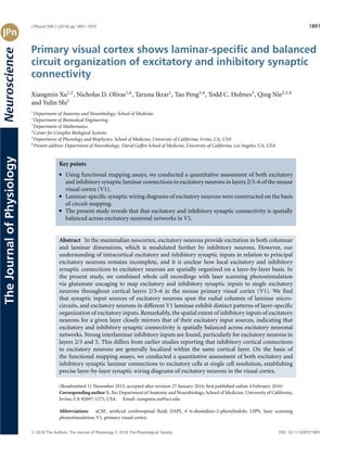
  • 1. J Physiol 594.7 (2016) pp 1891–1910 1891 TheJournalofPhysiologyNeuroscience Primary visual cortex shows laminar-specific and balanced circuit organization of excitatory and inhibitory synaptic connectivity Xiangmin Xu1,2 , Nicholas D. Olivas1,6 , Taruna Ikrar1 , Tao Peng3,4 , Todd C. Holmes5 , Qing Nie2,3,4 and Yulin Shi1 1 Department of Anatomy and Neurobiology, School of Medicine 2 Department of Biomedical Engineering 3 Department of Mathematics 4 Center for Complex Biological Systems 5 Department of Physiology and Biophysics, School of Medicine, University of California, Irvine, CA, USA 6 Present address: Department of Neurobiology, David Geffen School of Medicine, University of California, Los Angeles, CA, USA Key points r Using functional mapping assays, we conducted a quantitative assessment of both excitatory and inhibitory synaptic laminar connections to excitatory neurons in layers 2/3–6 of the mouse visual cortex (V1). r Laminar-specific synaptic wiring diagrams of excitatory neurons were constructed on the basis of circuit mapping. r The present study reveals that that excitatory and inhibitory synaptic connectivity is spatially balanced across excitatory neuronal networks in V1. Abstract In the mammalian neocortex, excitatory neurons provide excitation in both columnar and laminar dimensions, which is modulated further by inhibitory neurons. However, our understanding of intracortical excitatory and inhibitory synaptic inputs in relation to principal excitatory neurons remains incomplete, and it is unclear how local excitatory and inhibitory synaptic connections to excitatory neurons are spatially organized on a layer-by-layer basis. In the present study, we combined whole cell recordings with laser scanning photostimulation via glutamate uncaging to map excitatory and inhibitory synaptic inputs to single excitatory neurons throughout cortical layers 2/3–6 in the mouse primary visual cortex (V1). We find that synaptic input sources of excitatory neurons span the radial columns of laminar micro- circuits, and excitatory neurons in different V1 laminae exhibit distinct patterns of layer-specific organization of excitatory inputs. Remarkably, the spatial extent of inhibitory inputs of excitatory neurons for a given layer closely mirrors that of their excitatory input sources, indicating that excitatory and inhibitory synaptic connectivity is spatially balanced across excitatory neuronal networks. Strong interlaminar inhibitory inputs are found, particularly for excitatory neurons in layers 2/3 and 5. This differs from earlier studies reporting that inhibitory cortical connections to excitatory neurons are generally localized within the same cortical layer. On the basis of the functional mapping assays, we conducted a quantitative assessment of both excitatory and inhibitory synaptic laminar connections to excitatory cells at single cell resolution, establishing precise layer-by-layer synaptic wiring diagrams of excitatory neurons in the visual cortex. (Resubmitted 11 November 2015; accepted after revision 27 January 2016; first published online 4 February 2016) Corresponding author X. Xu: Department of Anatomy and Neurobiology, School of Medicine, University of California, Irvine, CA 92697-1275, USA. Email: xiangmin.xu@uci.edu Abbreviations aCSF, artificial cerebrospinal fluid; DAPI, 4 -6-diamidino-2-phenylindole; LSPS, laser scanning photostimulation; V1, primary visual cortex. C 2016 The Authors. The Journal of Physiology C 2016 The Physiological Society DOI: 10.1113/JP271891
  • 2. 1892 X. Xu and others J Physiol 594.7 Introduction The primary visual cortex (V1), similar to other cortical areas, contains excitatory and inhibitory cell types (Markram et al. 2004; Xu et al. 2010a). Excitatory neurons are a principal cell group; they account for ß80% of the whole cortical neuronal population and convey cortical excitation in both laminar and columnar dimensions. Given that cortical information processing is regulated by diverse types of inhibitory neurons (ß20% of the cortical neurons) and largely determined by local excitatory and inhibitory circuit interactions (Hasenstaub et al. 2005; Ikrar et al. 2012; Haider et al. 2013; D’Amour J & Froemke, 2015), understanding how cortical circuits operate requires the clarification of both excitatory and inhibitory circuit connectivity. Although laminar organization of cortical circuits and the flow of cortical excitation in V1 have been established using anatomical and physio- logical methods (Callaway, 1998; Douglas & Martin, 2004; Olivas et al. 2012), constructing layer-specific connectivity in cortical circuits based on identified neuronal types and their synaptic connections is much more difficult. Excitatory circuit connections to excitatory neurons in different V1 layers have been studied in vitro using physiological approaches such as paired intracellular recordingsofsynapticallyconnectedneurons(Thomson& Lamy, 2007) and laser scanning photostimulation (LSPS) in which wider input sources are mapped to intra- celluarly recorded neurons (Dantzker & Callaway, 2000; Yoshimura et al. 2005; Zarrinpar & Callaway, 2006). The data derived have added important information on local functional circuit connections. However, the knowledge of intracortical synaptic connections to principal excitatory neurons in V1 still remains incomplete because most studiesfocusonexcitatoryneuronsinasinglecorticallayer and there has yet to be a comprehensive and quantitative analysis that examines and compares excitatory synaptic connections to excitatory cell types across cortical layers 2/3–6. In addition, few studies have examined local laminar inhibitory connections to excitatory neurons in the sensory cortex (Xu & Callaway, 2009; Katzel et al. 2010) and it remains unclear how their excitatory and inhibitory synaptic connections are spatially arranged on a layer-by-layer basis across local cortical circuitry. Although excitatory cells receive dense inhibitory neuro- nal innervation in highly localized microcircuits (Fino & Yuste, 2011; Packer & Yuste, 2011), recent work suggests significant interlaminar or cross-laminar inhibitory connections to excitatory neurons (Kapfer et al. 2007; Silberberg & Markram, 2007; Apicella et al. 2012; Olsen et al. 2012; Harris & Shepherd, 2015; Pluta et al. 2015), prompting inhibitory cortical connections to be examined systematically using circuit mapping approaches. In the present study, we used LSPS combined with whole cell recordings (Xu & Callaway, 2009; Ikrar et al. 2011; Kuhlman et al. 2013) to characterize and compare localcircuitconnectivityoftheexcitatoryneuronsinlayers 2/3–6 in living mouse V1 slice preparations. We provide a quantitative assessment of the spatial distribution and input strength of excitatory and inhibitory connectivity with respect to individual pyramidal neurons across V1 laminar circuits, and construct laminar-specific synaptic wiring diagrams of excitatory neurons. The present study provides new knowledge on inhibitory laminar circuit connections and indicates that excitatory and inhibitory synaptic connectivity is spatially balanced onto V1 excitatory neurons. Methods Visual cortical slice preparations C57/B6 strain mice including C57/B6 back-crossed GIN (Oliva et al. 2000) and G42 mice (Chattopadhyaya et al. 2004) from either sex were used for the experiments, in which one animal yielded one or two high-quality V1 slices with clear laminar and cytoarchitectonic features. The GIN and G42 mice express green fluorescent protein in subsets of inhibitory cortical neurons, which facilitated targeted recordings of inhibitory neurons for calibration experiments. All animals were handled and the experiments conducted in accordance with procedures approved by the Institutional Animal Care and Use Committee at the University of California, Irvine. To prepare living brain slices, animals (postnatal days 17–21) were deeply anaesthetized with Nembutal (>100 mg kg−1 , I.P.), rapidly decapitated and their brains removed. The occipital lobe was dissected, and 400 μm thick visual cortical sections were cut in the coronal but slightly oblique plane (ß75° relative to the transverse) (Fig. 1) with a vibratome (VT1200S; Leica Microsystems, Wetzlar, Germany) in sucrose-containing artificial cerebrospinal fluid (aCSF) (in mM): 85 NaCl, 75 sucrose, 2.5 KCl, 25 glucose, 1.25 NaH2PO4, 4 MgCl2, 0.5 CaCl2 and 24 NaHCO3. This slice preparation pre- serves cell morphology and intracortical connections (Fig. 3). Slices were incubated for at least 30 min in sucrose containing aCSF at 32°C before being transferred into slice recording chambers with normal aCSF (in mM): 126 NaCl, 2.5 KCl, 26 NaHCO3, 2 CaCl2, 2 MgCl2, 1.25 NaH2PO4 and 10 glucose. Throughout the cutting, incubation and recording, solutions were continuously supplied with 95% O2/5% CO2. All steps were taken to use only healthy responsive slices. For a specific cut-off time, recordings were made within 5 h after the slice cutting. Electrophysiology and LSPS Our overall system of electrophysiological recordings, imaging and photostimulation was described in detail previously (Xu et al. 2010b). Slices were first placed C 2016 The Authors. The Journal of Physiology C 2016 The Physiological Society
  • 3. J Physiol 594.7 V1 synaptic circuit organization 1893 A A B C D E F G H I LM P V1 Pia LSPS Grid Layer L1 L2/3 L4 L5a L5b L6 1 3 2 2 3 2 3 1 2 3L L Direct Response within 10 ms 50 pA 50 pA 50 pA 50 pA 150 ms Synaptic Window 125 ± 6 325 ± 5 453 ± 7 571 ± 9 676 ± 10 887 ± 9 Distance from Pia (μm) L1 L4 L5a L5b L6 WM soma 250μm L2/3 L2/3 L4 L5a L5b L6pA 0 120 L2/3 L4 L5a L5b L6 pA 0 30 1 ms UV 100pA 100ms Figure 1. V1 slice preparation and LSPS circuit mapping A, schematic V1 slice preparation: slices are made from mouse primary visual cortex, cut at a 75o oblique angle relative to midline to preserve intracortical laminar connections. B, illustration of LSPS mapping of local cortical circuit input to single recorded cells. Excitatory neurons are recorded from binocular V1 region in whole cell mode, and the slice image is superimposed with a 16 × 16 LSPS mapping grid (blue dots, 65 µm2 spacing) centred around the cell soma (triangle) and is aligned to the pial surface. Laminar boundaries are determined by cytoarchitectonic landmarks in bright-field slice images, validated by the boundaries determined by post hoc DAPI staining. C, average depth of laminar boundaries measured from the pial surface to the bottom edge of each layer (n = 15 slices). D, representative LSPS excitatory input map from voltage clamping an L5a pyramidal neuron at −70 mV in response to spatially restricted glutamate uncaging in the mapping grid (B). Each trace is plotted at the LSPS location shown in (B). E, detailed view of evoked EPSCs measured from the L5a pyramidal neuron at three respective locations numbered in (D). Trace 1 demonstrates a large ‘direct response’ resulting from uncaging at the perisomatic region. Trace 2 provides an example of a relatively small direct response in L2/3 from uncaging at the apical dendrite coupled with overriding synaptic inputs (shown in green). Trace 3 illustrates synaptic inputs (EPSCs) measured from a L2/3 location. Note the difference of amplitudes and latencies of direct and synaptic input responses, thus allowing for functional characterization. Empirically, responses within the 10 ms window from laser onset are considered direct, and exhibit a distinct shape (shorter rise time) and occurred immediately after glutamate uncaging (shorter latency). Synaptic events (i.e. EPSCs) are measured with the analysis window of >10–160 ms after photostimulation (grey bar). For details, see Methods. F, colour-coded EPSC input map showing the overall spatial distribution and strength of excitatory inputs to the recorded L5a pyramidal cell. The map is constructed from the responses shown in (D); input responses per location are quantified in terms of average integrated EPSC strength within the analysis window, and colour coded according to the amplitude. G, representative LSPS inhibitory input map from voltage clamping an L5a pyramidal neuron at 5 mV in response to LSPS in the mapping grid similar to (D). H, examples of evoked IPSCs measured in an L5a pyramidal neuron at three respective locations numbered in (G). Trace 1 demonstrates large IPSCs measured near the cell soma. Traces 2 and 3 provide examples of interlaminar inhibition from L2/3. Consistent with excitatory inputs, IPSCs were measured with the analysis window of >10–150 ms after photostimulation (grey bar). I, colour-coded IPSC input map showing the overall spatial distribution and strength of inhibitory inputs made to the L5a pyramidal cell. C 2016 The Authors. The Journal of Physiology C 2016 The Physiological Society
  • 4. 1894 X. Xu and others J Physiol 594.7 Table 1. Electrophysiological properties of V1 excitatory neurons L2/3 L4 L5a L5b L6 AP threshold (mV) –40 ± 3.4 –36 ± 3 –36.2 ± 5.9 –36.9 ± 5.6 –31 ± 1.7 AP ht (mV) 73.8 ± 3.1 68 ± 1.8 78.9 ± 3.3 72.9 ± 3.3 66.6 ± 3.4 AP hw (ms) 2.9 ± 0.2 2.3 ± 0.1 2.2 ± 0.3 2.8 ± 0.5 2.1 ± 0.2 AHP (mV) 11.3 ± 0.9 11.1 ± 1 10.3 ± 2.1 12.1 ± 1.7 11.4 ± 1 AHP time (ms) 27.1 ± 2.8 22.4 ± 1.8 30 ± 0.01 26.7 ± 2.7 15.8 ± 2.5 AP rise time (ms) 1.2 ± 0.2 1 ± 0.04 0.9 ± 0.1 1 ± 0.1 1.1 ± 0.1 AP fall time (ms) 5.2 ± 0.4 4 ± 0.2 4.9 ± 0.7 5.4 ± 1 3.4 ± 0.3 Initial ISI (s) 0.04 ± 0.02 0.02 ± 0.004 0.04 ± 0.01 0.03 ± 0.001 0.03 ± 0.004 Spike rate (Hz) 15 ± 1.1 23 ± 1.8 19.2 ± 3 26.5 ± 10 22.1 ± 1.8 Spike adaptation index 4 ± 0.4 4 ± 0.3 4.6 ± 1 4.2 ± 0.4 3. 7 ± 0.6 The intrinsic physiological characteristics of individual pyramidal neurons recorded across V1 layers were quantitatively measured and grouped according to laminar location (L2/3–L6). Included in the measurements are action potential (AP) threshold, height (ht), half-width (hw), after hyperpolarization potential (AHP) and duration (AHP time), AP rise and fall time, the initial interspike interval (ISI) and spike rate at 200 pA of somatic current injection (Hz), and spike adaptation index. Data are presented as the mean ± SE. and carefully examined under a 4× objective of a DIC/fluorescencemicroscope(BX51WI;Olympus,Tokyo, Japan) for proper laminar targeting of excitatory neurons withinbinocularregionsofmouseV1usingthelandmarks defined in Antonini et al. (1999). To perform whole cell recordings, neurons were visualized at high magnification (60× objective, 0.9 NA; LUMPlanFl/IR; Olympus). Cell bodies of recorded neurons were at least 50 μm below the slice cutting surface and were initially targeted based upon the pyramidal appearance of the cell soma and thick apical dendrite when possible. Patch pipettes (4–6 M resistance) made of borosilicate glass were filled with an internal solution, containing (in mM) 126 K-gluconate, 4 KCl, 10 Hepes, 4 ATP-Mg, 0.3 GTP-Na and 10 phosphocreatine (pH 7.2; 300 mOsm), for examination of intrinsic electrophysiology in current clamp mode and measurement of EPSCs with the membrane potential voltage clamped at −70 mV. In separate recordings, potassium was replaced with caesium in the internal solution; the caesium-based internal solution, containing (in mM): 130 CsOH, 130 D-gluconic acid, 0.2 EGTA, 2 MgCl2, 6 CsCl, 10 Hepes, 2.5 ATP-Na, 0.5 GTP-Na and 10 phosphocreatine (pH 7.2; 300 mOsm), was used to hold excitatory neurons at membrane potentials (0–5 mV) in voltage clamp mode and measure IPSCs. The inter- nal solutions also contained 0.1% biocytin for post hoc cell labelling and further morphological identification. Oncestablewhole-cellrecordingswereachievedwithgood access resistance, basic electrophysiological properties were examined through hyperpolarizing and depolarizing current injections. Spike frequency and adaptation and spike shape analysis (Table 1) were conducted as described previously (Xu et al. 2006). Electrophysiological data were acquired with a Multiclamp 700B amplifier (Molecular Devices, Sunnyvale, CA, USA), data acquisition boards (models PCI MIO 16E-4 and 6713; National Instruments, Austin,TX,USA)and amodified versionofEphus,version 5 (https://openwiki.janelia.org). Data were digitized at 10 kHz and stored on a PC. Only neurons with an initial resting membrane potentials more negative than −55 mV and stable input resistances of <30 M were used. Any recordings in which the access resistance changed by >20% during the course of the experiment were excluded from analysis. During photostimulation experiments, the micro- scope objective was switched from 60× to 4× for delivery of the UV flash stimuli. A stock solution of MNI-caged-L-glutamate (Tocris Bioscience, St Louis, MO, USA) was added to 20 ml of circulating aCSF for a concentration of 0.2 mM caged glutamate. The cortical slice image was acquired at the 4× objective by a high-resolution digital CCD camera, which in turn was used for guiding and registering photostimulation sites. A laser unit (DPSS Lasers, Santa Clara, CA, USA) was used to generate 355 nm UV laser pulses for glutamate uncaging. Short pulses of laser flashes (1 ms, 15 mW) were controlled using an electro-optical modulator and a mechanical shutter. The laser beam formed uncaging spots, each approximating a Gaussian profile with a width of 50-100 μm laterally at the focal plane. Physiologically, under our experimental conditions, LSPS-evoked action potentials (APs) only occurred from stimulation locations within ß100 μm of targeted somata and occurred within ß10–150 ms after photostimulation. During the experiments mapping photostimulation-evoked spiking profiles (excitability profiles), individual excitatory neurons and inhibitory interneurons were recorded in whole cell mode and the spatial distribution of LSPS-evoked APs was measured relative to the cell soma. Our data show that, under our experimental conditions, LSPS had a sufficient resolution for V1 laminar circuit mapping (see below). C 2016 The Authors. The Journal of Physiology C 2016 The Physiological Society
  • 5. J Physiol 594.7 V1 synaptic circuit organization 1895 For our mapping experiments, a standard stimulus grid (16 × 16 stimulation sites, 65 μm2 spacing) was used to cover the V1 slices, spanning from pia to white matter. The LSPS site spacing was empirically determined to capture the smallest predicted distance in which photostimulation differentially activates adjacent neurons, and glutamate uncaging was delivered sequentially in a non-raster, non-random sequence, following a ‘shifting-X’ pattern designed to avoid revisiting the vicinity of recently stimulated sites (Shepherd & Svoboda, 2005). Because glutamate uncaging agnostically activates both excitatory and inhibitory neurons, we empirically determined the excitatory and inhibitory reversal potentials in excitatory pyramidal cells to properly iso- late EPSCs and IPSCs. During calibration experiments, excitatory neurons were voltage clamped at a range of holding potentials from −90 mV to −50 mV and GABA uncaging (6-cyano-7-nitroquinoxaline-2,3-dione caged GABA, 0.1 mM; Invitrogen, Carlsbad, CA, USA) was performed by delivering brief UV flashes (1 ms, 20 mW) to the perisomatic regions of recorded cells to determine the GABAergic reversal potential. The average GABAergic reversal potential ranged from −69 mV to −71 mV (n = 5 excitatory neurons) and so we voltage clamped the targeted pyramidal cells at −70 mV to detect EPSCs. Similarly, the holding potential (0–5 mV) for IPSC detection was determined using glutamate uncaging at a range of holding potentials (−20 mV to +10 mV) with the caesium-containing internal solution. Laminar circuit input analysis Photostimulationcaninducetwomajorformsofuncaging responses: (1) direct glutamate uncaging responses (direct activation of the glutamate receptors of the recorded neuron) and (2) synaptically mediated responses (either EPSCs or IPSCs) resulting from the suprathreshold activation of presynaptic neurons. Uncaging responses within the 10 ms window from laser onset were considered direct, exhibited a distinct shape often with large amplitudes and occurred immediately after glutamate uncaging, as demonstrated in Fig. 1. Synaptic currents with such short latencies are not possible because they would have to occur before the generation of APs in photo- stimulated neurons. Therefore, direct responses need to be excluded from the synaptic input analysis. However, at some locations, synaptic responses were overriding on the relatively small direct responses and were identified and included in synaptic input analysis (Fig. 1). For data map analysis, we implemented a new approach for the detection and extraction of photostimulation-evoked postsynaptic current responses (Shi et al. 2010), which allows detailed quantitative analyses of both EPSCs and IPSCs (amplitudes and the numbers of events across LSPS stimulation sites). LSPS-evoked EPSCs and IPSCs were first quantified across the 16 × 16 mapping grid for each map and two to four individual maps were averaged per recorded cell, reducing the effect of spontaneous synaptic events. The analysis window (>10 ms to 160 ms) after photostimulation was chosen because photostimulated neurons fired most of their APs during this time (Fig. 2). Averaged maps were analysed and response measurements were assigned to individual laminar locations according to slice cytoarchitectonic landmarks and cortical depths from the pia surface (see below). Laminar distributions, average integrated input strength and the numbers of EPSCs measured in excitatory neurons were quantified. Input maps were plotted with average integrated EPSC or IPSC amplitudes, as well as evoked EPSC and IPSC numbers per location. Because almost all layer 1 neurons are inhibitory cells, and pyramidal neurons with apical dendritic tufts in layer 1 could fire APs when their tufts were stimulated in layer 1 (Dantzker & Callaway, 2000), EPSCs detected after photo- stimulation in layer 1 were not included in the analyses. However, because layer 1 neurons can provide inhibition to layer 2/3 neurons, we analysed IPSCs detected after photostimulation in layer 1. Morphological examination and cell-type identification After physiological assays, brain slices were fixed in 4% paraformaldehyde overnight and then transferred to 30% sucrose solution in PBS. Slices were stained against biocytin with 1:500 Alexa Fluor 488-conjugated streptavidin (Jackson ImmunoResearch, West Grove, PA, USA) to visualize the morphology of the recorded cells. Sliceswerealsostainedfor4 -6-diamidino-2-phenylindole (DAPI) (Sigma-Aldrich, St Louis, MO, USA) to identify laminar boundaries. The cell morphology was visualized using a BX 61 (Olympus) epifluorescence microscope and the MetaMorph imaging suite (Molecular Devices). Based upon in vitro cytoarchitectonic landmarks observed in living slice images coupled with post hoc DAPI staining, the average depths of laminar boundaries for layers 1, 2/3, 4, 5a, 5b and 6 were determined to be 125 ± 6 μm, 325 ± 5 μm, 453 ± 7 μm, 571 ± 9 μm, 676 ± 10 μm and 887 ± 9 μm, respectively, from the pial surface (Fig. 1). Additionally, the somatic distance from pia was measured for each recorded neuron. These measurements enabled proper identification of the laminar locations of recorded cells and characterization of presynaptic input sources. Computational modelling We adopted a discrete dynamical model (Christley et al. 2009) with the inference of the connectivity among excitatory neurons at four different cortical layers to describe and simulate photostimulation mapped circuit C 2016 The Authors. The Journal of Physiology C 2016 The Physiological Society
  • 6. 1896 X. Xu and others J Physiol 594.7 activities. The input data of the model were derived from the temporal data based on the LSPS-mapped synaptic inputs (EPSCs and IPSCs) to the representative excitatory neurons in different cortical layers. We extracted the temporal data from LSPS-mapped synaptic inputs to excitatory neurons, with six representative neurons from layer 2/3, four neurons from layer 4, seven neurons from layer 5 and four neurons from layer 6. The integration data of synaptic inputs was extracted at each 10 ms window using the detection method described above. To set a cut-off threshold for background noises, the data points (10 ms per point) with their strengths less than 200 pA L2/3 PYR A B C E D F G H I L5 PYR L5 FS inhibitory neuron L4 PYR L6 PYR L2/3 non-FS inhibitory neurons 50 mV 20 mV 200 ms 100 mV 100 ms 1 ms UV 500 ms 100 pA –50 pA 100 pA 40 mV 1000 ms 50 pA –10 pA 150 pA 50 mV 1000 ms 100 pA –50 pA 150 pA 50 mV 1000 ms 50 pA –100 pA 400 pA 50 mV 1000 ms 250 pA –50 pA 400 pA 50 mV 1000 ms 250 pA –100 pA Pyramidal neurons Fast spiking (FS) inhibitory neurons Non-FS inhibitory neurons 100 120 Spikingdistance fromsoma(μm) Spikingdistance fromsoma(μm) Spikingdistance fromsoma(μm) 8 6 9 6 5 4 4 4 2 3 3 4 80 60 40 20 0 L2/3 neurons L4 L5a L5b L6 L2/3 neurons L4 L5a/b L6 L2/3 neurons L4 L5a/b 160 120 80 40 0 160 120 80 40 0 Figure 2. Spatial resolution of photostimulation A–F, examples of photostimulation-evoked excitability profiles of pyramidal and inhibitory interneurons in different visual cortical layers. A, current injection responses of an example L2/3 pyramidal neuron are shown on the left; the image of V1 slice where the cell was recorded in layer 2/3 is superimposed with photostimulation sites (∗, cyan dots, 65 µm2 spacing) (middle) and the photostimulation responses of the recorded neuron are plotted at the beginning of stimulation onset (right). The individual responses are plotted relative to their spatial locations in the mapping array shown in the middle. The small red circle indicates the somatic location of the recorded neuron. One response trace with photostimulation-evoked APs is indicated in red, and shown separately by the side. Laser flashes (1 ms, 15 mW) were applied for photostimulation mapping. The scale in (A) is 500 µm. B–F, similarly formatted as in (A), with example L4, L5 and L6 pyramidal neurons, and L5 fast spiking inhibitory neurons and L2/3 non-fast spiking inhibitory neurons, respectively. C, two response traces with photostimulation-evoked subthreshold depolarization (green, photostimulation at the apical dendrite) and suprathreshold APs (red, perisomatic region) are shown separately. G–I, spatial resolution of LSPS evoked excitability of pyramidal neurons, fast-spiking and non-fast spiking inhibitory neurons was determined by measuring the LSPS evoked spike distance relative to soma location. Note that the spiking distance is measured as the ‘vertical’ distance (perpendicular to cortical layers) above and below the cell body. The numbers of recorded neurons are shown at the bar graphs. Data are presented as the mean ± SE. C 2016 The Authors. The Journal of Physiology C 2016 The Physiological Society
  • 7. J Physiol 594.7 V1 synaptic circuit organization 1897 50 pA/10 ms were set to zero. For photostimulation in each and specific cortical layer (i.e. L2/3, L4, L5 and L6), the overall activation strength was calculated by summing the area under each curve of temporal input evolution across all layers, and the relative laminar activation was obtained by comparing its area under the curve with the overall activation across layers. Our model consists of the four cortical layers (L2/3, L4, L5 and L6) of excitatory neurons. In this model, a matrix, W = (Wij)n,n is used to represent the connectivity strength among different cortical layers. If Wij > 0, layer j receives excitatory input from layer i. If Wij < 0, layer j receives inhibitory input from layer i. If Wij = 0, there is no direct connection between layers i and j. Specifically, for the present study, the W = (Wij)4,4 has 16 entries, and the entries for L2/3 with L2/3, L4, L5 and L6 are W11, W12, W13 and W14. The entries for L4 with L2/3, L4, L5 and L6 are W21, W22, W23 and W24. The entries for L5 with L2/3, L4, L5 and L6 are W31, W32, W33 and W34. The entries for L6 with L2/3, L4, L5 and L6 are W41, W42, W43 and W44. The data from the mapping experiment were then used to fit the linear system to solve for W. The data fitting is further constrained by including the prior knowledge of the connectivity of cortical layers, as well as a term that controls the density of the connections, which may potentially remove very weak interlaminar connections. The model provides an optimal estimate for the connectivity strength matrix by minimizing the difference between the model-calculated signals and the measured experimental signals. Such an approach has been successfully used to obtain the gene regulatory network based on gene expression data (Christley et al. 2009). Mathematically, the objective function for fitting the model to the data is: W∗ = arg min W (g(W) + α||W||1 + β||W ◦ W0 ||1) where g(W) = p h=1 n k=1 m l=1 ||Wxl k,h − ul k,h ||2 2. In the objective function, α and β are the non-negative numbersandW0 isthepriorknowledgeoftheconnectivity strength of cortical layers (i.e. laminar relative activation; see above). The m (= 4) is the number of the given laminar photostimulation, the n (= 15) is the number of data pairs at each given photostimulation, the p (= 2) is the number of types of synaptic inputs (excitation and inhibition). If there is a connection from layer i to layer j, w0 ij = 0; otherwise, w0 ij = 1. ||W||1 is the term for controlling the sparseness of the network (i.e. the density of the connections), and ||W ◦ W0 ||1 is for incorporating the prior information where ◦ is the operation for the entry-wise multiplication. In the model, we used a simplified version of the network map as the prior information. The measured experimental data input, xl k,h , represents input strengths of L2/3, L4, L5 and L6 at time point k with a lth layer photostimulation (l = 1, 2, . . . , 4; photostimulation in L2/3, L4, L5 or L6), given hth type laminar photostimulation (for measuring excitation or inhibition), and ul k,h represents input strengths of L2/3, L4, L5 or L6 at time point k + 1 at the lth layer photostimulation given hth type laminar photo- stimulation as the corresponding output of xl k,h . Wxl k,h is the model-calculated input strengths of L2/3, L4, L5 or L6 at time point k + 1 at the lth given hth type laminar photostimulation. The objective of the modelling is to obtain the optimal connectivity strength matrix W∗ by minimizing the difference between the model-calculated input strengths Wxl k,h and the measured experimental input strengths ul k,h . For each given laminar photo- stimulation, there were 16 time points, including 15 data pairs (xk,h l ,uk,h l ), where k = 1, 2, . . . , 15, l = 1, 2, . . . , 4 (photostimulation in L2/3, L4, L5 or L6 respectively) and h = 1, 2 (measuring excitation or inhibition). Altogether, 120 data pairs (15 × 4 × 2) were employed to train the model. We used the standard 10-fold cross-validation technique from machine learning to determine the values of α and β. Then, the optimal connectivity matrix W∗ was calculated using the algorithm described in our previous study (Christley et al. 2009). Statistical analysis Data are presented as the mean ± SE. For statistical comparisons between groups, the data were checked for normality distribution and equal variance. If the criteria were met, a t test was performed to compare two groups; when the criteria were not met, a Mann–Whitney U test was used. For statistical comparisons across more than two groups, we used the Kruskal–Wallis test (non-parametric one-way ANOVA) and the Mann–Whitney U test for group comparisons. In all experiments, P < 0.05 was considered statistically significant. Results LSPS mapping of mouse V1 intracortical synaptic organization LSPS combined with whole cell recordings has been effectively applied for analysing cortical circuit organiza- tion (Schubert et al. 2003; Shepherd et al. 2003; Shepherd & Svoboda, 2005; Yoshimura et al. 2005; Zarrinpar & Callaway, 2006; Weiler et al. 2008; Xu & Callaway, 2009; Oviedo et al. 2010; Xu et al. 2010b; Hooks et al. 2011; Ikrar et al. 2011). It allows for high resolution mapping of presynaptic input sources to single neurons at a local C 2016 The Authors. The Journal of Physiology C 2016 The Physiological Society
  • 8. 1898 X. Xu and others J Physiol 594.7 circuit level, and the simultaneous recording from a postsynaptic neuron with sequential photostimulation of clusters of presynaptic neurons at many different locations provides a quantitative measure of the spatial distribution of excitatory or inhibitory inputs in the broad range of the photostimulation field. As shown in Fig. 1, the LSPS approach involves recording from single neurons, then stimulating at surrounding sites on the LSPS mapping grid to generate APs from neurons in those sites, thus providing a map for the recorded neuron based on activation of presynaptic inputs. With subsequent anatomical characterization of the postsynaptic neuron, as well as its functional properties, the cell type can be classified, allowing a map of input sources to be generated for a known cell type. Tointerpretinputmaps,itisfirstnecessarytodetermine the spatial extent or precision with which neurons at the stimulation site respond. As shown in Fig. 2, individual excitatory neurons (n = 34 across the layers) and inhibitory interneurons (14 fast-spiking inhibitory neurons and 10 non-fast spiking inhibitory neurons) were recorded in whole cell mode and the spatial pre- cision/resolution of photostimulation was assessed by the photostimulation-evoked spiking distance between the recorded neuron and the stimulation site. The distance is expressed as the ‘vertical’ distance (perpendicular to cortical layers) above and below the cell body because this is the measurement that is relevant for assignment of stimulation sites to correct layers. Recordings of fast spiking inhibitory neurons (Fig. 2E and H) and non-fast spiking inhibitory neurons (Fig. 2F and I) were facilitated by identification of EGFP expression in these cell types using G42 and GIN mice (Xu & Callaway, 2009). Overall, photostimulation-evoked neuronal excitability profiles indicate that LSPS has a sufficient resolution for V1 laminar circuit mapping. The mean ± SE distances of photostimulation-evoked APs from the recorded cell bodies for layer 2/3 (L2/3), layer 4 (L4), layer 5a (L5a), layer 5b (L5b) and layer 6 (L6) pyramidal neurons were 101.3 ± 12.8 μm, 63.3 ± 14.6 μm, 88.6 ± 13 μm, 89.2 ± 14.7 μm and 89 ± 25.2 μm, respectively (Fig. 2A–D and G). The numbers of APs evoked per spiking location were 1.9 ± 0.2 APs. The spiking distances for fast-spiking and non-fast spiking inhibitory interneurons appeared to be slightly larger than those of excitatory neurons, varying from ß90 μm to 160 μm in different cortical layers (Fig. 2E–F and H–I). The average distances of pooled inhibitory neurons were 117.5 ± 8.5 μm, and the numbers of evoked spikes measured per spiking location were 2.2 ± 0.5 APs. These excitability profiles further indicate that photostimulation-evoked spiking occurs upon direct stimulation and not through excitation of synaptically coupled neurons. Note that we could have restricted analyses to stimulation sites at least 65 μm from the laminar boundaries to further reduce ambiguity in determining laminar locations of presynaptic neurons (e.g. L4 vs. L5a, or L5b vs. L6), although our pre- liminary measurements showed no significant differences in the quantification of the laminar input patterns of cells compared to those not excluding the sites at laminar boundaries. Taken together, these data validate the use of LSPS for laminar circuit mapping of direct excitatory and inhibitory synaptic connections to V1 excitatory neurons. Werecordedfrom66excitatoryneurons(N=14,15,10, 11 and 16 cells for L2/3, L4, L5a, L5b and L6, respectively) that were morphologically and physiologically identified as excitatory pyramidal neurons (Fig. 3), and their excitatory synaptic connections from local circuits were assayed quantitatively. In terms of intrinsic electro- physiology, these excitatory cells exhibited relatively uniform properties associated with spike frequency and adaptation and spike shapes in response to intrasomatic current injection (Table 1). In addition, another 57 excitatory neurons (n = 10, 17, 10, 10 and 10 cells for L2/3, L4, L5a, L5b and L6, respectively) were separately recorded to map inhibitory synaptic connections in local V1 circuits. Laminar excitatory inputs to V1 excitatory cells Excitatory neurons in different V1 laminae exhibit distinct patterns of laminar-specific organization of excitatory inputs (Fig. 4). Neurons were binned according to their laminarlocationandtheaggregatesynapticinputmapsare L1 A B C D E F G L4 L5a L5b L6 200µm L2/3 Figure 3. Morphological profiles of V1 excitatory neurons Neurons recorded for laminar synaptic input analyses were morphologically examined via biocytin staining. DAPI counterstain was used to establish layer identity. A, representative upper L2/3 pyramidal neuron with truncated apical dendrites. B, excitatory neurons in deeper L2/3 exhibiting longer apical dendrites projecting toward the pial surface. C, almost all L4 excitatory cells recorded confirmed pyramidal cells with apical dendrites projecting toward the pial surface. D–E, L5a and L5b excitatory cells exhibiting typical tall pyramidal cell morphology. F, long L6 pyramidal cell with apical dendrites projecting into L5 and L4. G, example of a L6 pyramidal neuron with short apical dendrites terminating in L5. C 2016 The Authors. The Journal of Physiology C 2016 The Physiological Society
  • 9. J Physiol 594.7 V1 synaptic circuit organization 1899 displayed as averaged maps corresponding to postsynaptic locations of recorded excitatory cells. L2/3 pyramidal neurons receive the strongest excitatory input from L4 and L5a, weaker excitatory inputs from L2/3 and L5b, and minimal input from L6 (Fig. 4A and C and Table 2). We also normalized laminar input strength as the percentage evoked input by expressing the average evoked input from each layer as a percentage of the summed input from all layers (Table 2) (Xu & Callaway, 2009). The numbers of LSPS-evoked EPSCs per location are also L2/3 A B C D L2/3 L4 L5a L5b L6 L2/3 L4 L5a L5b L6 (n=14) (n=15) (n=10) (n=11) (n=16) (n=14) (n=15) (n=10) (n=11) (n=16) 0.2 mm 36 EPSCAmplitude(pA)NumberofEPSCs 18 0 3 1.5 0 L5a L5b L6 L2/3 L4 L4 L5a L5b L6 260 520 780 1040 DistancefromPia(µm)DistancefromPia(µm) 0 0 1 2 3 0 1 2 0 0 0 0.4 0.8 1.2 1.61 12 23 10 20 30 0 10 20 30 0 EPSC Amplitude (pA) EPSC Number 0 0 4 8 1210 1020 2030 40 260 520 780 1040 L2/3 L4 L5a L5b L6 L2/3 L4 L5a L5b L6 Figure 4. Local excitatory inputs to V1 pyramidal neurons A, group averaged excitatory input maps for excitatory pyramidal cells for each cortical lamina (L2/3, L4, L5a, L5b and L6). Each colour coded pixel represents the average evoked EPSC amplitude across the LSPS mapping grid; white triangles represent the cell body locations of recorded pyramidal neurons. The left most map charts laminar V1 boundaries relative to the 16 × 16 mapping grid based on post hoc DAPI counterstaining. B, numbers of EPSCs per LSPS location are plotted from the averaged maps presented in (A). For normalization, colour-coded laminar excitatory input maps are scaled to the highest input value across groups. C, EPSC amplitudes are quantified across the radial vector corresponding to the columnar position of recorded cells across layers. D, laminar profiles of EPSC numbers are quantified across the radial vector corresponding to the columnar position of recorded cells throughout the cortical depth, similar to (C). Data are presented as the mean ± SE. For the L2/3 pyramidal cells examined, the average numbers of evoked EPSCs per photostimulation are 1.1 ± 0.3, 1.8 ± 0.4, 1.5 ± 0.5, 0.5 ± 0.3 and 0.2 ± 0.1 for L2/3, L4, L5a, L5b and L6, respectively. The average numbers of evoked EPSCs per photostimulation for L4 pyramidal neurons are 0.5 ± 0.2, 2.2 ± 0.7 and 1.3 ± 0.5 in L2/3, L4 and L5a, respectively. The average numbers of evoked EPSCs per photostimulation for L5 pyramidal neurons are 1.4 ± 0.2, 1.5 ± 0.8, 1.5 ± 0.7, 0.8 ± 0.1 and 0.3 ± 0.2 from L2/3, L4, L5a, L5b and L6, respectively, whereas L5b cell values are 1.0 ± 0.3, 1.3 ± 0.7, 1.2 ± 0.6, 0.6 ± 0.1 and 0.7 ± 0.2 from L2/3, L4, L5a, L5b and L6, respectively. The average numbers of evoked EPSCs per photostimulation for L6 cells are 0.2 ± 0.6, 0.3 ± 0.5, 0.5 ± 0.1 and 1.1 ± 0.1 for L4, L5a, L5b and L6, respectively. C 2016 The Authors. The Journal of Physiology C 2016 The Physiological Society
  • 10. 1900 X. Xu and others J Physiol 594.7 Table2.LSPSevokedinput(EI)andpercentageevokeinput(%EI)fromeachcorticallayertotargetedexcitatorycellsacrosslocalV1circuits L2/3cellsL4cellsL5acellsL5bcellsL6cells InputlayerEI(pA)%EIEI(pA)%EIEI(pA)%EIEI(pA)%EIEI(pA)%EI Excitatoryinput L2/35.9±1.928.8±9.12.1±1.013.3±5.621.3±9.646.4±11.89.3±3.631.3±11.10.6±0.35.5±3.0 L414.3±3.135.4±7.612.9±3.752.2±10.515.3±6.118.6±6.014.6±3.927.6±6.01.7±0.78.2±4.4 L5a13.9±4.126.7±6.912.8±5.826.6±7.717.6±5.722.8±6.810.2±3.416.6±4.81.7±0.812±5.0 L5b4.5±2.13.8±1.90.58±0.51.3±1.05.5±2.04.1±1.14.2±1.04.7±4.72.6±0.96.5±1.6 L61.7±0.54.6±1.81.0±0.75.0±3.51.7±0.84.4±2.16.9±3.018.5±7.66.6±1.756.6±12.2 Inhibitoryinput L112.3±9.25.9±3.70.27±0.10.6±0.40.4±0.20.4±0.20.4±0.30.6±0.50.1±0.10.2±0.2 L2/345.7±11.751.8±9.48.4±2.427.1±7.611.8±3.419.5±5.024.8±13.016.9±7.00.9±0.51.7±1.0 L451±11.429.0±4.535.2±11.543.4±9.027.5±9.421.0±6.326.8±9.911.7±3.46.9±4.63.5±2.1 L5a15.2±3.69.55±2.414.1±5.123.3±6.455±10.044.0±4.639±10.019.5±5.47.2±3.75.0±1.9 L5b5.2±1.61.9±0.74.2±2.61.6±0.624.9±7.69.0±2.444.8±14.111.4±2.218.2±5.87.3±1.5 L61.1±0.41.3±0.52.0±1.13.4±1.65.7±2.66.0±2.251.6±15.032.9±8.141.3±11.079.0±12.8 Excitatoryinput:laminarexcitatorysynapticinputsmadeontoV1excitatorycellsoflayers2/3through6(columns)mappedbyLSPS.Notetheabsenceofexcitatoryneuronsin L1andhencenoexcitatoryinputsfromL1.LSPSevokedinputsaredisplayedasaverageexcitatoryevokedinputsinunitsofpAfromeachcorticallayer,andthepercentage ofexcitatoryevokedinput,whichshowstherelativecontributionofsynapticinputfromeachcorticallayertorecordedcells.Inhibitoryinputs:laminarinhibitoryinputsmade ontoV1excitatorycellsoflayers2/3through6(columns).LSPSevokedinputsaredisplayedasaveragedinhibitoryevokedinputsinunitsofpAandthepercentageofinhibitory evokedinput(%EI),whichshowstherelativecontributionofsynapticinputfromeachcorticallayertorecordedcells.Averageevokedinputsmeasuredfromeachlayerduring theanalysiswindowafterphotostimulationwerecomparedwithbaselinespontaneousactivityof100mspriortophotostimulation.Redvaluesindicatenon-significantlaminar inputsinferredfromsuchstatisticalcomparisons(P>0.05).Dataarepresentedasthemean±SE. C 2016 The Authors. The Journal of Physiology C 2016 The Physiological Society
  • 11. J Physiol 594.7 V1 synaptic circuit organization 1901 measured independent of EPSC strength (Fig. 4B and D), which provides a different means of evaluating excitatory synaptic connectivity across V1 laminar circuits. L4 pyramidal neurons have a laminar input pattern distinguishable from L2/3 cells because they receive localized excitatory input mostly from L4 and L5a (Fig. 4 and Table 2). Although L4 cells receive some L2/3 input, they do not receive significant evoked input from L5b or L6 compared to baseline spontaneous activity. The EPSC input amplitudes from L5b and L6 during photo- stimulation do not differ significantly from those of spontaneously occurring EPSCs. Among all the cortical cells in different laminar locations, L5 pyramidal cells have the most extensive input from the radial columns of laminar circuits and exhibit sublayer-specific connectivity to upper layer 5 (L5a) and lower layer 5 (L5b) pyramidal cells (Fig. 4 and Table 2). L5a and L5b are differentiated based upon cytoarchitectonic landmarks and cortical depths of the laminar positioning of the recorded cell somata. L5a pyramidal cells receive strong excitatory inputs from L2/3, L4 and L5a, and much weaker inputs from L5b and L6 (Table 2). In comparison, L5bpyramidalcellsreceiveweakexcitatoryinputsfromthe radial columns with relatively concentrated inputs from L2/3 and L4 (Table 2). The average numbers of evoked EPSCs per photostimulation for L5a and L5b cells follow the trend of their laminar input amplitudes. Compared with other cortical cells, L6 pyramidal cells receive generally weak and primarily localized inputs spanning a wide horizontal domain from within deep cortical layers (Fig. 4 and Table 2). L6 pyramidal neurons do not receive significant L2/3 input compared to baseline activity (Table 2). Laminar inhibitory inputs to V1 excitatory neurons Systematic examinations of inhibitory synaptic connec- tivity to excitatory neurons and comparisons of their excitatory connectivity indicate that the spatial distri- bution of local inhibitory inputs to V1 pyramidal neurons generally matches that of excitatory input sources for each cortical layer. The presence of inhibitory neurons in L1 prompted investigation of the extent of L1 inhibitory inputs to pyramidal neurons in L2/3, L4 and L5 because these cells have apical dendrites extended into L1 (Fig. 3). L2/3 pyramidal neurons receive a great majority of inhibitory inputs from L1, L2/3, L4 and L5a (Fig. 5 and Table 2). Compared to baseline spontaneous activity, L2/3 pyramidal neurons do not receive significant inhibitory input from L5b or L6, indicating a lack of direct inhibitory innervation from deep infragranular layers. L4 pyramidal cells receive dominant inhibitory inputs from within L4 with some interlaminar inhibitory input from upper L5 (Fig. 5 and Table 2). L4 pyramidal neurons do not receive significant inhibitory inputs from L1, L2/3, L5b and L6. Notably, L4 received the fewest number of LSPS-evoked IPSCs compared to other layers (Fig. 5). L5 excitatory pyramidal cells receive extensive inhibitory input from columnar circuits; these cells receive inhibitory input from both upper and deeper cortical layers (Fig. 5 and Table 2). Inhibitory inputs to L5b cells span the radial column of V1 with strongest input from within L5b (Fig. 5 and Table 2). LSPS-evoked inhibitory inputs to L5a or L5b cells from L1 are not statistically different from baseline activity. There is dense inhibitory innervation onto L5 pyramids from L5 and L6. L6 pyramidal cells receive extensive inhibitory inputs from within L6 and slight inhibitory input from the upper cortical layers (Fig. 5 and Table 2). L1 and L2/3-evoked IPSCs measured in L6 pyramidal neurons are not statistically different from baseline activity. Comparative excitatory and inhibitory laminar inputs across V1 To examine whether excitatory and inhibitory synaptic connectivity spatially overlaps, the spatial distributions of both excitatory and inhibitory synaptic input sources were compared for each layer across V1 laminar circuits. To directly compare excitatory and inhibitory input maps, aggregate synaptic input maps were normalized and coloured-coded on separate channels (Fig. 6A and B), and merged (Fig. 6C). Synaptic input fields of excitatory neurons span horizontally within laminar microcircuits, and cover 400–500 μm across the columnar microcircuits. To a large degree, excitatory neurons in each cortical layer appear to have matched excitatory and inhibitory synaptic input locations across both horizontal and vertical dimensions of columnar microcircuits. In validation of this balanced overlap, we further quantitatively compared the horizontal and vertical synaptic input profiles of excitatory and inhibitory synaptic connectivity (Fig. 6D and E). Statistical analyses indicate that the distributions of excitatory vs. inhibitory synaptic connectivity for excitatory neurons at a given layer do not differ significantly. A separate analysis was used to measure the degree of matched spatial distribution of synaptic inputs across V1 laminar circuits. Average excitatory and inhibitory input maps were converted to binary matrices and synaptic inputs measured across the LSPS mapping grid were identified as either all or nothing per LSPS location. This method allowed direct evaluation of whether excitatory and inhibitory responses are either present or absent at a given spatial location. The number of matched pixels per group of neurons in distinct layers was then measured relative to the total locations surveyed by LSPS. The high degree of matched excitatory and inhibitory circuit topography across layers is found to be 81%, C 2016 The Authors. The Journal of Physiology C 2016 The Physiological Society
  • 12. 1902 X. Xu and others J Physiol 594.7 95%, 89%, 82% and 81% for layers 2/3, 4, 5a, 5b and 6, respectively. This analysis reveals that pyramidal neurons receive closely matched distributions of excitatory and inhibitory connections across V1 laminar circuits. Functional synaptic connectivity of mouse V1 laminar circuitry Based upon our quantitative assessment of both excitatory and inhibitory synaptic laminar connections to excitatory L1 A B C D 80 40 0 4.5 2.25 0 L6L5bL5a IPSC Amplitude (pA) IPSCAmplitude(pA)NumberofIPSCs IPSC Number L4L2/3 L6L5bL5aL4L2/3 260 520 780 1040 DistancefromPia(µm)DistancefromPia(µm) 0 20 40 60 0 20 40 60 0 20 40 60 00 2020 4040 6060 8080 0 1 2 0 0 2 41 23 4 0 1 2 3 4 0 1 2 3 4 260 520 780 1040 L2/3 L2/3 (n=10) (n=17) (n=10) (n=10) (n=10) (n=10) (n=17) (n=10) (n=10) (n=10) 0.2 mm L4 L5a L5b L6 L2/3 L4 L5a L5b L6 L4 L5a L5b L6 L1 L2/3 L4 L5a L5b L6 Figure 5. The spatial extent of local inhibitory inputs to V1 pyramidal neurons generally matches that of excitatory input sources for each cortical layer A, group averaged inhibitory synaptic input maps for pyramidal neurons across V1 laminae. Each colour coded pixel represents the average IPSC amplitude across the LSPS mapping grid; black triangles represent the locations of recorded pyramidal neurons. The left most map charts V1 laminar boundaries relative to the LSPS mapping grid. B, numbers of IPSCs per LSPS location are plotted from the averaged maps presented in (A). For normalization, colour-coded laminar inhibitory input maps are scaled to the highest input value across groups. C, laminar profiles of evoked inhibitory synaptic input with average IPSC amplitudes quantified across the radial vector corresponding to the columnar position of recorded cells throughout the cortical depth. D, laminar profiles of evoked IPSC numbers quantified across the radial vector corresponding to the columnar position of recorded cells throughout the cortical depth, as in (C). Data are presented as the mean ± SE. The average numbers of evoked IPSCs per stimulation for L2/3 pyramidal cells are 0.6 ± 0.01, 2.6 ± 0.2, 2.6 ± 0.8 and 1.2 ± 0.7 for L1, L2/3, L4 and L5a, respectively (B and D). The average numbers of evoked IPSCs per stimulation for L4 pyramidal cells are 1.8 ± 0.6 and 1.18 ± 0.5 for L4 and L5a. The average numbers of evoked IPSCs per stimulation for L5a pyramidal cells are 1.1 ± 0.3, 1.7 ± 0.8, 3.9 ± 0.7, 2.2 ± 0.1 and 0.8 ± 0.2 from L2/3, L4, L5a, L5b and L6, respectively, whereas the values for L5b pyramidal cells are 1.0 ± 0.28, 1.5 ± 0.8, 2.4 ± 0.7, 2.8 ± 0.1 and 2.8 ± 0.2 from L2/3, L4, L5a, L5b and L6, respectively. The average numbers of evoked IPSCs per stimulation for L6 pyramidal cells are 0.4 ± 0.8, 0.7 ± 0.7, 1.4 ± 0.1 and 3.4 ± 0.2 from L4, L5a, L5b and L6, respectively. C 2016 The Authors. The Journal of Physiology C 2016 The Physiological Society
  • 13. J Physiol 594.7 V1 synaptic circuit organization 1903 L2/3 A B C D E ExcitatoryinputInhibitoryinput DistancefromPia(µm)Normalized SynapticInput L2/3 (n=14) 200 µm (n=15) (n=10) (n=11) (n=16) (n=17) (n=10)(n=10) (merged) (merged) (merged) (merged) (merged) (n=10)(n=10) L4 L5a L5b L6 L2/3 L4 L5a L5b L6 L2/3 L4 L5a L5b L6 <90% <90% >10% >10% L5a L5b L6 L2/3 L4 L4 L5a L5b L6 L2/3 L4 L5a L5b L6 1 0 260 520 780 1040 –1 –1 0 1 –1 0 1 –1 0 1 –1 0 1 –1 0 1 –290 Horizontal Distance from soma (µm) Normalized Synaptic Input –160 160 290 –290 –160 160 290 –290 –160 160 290 –290 –160 160 290 –290 –160 160 290 L2/3 E E I I L4 L5a L5b L6 L2/3 L4 L5a L5b L6 Figure 6. Excitatory and inhibitory synaptic connectivity spatially overlap in a balanced manner for each layer across V1 circuits A, excitatory synaptic input maps are normalized within respective postsynaptic layers and re-plotted from black to blue. Dominant local pathways are displayed by filtering the lower 10% of excitatory inputs based upon EPSC amplitude and scaling the respective laminar maps to 90% of the largest excitatory synaptic inputs measured. B, inhibitory synaptic input maps are normalized within respective postsynaptic layers and plotted across lamina, as in (A) but colour coded from black to red. Consistent with (A), dominant local pathways are displayed from filtering the lower 10% of excitatory inputs based upon IPSC amplitude and scaling the respective maps to 90% of the largest inhibitory synaptic inputs measured within respective postsynaptic layers. C, excitatory and inhibitory normalized input maps are merged according to the laminar position of recorded postsynaptic neurons; violet pixels represent locations of matched synaptic input from both presynaptic excitatory and inhibitory input domains. D, horizontal distribution of normalized excitatory inputs (blue) and inhibitory inputs (red) are plotted for inputs measured across the vertical vector on the LSPS mapping grid to the recorded postsynaptic neurons. E, vertical distribution of normalized synaptic inputs are plotted for inputs measured across the horizontal vector on the LSPS mapping grid to the recorded postsynaptic neurons. C 2016 The Authors. The Journal of Physiology C 2016 The Physiological Society
  • 14. 1904 X. Xu and others J Physiol 594.7 cells in each cortical layer, we constructed layer-by-layer synaptic wiring diagrams indicating the comparative strength of excitatory and inhibitory inputs to excitatory neurons in the visual cortex (Fig. 7). Using the quantitative data shown in Table 2, the average excitatory and inhibitory input strength of specific cortical laminae to targeted postsynaptic excitatory cells was visualized in the matrices of Fig. 7A and B, respectively. Several features merged from functional wiring diagrams of excitatory neurons are summarized in Fig. 7C for which matched excitatory and inhibitory circuit topography is clearly seen. Thestrengthofexcitatoryandinhibitoryconnectionsto L2/3, L4 and L5a pyramidal cells exceeded the connection strength of L5b and L6 pyramids. Although strong inter- laminarexcitatoryconnectionsareknowntoexistbetween excitatory cells (e.g. L4 → L2/3; L2/3 ↔ L5), in the present study, we unravelled robust interlaminar inhibitory inputs totheseexcitatoryneurons,whichindicatesthatinhibitory cortical connections are not necessarily limited in highly localized dimensions as generally described (Binzegger et al. 2004; Thomson & Lamy, 2007; Katzel et al. 2011). In addition, L4 and L5a excitatory cells have stronger intra- laminarexcitatoryconnectionsthanL2/3,L5bandL6cells; L4 cells, which comprise the major thalamocortical input layer, have weaker intralaminar inhibitory connections than the cells in other cortical layers. Only L2/3 cells have significant inhibitory input from L1. L2/3 25 60 30 0 12 0 L2/3 Targeted cell location A B C Local Excitatory Inputs Descending Ascending Descending Ascending Local Inhibitory Inputs PrestnapticInputLayer PrestnapticInputLayer Targeted cell location InhibitorySynapticInput(pA) ExcitatorySynapticInput(pA) L4 L4 L5a L5a L5b L5b L6 L1 L2/3 L4 L5a L5b L6 L6 L2/3 L4 L5a L5b L6 L2/3 L4 L5a L5b L6 Figure 7. Organization of the comparative strength of excitatory and inhibitory inputs between cortical layers in local V1 circuits A, input layer → single neuron connectivity matrix showing average excitatory inputs from specific cortical laminae to targeted postsynaptic pyramidal cells (for detailed quantitative results, see Table 2). B, input layer → single neuron connectivity matrix showing average inhibitory inputs from specific cortical laminae to targeted post- synaptic pyramidal cells (for detailed quantitative results, see Table 2). C, functional wiring diagram of excitatory neurons constructed based upon the quantitative assessments shown in (A) and (B). Left: excitatory laminar inputs to pyramidal cells across V1 local circuits. Descending interlaminar excitatory inputs are indicated in cyan and ascending interlaminar excitatory inputs are indicated in blue. Intralaminar inputs are shown within each targeted layer. Note that the thickness of arrows indicates the relative strength of synaptic connectivity from the input layer to the recipient neurons. Right: inhibitory laminar inputs to pyramidal cells. Descending interlaminar inhibitory inputs are indicated in violet and ascending interlaminar inhibitory inputs are indicated in red. C 2016 The Authors. The Journal of Physiology C 2016 The Physiological Society
  • 15. J Physiol 594.7 V1 synaptic circuit organization 1905 2000 A B C D E F G H L2/3 L4 L5 L6 L2/3 L4 L5 L6 L2/3 L4 L5 L6 L2/3 L4 L5 L6 L2/3 L4 L5 L6 L2/3 L4 L5 L6 L23 L4 L5 L6 Excitation from layer 2/3 L2/3 L4 L5 L6 Inhibition from layer 2/3 Excitation from layer 4 Inhibition from layer 4 Excitation from layer 5 Inhibition from layer 5 Excitation from layer 6 Inhibition from layer 6 Strength(pA/10ms) Strength(pA/10ms) StrengthStrength StrengthStrengthStrength Strength 3000 2500 2000 1500 1000 500 0 3000 2500 2000 1500 1000 500 0 3000 2500 2000 1500 1000 500 0 3000 2500 2000 1500 1000 500 0 1500 1000 500 0 20 40 60 80 100 120 140 160 Time (ms) 20 40 60 80 100 120 140 160 Time (ms) 20 40 60 80 100 120 140 160 Time (ms) 20 40 60 80 100 120 140 160 Time (ms) 20 40 60 80 100 120 140 160 Time (ms) 20 40 60 80 100 120 140 160 Time (ms) 2000 20 40 60 80 100 120 140 160 Time (ms) 20 40 60 80 100 120 140 160 Time (ms) 1500 1000 500 0 2000 1500 1000 500 0 2000 1500 1000 500 0 Figure 8. The temporal evolution and laminar distributions of local V1 circuit inputs to excitatory pyramidal neurons across different cortical layers in response to layer-specific photostimulation A, C, E and G, each showing excitatory inputs to L2/3, L4, L5 and L6 excitatory neurons in response to photo- stimulation in L2/3, L4, L5 and L6, respectively. B, D, F and H, each showing inhibitory inputs to L2/3, L4, L5 and L6 excitatory neurons in response to photostimulation in L2/3, L4, L5 and L6, respectively. The x-axis represents the time (ms) and the y-axis represents the input strength (integrated synaptic input strength, pA/10 ms). Lines with different colours indicate the plots of inputs to the specified cortical layers. C 2016 The Authors. The Journal of Physiology C 2016 The Physiological Society
  • 16. 1906 X. Xu and others J Physiol 594.7 In addition, we examined the temporal features of laminar distributions of local V1 circuit inputs to excitatory pyramidal neurons. To complement the static wiring (Fig. 7), the temporal evolution of excitatory and inhibitory synaptic inputs to excitatory neurons in different cortical layers is shown in Fig. 8. Based on the data derived from a typical subset of sampled neurons, a great majority of inputs are observed to occur within 100 ms of layer-specific photostimulation, with the peak input strengths located between 20 and 40 ms. In Fig. 8A, the L5 excitation in response to L2/3 photostimulation exhibits a single peak that is temporally correlated with the L2/3 excitation, and the L2/3 → L5 input quickly falls off to the baseline at ß50 ms post-photostimulation. If there were significant polysynaptic activation, the L5 excitation should have been broader with multiple peaks, and the L4 excitation would be much stronger. In Fig. 8C, L4 → L2/3 or L5 excitation peaks earlier than the L4 excitation. Their excitation falls rapidly, as predicted by the time course of the direct inputs. In addition, layer-specific inhibition generally decays quickly (Fig. 8B, D, F and H), matching the time course of layer-restricted excitation (Fig. 8A, C, E and G). Taken together, this temporal analysis supports the conclusion that LSPS maps direct synaptic inputs, and argues strongly against the possibility that feed-forward synaptically driven events could account for most of the input mapping responses measured. A discrete dynamical model (Christley et al. 2009) was used to infer the connectivity among excitatory neurons at four different cortical layers. As shown in Fig. 9, the photostimulation-evoked circuit input activities are simulated well by the discrete dynamical model. The model simulations further support that the temporal evolution of excitatory and inhibitory synaptic inputs to excitatory neurons is laminar-specific and balanced in the visual cortex. The proof-of-principle demonstration indicates that our photostimulation-based experiments arecapableofgeneratingeffectivespatiotemporaldatathat can be directly used through computational modelling to predict the cortical circuit operations. Discussion In the present study, we have used the LSPS mapping approach to assess connectivity of excitatory cells → excitatory cells and inhibitory cells → excitatory cells on a layer-by-layer basis in local mouse V1 circuitry. The comprehensive and quantitative analysis allows for the construction of precise synaptic wiring diagrams of excitatory neurons in the visual cortex, which demonstrates the preserved excitatory laminar connectivity features reported previously in mouse primary somatosensory (S1), secondary somatosensory (S2) and primary motor cortex (M1) (Weiler et al. 2008; Hooks et al. 2011). This finding is consistent with the general notion that cortical organization provides a common computation platform with shared features between different cortical areas. We show that excitatory and inhibitory synaptic connectivity in V1 is spatially balanced across excitatory neuronal networks for all cortical layers, which is striking when considering that excitatory cells greatly outnumber the inhibitory cells in the cortex. The present study also provides a high resolution map of local V1 inhibitory circuit connections. Until now, the synaptic organization of V1 excitatory neurons has not been examined comprehensively across all cortical layers at single cell resolution using LSPS, as in S1, S2 and M1 (Shepherd & Svoboda, 2005; Weiler et al. 2008; Hooks et al. 2011). Although we have mapped inter- laminar functional connectivity and circuit dynamics at the neuronal population level in mouse V1 through fast voltage-sensitive dye imaging and photostimulation (Xu et al. 2010b; Olivas et al. 2012), the published studies have limitations in that voltage-sensitive dye imaging does not have a single cell resolution and do noy allow us to map circuit inhibition independently. Because detailed anatomical studies do not address the magnitude of functional synaptic connections (Burkhalter, 1989; Binzegger et al. 2004) and paired intracellular recordings only provide limited circuit connectivity profiles at a microscopic level, the present study complements our previous imaging studies with mapping local circuit connections to single neurons located in specific V1 layers with LSPS and whole cell recordings. Although both excitatory cell subtypes, spiny stellates and pyramidal cells, have been reported in layer 4 of monkey and cat V1 (Lund et al. 1979; Saint Marie & Peters, 1985; Anderson et al. 1994; Stratford et al. 1996), as well as in rat and mouse S1 (Schubert et al. 2003; Feldmeyer et al. 2005), almost all the recorded L4 cells in our sample were morphologically identified as pyramidal with apical dendrites spanning to the pial surface, as also reported in guinea-pig V1 L4 excitatory cells (Saez & Friedlander, 2009). This is probably a result of whole-cell recording selection bias partly attributed to the use of DIC aided visualization. The excitatory laminar connectivity of mouse V1 circuits shares some common features with other cortical regions, including mouse and rat primary somatosensory, mouse secondary somatosensory, and mouse primary motor cortex and primary auditory cortex (A1) (Shepherd & Svoboda, 2005; Weiler et al. 2008; Hooks et al. 2011). Note that these pre- vious studies did not map inhibitory connections. Strong excitatory connections include the L4 projections to L2/3 and L5 and the reciprocal projections between L2/3 and L5. L2/3 excitatory cells receiving strong projections from L4 and L5a have also been demonstrated in S1 and A1 of the rat and mouse (Shepherd & Svoboda, 2005; Xu & Callaway, 2009; Oviedo et al. 2010). In terms of local cortical connectivity, mouse V1 L4 pyramidal cells receive C 2016 The Authors. The Journal of Physiology C 2016 The Physiological Society
  • 17. J Physiol 594.7 V1 synaptic circuit organization 1907 dominant inputs from L4 and L5a but with some input from L2/3, differing from rat S1 L4 spiny stellate cells that have circuit connections almost exclusively from within the same barrel in L4 (Schubert et al. 2003). In mouse V1, there are sublayer-specific connections with L5 pyramidal cells where inputs from L2/3→L5a are stronger than L2/3→L5b, generally similar to that observed in mouse motor and somatosensory regions (Anderson et al. 2010; Hooks et al. 2011). Note that Anderson et al. (2010) further showed pathway-specific L2/3 → L5b projections because L2/3 inputs to L5B corticospinal neurons are very robust but L2/3 inputs to L5B corticostriatal neurons are significantly less so. However, in the present study, we found that L5 excitatory pyramidal cells have extensive excitatory connections throughout the cortical columns (L2/3–L6), which appears to be different from mouse S1 (Hooks et al. 2011; but see rat S1 in Schubert et al. 2001) and mouse M1 (Weiler et al. 2008; Anderson et al. 2010), but more similar to L5 neurons in monkey V1 (Briggs & Callaway, 2005). This suggests that L5 cells functionally integrate information from a broad range of V1 circuit connections. L6 interlaminar input and output connections are weak, as seen in rat V1 (Zarrinpar & Callaway, 2006) and in mouse motor and somatosensory areas (Hooks et al. 2011). The absence of L6→L4connectivityasinferredfromascendingL6axonal projections to L4 in highly visual mammals, such as cats and monkeys, may represent a species-specific feature not found in rodents. This potential species difference suggests that there may be L6 projections to non-excitatory cells in rodent L4 that have not yet been examined thoroughly. Compared with connectivity of excitatory cells → excitatory cells, inhibitory synaptic connections across V1 laminar circuitry are much less understood (Thomson & Lamy, 2007). Although cell-type specificity has emerged as a common feature of microcortical connections, recent studies highlightahigh density ofpromiscuous,unspecific inhibitory connectivity onto excitatory neurons, which can provide a global ‘blanket of inhibition’ to nearby excitatory neurons (Fino & Yuste, 2011; Packer & Yuste, 2011; Fino et al. 2013; Packer et al. 2013). Whether specific inhibitory neurons are wired specifically in different scales of cortical circuits remains to be investigated (Otsuka & Kawaguchi, 2009). The present study has provided new information on inhibitory synaptic connections to excitatory neurons on a layer-by-layer basis across local cortical circuitry and indicates that the laminar organization of inhibitory inputs is more specific than pre- viously assumed. In addition, the prominent interlaminar inhibitory connections (as seen in L4 → L2/3, between L2/3 L2/3 InputtoInputto L2/3 L4 L4 L4 Photostimulation Photostimulation Activity Strengths Calculated from Experimental Data Activity Strengths Calculated by the Model B C A Inhibition Excitation 0.5 0.5 1 –0.5 –0.5 –1 0 0 L6 L6 L6 L5 L5 L5 L2/3 L4 L6L5 L5 L2/3 L4 L6 Figure 9. Photostimulation mapped circuit activities are simulated by the discrete dynamical model A simplified laminar connectivity map (A) and the temporal evolution data across layers are used for the prior information in the model. B, synaptic input strengths at given time points in different cortical layers simulated by the discrete dynamical model with the optimal connectivity matrix (see Methods) [0.7818, −0.04, −0.0049, 0; 0, 0.6933, 0.0129, 0; 0.2200, 0, 0.6087, 0; 0, 0, 0.1086, 0.3108]. C, synaptic input strengths at given time points in different layers observed by experiments. The x-axis of (B) and (C) represents the conditions of photostimulation in L2/3, L4, L5 and L6. For each photostimulation in a specified layer, the dark zone indicates the temporal domain (150 ms post-photostimulation) of excitatory inputs evoked by photostimulation, whereas the grey zone indicates the temporal domain (150 ms post-photostimulation) of inhibitory inputs. The y-axis of (B) and (C) represents the input strengths for L2/3, L4, L5 and L6, according to the colour scales, in which the relative activation strengths are coded at given time points. C 2016 The Authors. The Journal of Physiology C 2016 The Physiological Society
  • 18. 1908 X. Xu and others J Physiol 594.7 L2/3 and L5, L6 → L5) can help to explain V1 circuit interaction and physiology both in vitro and in vivo (Olsen et al. 2012). Laminar inhibitory connections to excitatory cells in primary motor, somatosensory and visual cortex have been examined using genetically targeted photo- stimulation in a mouse knock-in line that conditionally expresses channelrhodopsin-2 in GABAergic neurons (Katzel et al. 2011). Despite technical considerations concerning varied channelrhodopsin-2 expression levels in inhibitory neurons and partial targeting of all inhibitory cells, Katzel et al. (2011) reported that inhibitory inputs to excitatory neurons derive largely from the same cortical layer, with subsets of pyramidal cells in layers 2/3 and 5B receiving extensive interlaminar inhibition. As a result of the established robustness of glutamate uncaging, the present study shows more extensive interlaminar inhibitory circuit connections to excitatory neurons in all cortical layers in addition to intralaminar inhibitory connections. The LSPS approach can effectively map the overall inhibitory connections from diverse inhibitory cell types to targeted cell types, although it is not possible to clarify the specific contributions of precise inhibitory cell types with respect to the total inhibitory synaptic inputtotherecordedneuronsbecauseglutamateuncaging activates different types of inhibitory neurons agnostically. Thus, although it is important to examine overall inhibitory inputs first using LSPS, we plan to map cell type-specific inhibitory connections using optogenetic methods that genetically target and selectively photo- activate specific subsets of inhibitory cortical neurons providing inputs to excitatory neurons. Clearly, the cell-type specific input mapping study will require a large investmentofourefforttoextendthepresentfundamental findings described in the present study. On the basis of the functional circuit mapping, the present study has provided important information for further computational modelling analysis. In particular, through in silico perturbation of circuit nodes in the model, dynamic network characteristics beyond the direct laminarcircuitconnectionsmaybeobtained.Forexample, an early initiating event in visual critical period plasticity is disinhibition in L2/3. One day of monocular deprivation during the critical period reduces excitatory drive onto parvalbumin-positive interneurons in binocular V1. This decrease in cortical inhibition is permissive for synaptic competition between excitatory inputs from each eye and is sufficient for subsequent shifts in excitatory neuronal ocular dominance (Kuhlman et al. 2013). Although the impact in L2/3 has been directly assessed, whether the effects of disinhibition may be expanded to other cortical layers remains to be explored. To address this and other related questions, we aim to test the circuit model in the future by blocking inhibitory projections from L2/3 inhibitory neurons to excitatory neurons. Reciprocally, physiological mapping experiments can be designed to test the predictions made by the model. Given that L2/3 neurons send strong projections to L5, we predict that the disinhibition effect would propagate to L5 for the laminar shift of cortical plasticity. Taken together, the interplaybetweenmodellingandexperimentwillprobably provide new insights that could not be obtained by the experimental approach alone because the model drives the experimental design. References Anderson CT, Sheets PL, Kiritani T & Shepherd GM (2010). Sublayer-specific microcircuits of corticospinal and corticostriatal neurons in motor cortex. Nat Neurosci 13, 739–744. Anderson JC, Douglas RJ, Martin KA & Nelson JC (1994). Synaptic output of physiologically identified spiny stellate neurons in cat visual cortex. J Comp Neurol 341, 16–24. Antonini A, Fagiolini M & Stryker MP (1999). Anatomical correlates of functional plasticity in mouse visual cortex. J Neurosci 19, 4388–4406. Apicella AJ, Wickersham IR, Seung HS & Shepherd GM (2012). Laminarly orthogonal excitation of fast-spiking and low-threshold-spiking interneurons in mouse motor cortex. J Neurosci 32, 7021–7033. Binzegger T, Douglas RJ & Martin KA (2004). A quantitative map of the circuit of cat primary visual cortex. J Neurosci 24, 8441–8453. Briggs F & Callaway EM (2005). Laminar patterns of local excitatory input to layer 5 neurons in macaque primary visual cortex. Cereb Cortex 15, 479–488. Burkhalter A (1989). Intrinsic connections of rat primary visual cortex: laminar organization of axonal projections. J Comp Neurol 279, 171–186. Callaway EM (1998). Local circuits in primary visual cortex of the macaque monkey. Annu Rev Neurosci 21, 47–74. Chattopadhyaya B, Di Cristo G, Higashiyama H, Knott GW, Kuhlman SJ, Welker E & Huang ZJ (2004). Experience and activity-dependent maturation of perisomatic GABAergic innervation in primary visual cortex during a postnatal critical period. J Neurosci 24, 9598–9611. Christley S, Nie Q & Xie X (2009). Incorporating existing network information into gene network inference. PLoS ONE 4, e6799. D’Amour JA & Froemke RC (2015). Inhibitory and excitatory spike-timing-dependent plasticity in the auditory cortex. Neuron 86, 514–528. Dantzker JL & Callaway EM (2000). Laminar sources of synaptic input to cortical inhibitory interneurons and pyramidal neurons. Nat Neurosci 3, 701–707. Douglas RJ & Martin KA (2004). Neuronal circuits of the neocortex. Annu Rev Neurosci 27, 419–451. Feldmeyer D, Roth A & Sakmann B (2005). Monosynaptic connections between pairs of spiny stellate cells in layer 4 and pyramidal cells in layer 5A indicate that lemniscal and paralemniscal afferent pathways converge in the infra- granular somatosensory cortex. J Neurosci 25, 3423–3431. C 2016 The Authors. The Journal of Physiology C 2016 The Physiological Society
  • 19. J Physiol 594.7 V1 synaptic circuit organization 1909 Fino E, Packer AM & Yuste R (2013). The logic of inhibitory connectivity in the neocortex. Neuroscientist 19, 228–237. Fino E & Yuste R (2011). Dense inhibitory connectivity in neocortex. Neuron 69, 1188–1203. Haider B, Hausser M & Carandini M (2013). Inhibition dominates sensory responses in the awake cortex. Nature 493, 97–100. Harris KD & Shepherd GM (2015). The neocortical circuit: themes and variations. Nat Neurosci 18, 170–181. Hasenstaub A, Shu Y, Haider B, Kraushaar U, Duque A & McCormick DA (2005). Inhibitory postsynaptic potentials carry synchronized frequency information in active cortical networks. Neuron 47, 423–435. Hooks BM, Hires SA, Zhang YX, Huber D, Petreanu L, Svoboda K & Shepherd GM (2011). Laminar analysis of excitatory local circuits in vibrissal motor and sensory cortical areas. PLoS Biol 9, e1000572. Ikrar T, Olivas ND, Shi Y & Xu X (2011). Mapping inhibitory neuronal circuits by laser scanning photostimulation. J Vis Exp 56, 3109. Ikrar T, Shi Y, Velasquez T, Goulding M & Xu X (2012). Cell-type specific regulation of cortical excitability through the allatostatin receptor system. Front Neural Circuits 6, 2. Kapfer C, Glickfeld LL, Atallah BV & Scanziani M (2007). Supralinear increase of recurrent inhibition during sparse activity in the somatosensory cortex. Nat Neurosci 10, 743–753. Katzel D, Zemelman BV, Buetfering C, Wolfel M & Miesenbock G (2010). The columnar and laminar organization of inhibitory connections to neocortical excitatory cells. Nat Neurosci 14, 100–107. Katzel D, Zemelman BV, Buetfering C, Wolfel M & Miesenbock G (2011). The columnar and laminar organization of inhibitory connections to neocortical excitatory cells. Nat Neurosci 14, 100–107. Kuhlman SJ, Olivas ND, Tring E, Ikrar T, Xu X & Trachtenberg JT (2013). A disinhibitory microcircuit initiates critical- period plasticity in the visual cortex. Nature 501, 543–546. Lund JS, Henry GH, MacQueen CL & Harvey AR (1979). Anatomical organization of the primary visual cortex (area 17) of the cat. A comparison with area 17 of the macaque monkey. J Comp Neurol 184, 599–618. Markram H, Toledo-Rodriguez M, Wang Y, Gupta A, Silberberg G & Wu C (2004). Interneurons of the neocortical inhibitory system. Nat Rev Neurosci 5, 793–807. Oliva AA, Jr., Jiang M, Lam T, Smith KL & Swann JW (2000). Novel hippocampal interneuronal subtypes identified using transgenic mice that express green fluorescent protein in GABAergic interneurons. J Neurosci 20, 3354–3368. Olivas ND, Quintanar-Zilinskas V, Nenadic Z & Xu X (2012). Laminar circuit organization and response modulation in mouse visual cortex. Front Neural Circuits 6, 70. Olsen SR, Bortone DS, Adesnik H & Scanziani M (2012). Gain control by layer six in cortical circuits of vision. Nature 483, 47–52. Otsuka T & Kawaguchi Y (2009). Cortical inhibitory cell types differentially form intralaminar and interlaminar subnet- works with excitatory neurons. J Neurosci 29, 10533–10540. Oviedo HV, Bureau I, Svoboda K & Zador AM (2010). The functional asymmetry of auditory cortex is reflected in the organization of local cortical circuits. Nat Neurosci 13, 1413–1420. Packer AM, McConnell DJ, Fino E & Yuste R (2013). Axo-dendritic overlap and laminar projection can explain interneuron connectivity to pyramidal cells. Cereb Cortex 23, 2790–2802. Packer AM & Yuste R (2011). Dense, unspecific connectivity of neocortical parvalbumin-positive interneurons: a canonical microcircuit for inhibition? J Neurosci 31, 13260–13271. Pluta S, Naka A, Veit J, Telian G, Yao L, Hakim R, Taylor D & Adesnik H (2015). A direct translaminar inhibitory circuit tunes cortical output. Nat Neurosci. Saez I & Friedlander MJ (2009). Synaptic output of individual layer 4 neurons in guinea pig visual cortex. J Neurosci 29, 4930–4944. Saint Marie RL & Peters A (1985). The morphology and synaptic connections of spiny stellate neurons in monkey visual cortex (area 17): a Golgi-electron microscopic study. J Comp Neurol 233, 213–235. Schubert D, Kotter R, Zilles K, Luhmann HJ & Staiger JF (2003). Cell type-specific circuits of cortical layer IV spiny neurons. J Neurosci 23, 2961–2970. Schubert D, Staiger JF, Cho N, Kotter R, Zilles K & Luhmann HJ (2001). Layer-specific intracolumnar and transcolumnar functional connectivity of layer V pyramidal cells in rat barrel cortex. J Neurosci 21, 3580–3592. Shepherd GM, Pologruto TA & Svoboda K (2003). Circuit analysis of experience-dependent plasticity in the developing rat barrel cortex. Neuron 38, 277–289. Shepherd GM & Svoboda K (2005). Laminar and columnar organization of ascending excitatory projections to layer 2/3 pyramidal neurons in rat barrel cortex. J Neurosci 25, 5670–5679. Shi Y, Nenadic Z & Xu X (2010). Novel use of matched filtering for synaptic event detection and extraction. PLoS ONE 5, e15517. Silberberg G & Markram H (2007). Disynaptic inhibition between neocortical pyramidal cells mediated by Martinotti cells. Neuron 53, 735–746. Stratford KJ, Tarczy-Hornoch K, Martin KA, Bannister NJ & Jack JJ (1996). Excitatory synaptic inputs to spiny stellate cells in cat visual cortex. Nature 382, 258–261. Thomson AM & Lamy C (2007). Functional maps of neocortical local circuitry. Front Neurosci 1, 19–42. Weiler N, Wood L, Yu J, Solla SA & Shepherd GM (2008). Top-down laminar organization of the excitatory network in motor cortex. Nat Neurosci 11, 360–366. Xu X & Callaway EM (2009). Laminar specificity of functional input to distinct types of inhibitory cortical neurons. J Neurosci 29, 70–85. Xu X, Roby KD & Callaway EM (2010a). Immunochemical characterization of inhibitory mouse cortical neurons: three chemically distinct classes of inhibitory cells. J Comp Neurol 518, 389–404. C 2016 The Authors. The Journal of Physiology C 2016 The Physiological Society
  • 20. 1910 X. Xu and others J Physiol 594.7 Xu X, Olivas ND, Levi R, Ikrar T & Nenadic Z (2010b). High precision and fast functional mapping of cortical circuitry through a combination of voltage sensitive dye imaging and laser scanning photostimulation. J Neurophysiol 103, 2301–2312. Xu X, Roby KD & Callaway EM (2006). Mouse cortical inhibitory neuron type that coexpresses somatostatin and calretinin. J Comp Neurol 499, 144–160. Yoshimura Y, Dantzker JL & Callaway EM (2005). Excitatory cortical neurons form fine-scale functional networks. Nature 433, 868–873. Zarrinpar A & Callaway EM (2006). Local connections to specific types of layer 6 neurons in the rat visual cortex. J Neurophysiol 95, 1751–1761. Additional information Competing interests The authors declare that they have no competing interests. Funding This work was funded by the US National Institutes of Health (NIH) grants NS078434 and MH105427 to XX. The computational analysis work was supported by a seed fund from the NIH Center of Excellence Grant P50GM76516. QN was partially supported by National Science Foundation grant DMS-1161621, and NIH grants R01GM107264, R01NS095355, R01ED023050. Author contributions NDO,TIandTPconductedtheelectrophysiologicalexperiments and computational analysis. NDO, TP, YS, QN, MF and XX performed data analysis. XX designed the project, wrote the manuscript and completed the revisions. All authors declare that they have no conflicts of interest. All authors have approved the final version of the manuscript and agree to be accountable for all aspects of the work. All persons designated as authors qualify for authorship, and all those who qualify for authorship are listed. C 2016 The Authors. The Journal of Physiology C 2016 The Physiological Society