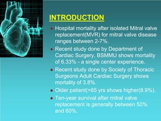
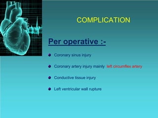
COMPLICATION OF MITRAL
VALVE REPLACEMENT
SURGERY
Dr. Bijay Kumar Sah
Phase B Resident, MS, CVTS
Department of Cardiac Surgery
BSMMU, Dhaka, Bangladesh
INTRODUCTION
Hospital mortality after isolated Mitral valve
replacement(MVR) for mitral valve disease
ranges between 2-7%.
Recent study done by Department of
Cardiac Surgery, BSMMU shows mortality
of 6.33% - a single center experience.
Recent study done by Society of Thoracic
Surgeons Adult Cardiac Surgery shows
mortality of 3.8%.
Older patient(>65 yrs shows higher(8.9%).
Ten-year survival after mitral valve
replacement is generally between 50%
and 60%.
COMPLICATION
Per operative :-
Coronary sinus injury
Coronary artery injury mainly left circumflex artery
Conductive tissue injury
Left ventricular wall rupture
Post operative:-
Thromboembolic Events
Acute Valve Thrombosis
Hemorrhagic Events from Anticoagulation
Structural Valve Degeneration
Prosthetic valve endocarditis
Periprosthetic leakage
Chronic Haemolysis
LEFT VENTRICULAR WALL RUPTURE
• Massive intrapericardial hemorrhage may
occur shortly after discontinuing CPB or in
the intensive care unit a few hours later.
• Nearly all patients die when the rupture
occurs postoperatively, but some may be
saved when it occurs while the chest is
open.
• LV rupture with hemorrhage is more likely
to occur in women and in patients with
small LVs.
TYPES:-
• Type 1- ruptures occur at the level of the
anulus.
• Type 2- rupture originates at the base of
an excised papillary muscle.
• Type 3- rupture(mid-ventricular rupture)
originates at mid-distance between the
stumps of papillary muscles and the
anulus.
CAUSES / CONTRIBUTING FACTORS:-
(1) Undue traction on the anulus during
excision of the mitral valve or insertion of
the prosthesis.
(2) Tearing of the anulus by sutures already
placed when the heart is manually tilted
up after the mitral prosthesis is in place.
(3) Penetration of stitches into the left AV
groove posteriorly.
(4) Perforation of LV wall as papillary muscle
is excised.
(5) Perforation of the AV groove as a calcific
deposit is being removed
MANAGEMENT:-
Once massive hemorrhage has occurred,
1)CPB must be reinitiated as quickly as
possible while the hemorrhage is
controlled digitally to the extent possible.
2) A premature attempt to suture it will surely
end in death, and even with proper
management, the risk of death is great.
3) After establishing CPB, clamping the
aorta, and administering cardioplegic
solution, the left atrium is reopened and
the valve removed.
CONT:-
4) An appropriately shaped piece of
pericardium is fashioned and positioned as
an onlay patch on the inside of the heart
over the presumed or identified area of
rupture, using multiple large, felt-pledgeted
sutures.
5) Even though every attempt is made to
avoid the proximal circumflex artery, it may
be compromised by these sutures, so the
area of the circumflex artery must be
carefully inspected before reinserting the
valve.
CONT:-
6) If it is compromised, a saphenous vein
bypass graft to a large marginal branch
should be placed.
7) The valve is then reinserted and the
remainder of the procedure completed as
usual.
8) Great care must be taken with myocardial
management throughout this procedure.
PREVENTION:-
Because of potential LV rupture,
1. The surgeon must be very gentle in all
maneuvers during mitral valve
replacement.
2. The heart should not be tipped up for air
evacuation or ligation of the left atrial
appendage or for routine inspection of the
back of it after the prosthesis has been
inserted.
3. Excising only the chordae tendineae
rather than the whole papillary muscle
CONT:-
4) Simply leaving in place deeply embedded
calcific deposits in the anulus and placing
sutures around them or only on their atrial
side, should eliminate these causes of LV
rupture.
5) Alternatively, a chordal-sparing mitral
valve replacement technique can be
performed.
Thromboembolic Events
• Thromboembolism is one of the more
common complications of mitral
prostheses, but the incidence is somewhat
less in patients with biologic valves.
• Life-long warfarin therapy is required for all
patients with mechanical valves.
• An INR of 2.5 to 3.5 is recommended.
• Warfarin therapy for 3 months
postoperatively and aspirin indefinitely are
as effective as long-term warfarin therapy
in patients with a biologic mitral prosthesis
CONT:-
• Patients with a bioprosthetic mitral valve
and risk factors of atrial fibrillation, prior
thromboembolism, LV dysfunction, or
hypercoagulable conditions, warfarin (in
addition to aspirin) is indicated to keep the
INR at 2.0 to 3.0.
• When warfarin is indicated for mechanical
or bioprosthetic valves and the patient
cannot take warfarin, aspirin is indicated at
a dose of 81 to 325 mg daily.
CONT:-
• Among patients who experience a
thromboembolic event while on
recommended antithrombotic therapy, a
more intensive regimen of warfarin or
aspirin is advisable.
• INR should be increased to 3.5 to 4.5
unless bleeding issues contraindicate this
level of anticoagulation.
Acute Valve Thrombosis
• Uncommon but devastating.
• Occurs more frequently with a mechanical
device than a biologic valve.
• Occurs almost exclusively in the setting of
suboptimal anticoagulation.
• Easily diagnosed by Fluoroscopy and
Echocardiography.
CONT:-
It is ususally more acute, manifesting as
“sudden” valve dysfunction and clinically
characterized by:-
• low cardiac output (shock)
• pulmonary venous hypertension
• Pulmonary edema
• Loss of valve click sounds in mechanical
valves and muffled sounds in
bioprosthesis.
CONT:-
• If the patient is not in cardiogenic shock,
for patients in NYHA class I/II heart failure
or if surgery is not available then it can be
treated with thrombolytic agents like
urokinase, streptokinase, or recombinant
tissue plasminogen activator.
• Hemodynamic instability warrants
emergency operation, and surgical
thrombectomy typically achieves a good
result.
Hemorrhagic Events from Anticoagulation
• Usually occurs in the gastrointestinal,
urogenital, and central nervous systems.
• Severity is proportional to the INR.
• Advancement in the valve decrease this
event to some extent.
• INR required for ball-and-cage valves was
3.5 to 4.5 compared to 2.5 to 3.5 for
current mechanical prostheses.
• In the absence of bleeding, simply
withholding warfarin until the INR is again
therapeutic is usually sufficient.
CONT:-
• If more aggressive reversal is desirable,
oral vitamin K should be administered with
caution because overcorrection may lead
to a hypercoagulable state.
• Low-dose oral (1-2.5 mg) or intravenous
(1 mg) vitamin K appears generally safe in
this situation.
• In emergency situations, administration of
fresh frozen plasma is preferable to
highdose vitamin K.
Structural Valve Degeneration
• principle complication of biologic
prostheses.
• Rates of biologic valve failure are higher in
the mitral position than in the aortic
position.
• Significantly higher in younger individuals
(<65 years).
• Uncommon in the first 5 years but begins
to increase thereafter with 15-year
freedom.
• This incidence is almost 0 for bileaflet,
tilting-disk,and ball-and-cage mechanical
valves
Prosthetic Valve Endocarditis
• Prosthetic mitral valve endocarditis is
significantly less common than prosthetic
valve endocarditis in the aortic position.
• However, it remains a serious complication
that results in the death of more than half
of those affected.
• Prosthetic valve endocarditis that occurs
within 6 months of the primary operation is
usually due to contamination in the
operating theater and later is generally the
result of a new bacteremia.
CONT:-
CAUSATIVE ORGANISM:
• Staphylococcus epidermidis(within 2
months of operation)
• Staphylococcus aureus(most virulent)
• Viridans streptococci
• Enterococcal like Enterrococcus faecalis
or Enterococcus faccium (mainly in elderly
people or with malignancy)
MANAGEMENTS:-
• Prosthetic mitral valve endocarditis that
occurs early after surgery greatly
increases the risk of dehiscence and
death.
• Broad-spectrum antibiotic therapy should
be initiated until sensitivities are identified.
• After culture and sensitivity, specific
antibiotics is given as listed in table below.
• In most cases, early prosthetic valve
replacement is required.
Periprosthetic Leakage
• Periprosthetic leakage is an uncommon
complication following mitral valve
replacement that is generally caused by
technical errors.
• In the current era, the incidence of
periprosthetic leakage approaches zero in
uninfected patients.
• Preoperative infective endocarditis and
mitral annular calcification increase the
risk of this complication.
CONT:-
• Surgery is indicated in all symptomatic
patients.
• Alternatively, offlabel application of
transcatheter septal defect closure devices
for these leaks has been successful.
Chronic Hemolysis
• Well-functioning prostheses used in the
current era rarely produce more than mild
chronic hemolysis unless periprosthetic
leakage is present.
• With periprosthetic leakage, hemolysis
may be severe with any device.
• A mechanical prosthesis of small size
relative to body size and hemodynamics
may result in severe hemolysis.
• Surgery is indicated in all symptomatic
patients and in those with minimal
symptoms that require transfusion.
TAKE HOME MESSAGE
• As all the complication isn’t reversible and
may lead to death so, the surgeon most be
alert enough to prevent such condition and
expert enough to tackle such situition if
arise.
• mitral valve surgery is associated with a potential risk of
iatrogenic myocardial ischemia. In order to avoid this
sequela, it is mandatory to perform coronary angiography
in every patient who undergoes mitral valve surgery, to
verify the course of the Cx and thereby avoid maneuvers
that increase the risk of Cx lesion and embolism.
• The risk of damaging the circumflex coronary artery
depends mainly upon the proximity of that vessel to the
posterior segment of the mitral annulus, and this varies
from patient to patient.

Complication of Mitral valve replacement surgery

  • 1.
    COMPLICATION OF MITRAL VALVEREPLACEMENT SURGERY Dr. Bijay Kumar Sah Phase B Resident, MS, CVTS Department of Cardiac Surgery BSMMU, Dhaka, Bangladesh
  • 2.
    INTRODUCTION Hospital mortality afterisolated Mitral valve replacement(MVR) for mitral valve disease ranges between 2-7%. Recent study done by Department of Cardiac Surgery, BSMMU shows mortality of 6.33% - a single center experience. Recent study done by Society of Thoracic Surgeons Adult Cardiac Surgery shows mortality of 3.8%. Older patient(>65 yrs shows higher(8.9%). Ten-year survival after mitral valve replacement is generally between 50% and 60%.
  • 3.
    COMPLICATION Per operative :- Coronarysinus injury Coronary artery injury mainly left circumflex artery Conductive tissue injury Left ventricular wall rupture
  • 4.
    Post operative:- Thromboembolic Events AcuteValve Thrombosis Hemorrhagic Events from Anticoagulation Structural Valve Degeneration Prosthetic valve endocarditis Periprosthetic leakage Chronic Haemolysis
  • 5.
    LEFT VENTRICULAR WALLRUPTURE • Massive intrapericardial hemorrhage may occur shortly after discontinuing CPB or in the intensive care unit a few hours later. • Nearly all patients die when the rupture occurs postoperatively, but some may be saved when it occurs while the chest is open. • LV rupture with hemorrhage is more likely to occur in women and in patients with small LVs.
  • 6.
    TYPES:- • Type 1-ruptures occur at the level of the anulus. • Type 2- rupture originates at the base of an excised papillary muscle. • Type 3- rupture(mid-ventricular rupture) originates at mid-distance between the stumps of papillary muscles and the anulus.
  • 7.
    CAUSES / CONTRIBUTINGFACTORS:- (1) Undue traction on the anulus during excision of the mitral valve or insertion of the prosthesis. (2) Tearing of the anulus by sutures already placed when the heart is manually tilted up after the mitral prosthesis is in place. (3) Penetration of stitches into the left AV groove posteriorly. (4) Perforation of LV wall as papillary muscle is excised. (5) Perforation of the AV groove as a calcific deposit is being removed
  • 8.
    MANAGEMENT:- Once massive hemorrhagehas occurred, 1)CPB must be reinitiated as quickly as possible while the hemorrhage is controlled digitally to the extent possible. 2) A premature attempt to suture it will surely end in death, and even with proper management, the risk of death is great. 3) After establishing CPB, clamping the aorta, and administering cardioplegic solution, the left atrium is reopened and the valve removed.
  • 9.
    CONT:- 4) An appropriatelyshaped piece of pericardium is fashioned and positioned as an onlay patch on the inside of the heart over the presumed or identified area of rupture, using multiple large, felt-pledgeted sutures. 5) Even though every attempt is made to avoid the proximal circumflex artery, it may be compromised by these sutures, so the area of the circumflex artery must be carefully inspected before reinserting the valve.
  • 10.
    CONT:- 6) If itis compromised, a saphenous vein bypass graft to a large marginal branch should be placed. 7) The valve is then reinserted and the remainder of the procedure completed as usual. 8) Great care must be taken with myocardial management throughout this procedure.
  • 11.
    PREVENTION:- Because of potentialLV rupture, 1. The surgeon must be very gentle in all maneuvers during mitral valve replacement. 2. The heart should not be tipped up for air evacuation or ligation of the left atrial appendage or for routine inspection of the back of it after the prosthesis has been inserted. 3. Excising only the chordae tendineae rather than the whole papillary muscle
  • 12.
    CONT:- 4) Simply leavingin place deeply embedded calcific deposits in the anulus and placing sutures around them or only on their atrial side, should eliminate these causes of LV rupture. 5) Alternatively, a chordal-sparing mitral valve replacement technique can be performed.
  • 13.
    Thromboembolic Events • Thromboembolismis one of the more common complications of mitral prostheses, but the incidence is somewhat less in patients with biologic valves. • Life-long warfarin therapy is required for all patients with mechanical valves. • An INR of 2.5 to 3.5 is recommended. • Warfarin therapy for 3 months postoperatively and aspirin indefinitely are as effective as long-term warfarin therapy in patients with a biologic mitral prosthesis
  • 14.
    CONT:- • Patients witha bioprosthetic mitral valve and risk factors of atrial fibrillation, prior thromboembolism, LV dysfunction, or hypercoagulable conditions, warfarin (in addition to aspirin) is indicated to keep the INR at 2.0 to 3.0. • When warfarin is indicated for mechanical or bioprosthetic valves and the patient cannot take warfarin, aspirin is indicated at a dose of 81 to 325 mg daily.
  • 15.
    CONT:- • Among patientswho experience a thromboembolic event while on recommended antithrombotic therapy, a more intensive regimen of warfarin or aspirin is advisable. • INR should be increased to 3.5 to 4.5 unless bleeding issues contraindicate this level of anticoagulation.
  • 16.
    Acute Valve Thrombosis •Uncommon but devastating. • Occurs more frequently with a mechanical device than a biologic valve. • Occurs almost exclusively in the setting of suboptimal anticoagulation. • Easily diagnosed by Fluoroscopy and Echocardiography.
  • 17.
    CONT:- It is ususallymore acute, manifesting as “sudden” valve dysfunction and clinically characterized by:- • low cardiac output (shock) • pulmonary venous hypertension • Pulmonary edema • Loss of valve click sounds in mechanical valves and muffled sounds in bioprosthesis.
  • 18.
    CONT:- • If thepatient is not in cardiogenic shock, for patients in NYHA class I/II heart failure or if surgery is not available then it can be treated with thrombolytic agents like urokinase, streptokinase, or recombinant tissue plasminogen activator. • Hemodynamic instability warrants emergency operation, and surgical thrombectomy typically achieves a good result.
  • 19.
    Hemorrhagic Events fromAnticoagulation • Usually occurs in the gastrointestinal, urogenital, and central nervous systems. • Severity is proportional to the INR. • Advancement in the valve decrease this event to some extent. • INR required for ball-and-cage valves was 3.5 to 4.5 compared to 2.5 to 3.5 for current mechanical prostheses. • In the absence of bleeding, simply withholding warfarin until the INR is again therapeutic is usually sufficient.
  • 20.
    CONT:- • If moreaggressive reversal is desirable, oral vitamin K should be administered with caution because overcorrection may lead to a hypercoagulable state. • Low-dose oral (1-2.5 mg) or intravenous (1 mg) vitamin K appears generally safe in this situation. • In emergency situations, administration of fresh frozen plasma is preferable to highdose vitamin K.
  • 21.
    Structural Valve Degeneration •principle complication of biologic prostheses. • Rates of biologic valve failure are higher in the mitral position than in the aortic position. • Significantly higher in younger individuals (<65 years). • Uncommon in the first 5 years but begins to increase thereafter with 15-year freedom. • This incidence is almost 0 for bileaflet, tilting-disk,and ball-and-cage mechanical valves
  • 22.
    Prosthetic Valve Endocarditis •Prosthetic mitral valve endocarditis is significantly less common than prosthetic valve endocarditis in the aortic position. • However, it remains a serious complication that results in the death of more than half of those affected. • Prosthetic valve endocarditis that occurs within 6 months of the primary operation is usually due to contamination in the operating theater and later is generally the result of a new bacteremia.
  • 23.
    CONT:- CAUSATIVE ORGANISM: • Staphylococcusepidermidis(within 2 months of operation) • Staphylococcus aureus(most virulent) • Viridans streptococci • Enterococcal like Enterrococcus faecalis or Enterococcus faccium (mainly in elderly people or with malignancy)
  • 24.
    MANAGEMENTS:- • Prosthetic mitralvalve endocarditis that occurs early after surgery greatly increases the risk of dehiscence and death. • Broad-spectrum antibiotic therapy should be initiated until sensitivities are identified. • After culture and sensitivity, specific antibiotics is given as listed in table below. • In most cases, early prosthetic valve replacement is required.
  • 26.
    Periprosthetic Leakage • Periprostheticleakage is an uncommon complication following mitral valve replacement that is generally caused by technical errors. • In the current era, the incidence of periprosthetic leakage approaches zero in uninfected patients. • Preoperative infective endocarditis and mitral annular calcification increase the risk of this complication.
  • 27.
    CONT:- • Surgery isindicated in all symptomatic patients. • Alternatively, offlabel application of transcatheter septal defect closure devices for these leaks has been successful.
  • 28.
    Chronic Hemolysis • Well-functioningprostheses used in the current era rarely produce more than mild chronic hemolysis unless periprosthetic leakage is present. • With periprosthetic leakage, hemolysis may be severe with any device. • A mechanical prosthesis of small size relative to body size and hemodynamics may result in severe hemolysis. • Surgery is indicated in all symptomatic patients and in those with minimal symptoms that require transfusion.
  • 29.
    TAKE HOME MESSAGE •As all the complication isn’t reversible and may lead to death so, the surgeon most be alert enough to prevent such condition and expert enough to tackle such situition if arise.
  • 31.
    • mitral valvesurgery is associated with a potential risk of iatrogenic myocardial ischemia. In order to avoid this sequela, it is mandatory to perform coronary angiography in every patient who undergoes mitral valve surgery, to verify the course of the Cx and thereby avoid maneuvers that increase the risk of Cx lesion and embolism. • The risk of damaging the circumflex coronary artery depends mainly upon the proximity of that vessel to the posterior segment of the mitral annulus, and this varies from patient to patient.