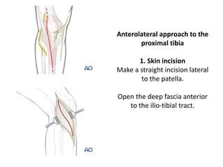
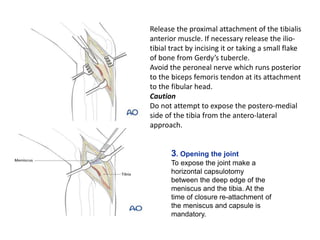
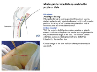
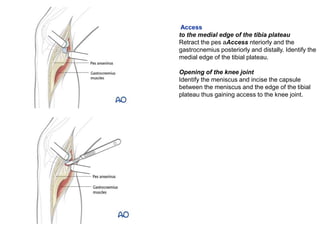
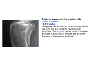
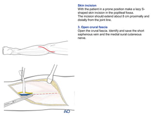
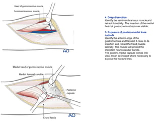
Tortora PRINCIPLES OF ANATOMY AND PHYSIOLOGY - Tortora - 14th Ed.pdf
proximal tibia approach.pptx
1. Anterolateral approach to the
proximal tibia
1. Skin incision
Make a straight incision lateral
to the patella.
Open the deep fascia anterior
to the ilio-tibial tract.
2. Release the proximal attachment of the tibialis
anterior muscle. If necessary release the ilio-
tibial tract by incising it or taking a small flake
of bone from Gerdy’s tubercle.
Avoid the peroneal nerve which runs posterior
to the biceps femoris tendon at its attachment
to the fibular head.
Caution
Do not attempt to expose the postero-medial
side of the tibia from the antero-lateral
approach.
3. Opening the joint
To expose the joint make a
horizontal capsulotomy
between the deep edge of the
meniscus and the tibia. At the
time of closure re-attachment of
the meniscus and capsule is
mandatory.
3. 4. Wound closure
Close the ilio-tibial band and if necessary reattach the
Gerdy’s tubercle. Do not close the fascia to avoid a
compartment syndrome. Close the remaining soft tissues in
a routine manner.
4. Medial/posteromedial approach to the
proximal tibia
Principles
Patient positioning
If the patient’s hip is normal, position the patient supine,
abduct and externally rotate the leg and put it in a figure of 4
position. If the hip is stiff position the patient in a lateral
decubitus with the involved limb down.
2. Skin incision
With the knee in slight flexion make a straight or slightly
curved incision running from the medial epicondyle towards
the posteromedial edge of the tibia. The incision can be
extended as needed both proximally and distally as
indicated by the dashed line.
Clinical image of the skin incision for the postero-medial
approach.
6. Access
to the medial edge of the tibia plateau
Retract the pes aAccess nteriorly and the
gastrocnemius posteriorly and distally. Identify the
medial edge of the tibial plateau.
Opening of the knee joint
Identify the meniscus and incise the capsule
between the meniscus and the edge of the tibial
plateau thus gaining access to the knee joint.
7. Exposure of the anteromedial part (medial column) of proximal tibia is possible by a subcutaneous
dissecting anteriorly. The pes can be retracted posteriorly when dealing with the fracture in this part
Clinical picture showing medial plateau and medial meniscus, pes anserinus, and distally the
gastrosoleus.
5. Wound closure
Close the capsule. If needed insert suction drains and close the soft tissues in a routine manner.
8. Posterior approach to the proximal tibia
Select a chapter
•1. Principles
The posteromedial side can be approached without
exposing and dissecting the neurovascular
structures. This approach allows repair of avulsion
fractures of the posterior cruciate and tangential
fractures of the proximal tibial head.
9. Skin incision
With the patient in a prone position make a lazy S-
shaped skin incision in the popliteal fossa.
The incision should extend about 8 cm proximally and
distally from the joint line.
3. Open crural fascia
Open the crural fascia. Identify and save the short
saphenous vein and the medial sural cutaneous
nerve.
10. 4. Deep dissection
Identify the semimembranosus muscle and
retract it medially. The insertion of the medial
head of gastrocnemius becomes visible.
5. Exposure of postero-medial knee
capsule
Identify the anterior edge of the
gastrocnemius and transect it close to its
insertion and retract the freed muscle
laterally. The muscle will protect the
important neurovascular bundle.
The postero-medial capsule comes into
view. It can be incised where necessary to
expose the fracture lines.
11. 6. Wound closure
Reattach the medial head of the
gastrocnemius. Place a deep
suction drain. Carry out a routine
closure of the soft tissues.