
Recommended
More Related Content
What's hot
What's hot (20)
Similar to Salivary glands
Similar to Salivary glands (20)
Recently uploaded
Recently uploaded (20)
Salivary glands
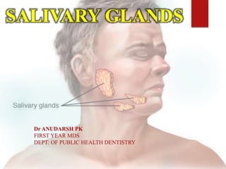
- 1. SALIVARY GLANDS Dr ANUDARSH PK FIRST YEAR MDS DEPT: OF PUBLIC HEALTH DENTISTRY
- 2. INTRODUCTION • Salivary glands are compound tubuloacinar, exocrine gland and the ducts opens in the oral cavity. • Salivary glands secretes a fluid called saliva that coats the teeth and the mucosa. • Saliva is a complex fluid, produced by the salivary glands, the most important function of which is to maintain the well- being of mouth. • Individuals with a deficiency of salivary secretion experience difficulty in eating, speaking, and swallowing and become prone to mucosal infections and dental caries.
- 3. Development of salivary glands occurs in six stages: 1) Bud Formation 2) Formation And Growth Of Epithelial Cords 3) Branching Of Cords 4) Dichotomous Branching 5) Canalization 6) Cyto- differentiation. DEVELOPMENT OF SALIVARY GLANDS
- 4. STAGE 1-BUD FORMATION The underlying mesenchyme induces the overlying epithelium to proliferate The epithelium thus forms a bud.
- 5. STAGE 2 -FORMATION AND GROWTH OF EPITHELIAL CORD • The epithelial bud proliferates to form a solid cord of cells. • The underlying mesenchymal condensation also proliferates.
- 6. STAGE 3-BRANCHING OF CORDS The epithelial cords proliferates rapidly and branches into terminal bulbs.
- 7. STAGE 4-DICHOTOMOUS BRANCHING The terminal end branches extensively forms numerous bulbs. The connective tissue below the epithelial cord forms a capsule and surrounds the entire glandular structure.
- 8. STAGE 5-CANALIZATION Extensive branching of the duct structure and growth of connective tissue septa continues in this stage First in distal ends of main cord & in branch cords then in proximal part of main cord & finally in central portion of main cord.
- 9. STAGE 6- CYTODIFFERENTIATION At last there is cytodifferentiation of the functional acini and intercalated ducts.
- 10. DEVELOPMENT OF SALIVARY GLAND IN INTRAUTERINE LIFE • Parotid Gland- 4th Week of intrauterine life • Submandibular Gland- 6th Week of intrauterine life • Sublingual Gland- 8th week of intrauterine life • Minor glands- 12th week of intrauterinelife.
- 11. CLASSIFICATION OF SALIVARY GLANDS 1) ACCORDING TO SIZE: MAJOR Parotid Submandibular Sublingual MINOR Glands of Lips Cheeks Hard Palate Soft palate Von Ebners Blandin&Nuhn.
- 12. 2) ACCORDING TO BRANCHING OF DUCTS: A) Compound- Major glands B) Simple- Minor glands
- 13. 3) ACCORDING TO SECRETION: SEROUS a)Parotid b)Von-ebner MUCOUS a)Palatine b)Post. lingual c) Glossopalatine MIXED a)Submandibular b)Sublingual c)Buccal d)Labial.
- 14. ANATOMY OF SALIVARY GLANDS PAROTID GLAND • Is the largest • Weight – 15gms • Is situated below the External acoustic meatus, b/w the ramus of the mandible and the sternocleidomastoid. • Secretion of parotid gland is serous. • Anteriorly , the gland overlaps the masseter muscle. A part of this forward extension is often detached – Accessory Parotid which lies b/w the zygomatic arch and the parotid duct.
- 16. PAROTID CAPSULE • The investing layer of deep cervical fascia forms the capsule for the gland. • The fascia splits to enclose the gland. • Superficial lamina – thick & adherent to the gland. • Deep lamina – thin – attached to the styloid process, the mandible and tympanic plate. • A portion of deep lamina, extending between the styloid process and the mandible – thickened to form Stylo mandibular Ligament – separates the Parotid gland from the Submandibular salivary gland.
- 17. SURFACES & BORDERS • The glands has four surfaces a) Superior b) Superficial c) Antero-medial d) Postero-medial • The surfaces are separated by three borders. a) Anterior b) Posterior c) Medial
- 18. 1. Superior surface related to Cartilaginous part of External acoustic meatus Superficial temporal vessels Posterior surface of TMJ Auriculotemporal nerve
- 19. 2. Superficial surface (largest) A. Skin B. Superficial fascia C. Great auricular nerve D. Pre-auricular lymph node E. Posterior fibers of platysma F. Parotid fascia G. Deep parotid lymph nodes
- 20. 3. Anteromedial surface related to Masseter Lateral surface of TMJ Posterior border of the mandible Medial pterygoid Terminal branches of facial nerve
- 22. 4 Posteromedial surface Mastoid process Sternocleidomastoid Posterior belly of the digastric Styloid process
- 23. BORDERS ANTERIOR BORDER : – Separates the superficial surface from the Anteromedial surface – Extends from –anterior part of the superior surface to the apex – Structures emerging: Parotid duct Terminal branches of the facial nerve Transverse facial vessels
- 25. POSTERIOR BORDER : – Separated the superficial surface from the posteromedial surface – Overlaps the sternomastoid. MEDIAL BORDER : – Separates the Anteromedial surface from the posteromedial surface. – Related to the lateral wall of pharynx
- 26. STRUCTURES WITHIN THE PAROTID GLAND 1) Arteries ECA-external carotid artery Maxillary artery Superficial temporal vessels Posterior auricular artery
- 27. 2) VEINS Retromandibular vein divides into: 1. Posterior division drains into external jugular vein 2. Anterior division drains into common facial vein
- 28. 3) Nerves • Terminal branches of facial nerve a) Temporal b) Zygomatic c) Buccal d) Mandibular e) cervical 4) Parotid lymph nodes
- 29. NERVE SUPPLY TO PAROTID INFERIOR SALIVATORY NUCLEUS AURICULOTEMPORAL NERVE TYMPANIC BRANCH LESSER PETROSAL NERVE RELAY IN OTIC GANGLION POST GANGLIONIC FIBRES THROUGH AURICULOTEMPORAL NERVE PAROTID
- 32. PAROTID DUCT • Stensen’s duct-5cm long • Emerges from the substance of the gland to course anteriorly until it reaches the anterior border of the masseter muscle at a point of the upper and middle third. • When it crosses the masseter muscle it receives the duct of the accessory lobe.
- 33. • Around the border of the masseter muscle-turns sharply medially, often embedded in the furrow of the protruding buccal fat pad. • In its medial course ,the duct reaches the outer surface of the buccinator muscle, where it perforates in the oblique direction anteriorly and medially. • Then runs for a short distance obliquely forward, between the buccinator and the mucous membrane – opens on the oral surface of the cheek
- 34. SUBMANDIBULAR GLAND • Situated in the anterior part of the Digastric triangle • Size that of a walnut • Is J-shaped • Secretion of submandibular gland is both serous and mucous. • Divided by the posterior border of the Mylohyoid Muscle into 1. Larger part superficial to the muscle • Covers the inferior surface of the gland • Attached to the base of the mandible
- 35. 2. Small part lying deep to the muscle • Covers the medial surface of the gland • Attached to the mylohyoid line of the mandible
- 36. Superficial part: – Fills the digastric triangle – It has a ) Inferior surface-covered by- Skin Platysma Cervical branch of facial nerve Deep fascia Facial vein Submandibular lymph nodes.
- 37. b) Lateral surface Submandibular fossa on the mandible Facial artery c) Medial surface divided into 1. Anterior part – related to the – Mylohyoid muscle – Nerve – Vessels
- 38. 2. Middle part- • Hyoglossus • Styloglossus • Lingual nerve • Hypoglossal nerve 3. Posterior part- • Styloglossus • Stylohyoid ligament • Glossopharengeal nerve • Wall of pharynx
- 40. • Called “WHARTONS” duct • 5 cm long • Emerges at the anterior end of deep part of the gland • Runs forwards on the hyoglossus ,between the lingual and the hypoglossal nerves • Opens in the floor of the mouth on the summit of the sublingual papilla, at the side of the frenum of the tongue
- 41. BLOOD SUPPLYAND LYMPHATIC DRAINAGE : • Facial artery • Veins drain into common facial vein or lingual vein • Lymph passes into the Submandibular lymph nodes NERVE SUPPLY : • Supplied by branches from Submandibular ganglion • These convey – Secretomotor fibers – Sensory fibers from lingual nerve – Vasomotor sympathetic fibers from facial nerve.
- 42. SUBLINGUAL GLAND
- 43. • Is the smallest • Almond shaped • 3-4gm weight • Secretion of sublingual gland is both serous and mucous. • Lies above the mylohyoid, below the mucosa of the floor of the mouth • Medial to the sublingual fossa
- 44. • Lateral to genioglossus • About 15 ducts emerge from the gland – Called ducts of rivinus BLOOD SUPPLY: • Lingual and submental arteries
- 46. • Present under epithelium of lips & cheeks • Mixed in nature • Intercalated duct are variable in length LABIAL & BUCCAL GLANDS
- 47. • Present at glandular region of hard palate i.e. posterolateral part of hard palate. • Pure mucous gland PALATINE GLANDS
- 48. GLANDS OF TONGUE Anterior Gland: • Near apex of tongue anterior lingual gland are present. These are glands of Blandin & Nuhn. • Mucous in nature • Ducts of these gland open on ventral surface of tongue near lingual frenum.
- 49. Posterior Glands: • Present on posterior part of tongue near suclcus terminalis • Mucous in nature • Ducts open at dorsal surface of tongue von Ebner’sgland: • Serous gland • Opening through vallate papilla.
- 50. COMMON SALIVARY GLAND DISORDERS SIALOLITHIASIS (salivary gland stones).Tiny, calcium-rich stones sometimes form inside the salivary glands with prevalence of 1.2%. The exact cause of these stones is unknown. Some stones may be related to: • Dehydration, which thickens the saliva • Decreased food intake, which lowers the demand for saliva • Medications that decrease saliva production, including certain antihistamines, blood pressure drugs and psychiatric medications Some stones sit inside the gland without causing any symptoms. In other cases, a stone blocks the gland's duct, either partially or completely. When this happens, the gland typically is painful and swollen, and saliva flow is partially or completely blocked.
- 51. The majority of calculi are found within the submandibular gland (80%–92%), likely due to the fact that the submandibular gland produces highly viscous saliva that needs to travel upward against gravity as it traverses Wharton’s duct. The remaining calculi are found in the parotid gland (6%–20%) and sublingual/minor salivary glands (1%–2%)
- 52. SIALADENITIS Sialadenitis is a general term used to denote an infectious or inflammatory process of the salivary glands. Sialadenitis can be due to a variety of causes including viral, bacterial, fungal, and granulomatous processes ,immunologically mediated disease, and, parasitic.
- 53. INFECTIOUS SIALADENITIS Worldwide, viral sialadenitis is most commonly due to mumps virus other viruses including parainfluenza, influenza, coxsackie, Epstein–Barr virus, herpes simplex virus, and HIV mumps sialadenitis begins with a prodromal period followed by acute bilateral salivary gland swelling typically affecting the parotid glands
- 54. In HIV patients it is most commonly due to the same viral/bacterial pathogens as non-HIV patients, they can develop a separate entity referred to as HIV-associated salivary gland disease. This is characterized by progressive, painless swelling of the bilateral salivary glands due to the formation of benign lymphoepithelial cysts (BLECs)
- 55. Bacterial sialadenitis presents with acute unilateral salivary gland swelling without a preceding prodromal period. The most common bacterial pathogens are Staphylococcus aureus and anaerobes
- 56. INFLAMMATORY SIALADENITIS Autoimmune sialadenitis refers to a group of noninfectious disorders that result in chronic inflammation and fibrosis of the salivary glands. Sjogren’s sialadenitis is a female predominant disorder (>90%) most commonly seen in the postmenopausal age group (50–70 years of age). Sarcoid sialadenitis refers to chronic inflammation of the salivary glands in patients with sarcoidosis. It is seen in 10%–30% of patients with sarcoidosis and patients typically present with painless bilateral parotid swelling.
- 57. POSTRADIATION SIALADENITIS Radiation-induced sialadenitis is frequently seen in patients who receive radiation treatment for oropharyngeal cancer. The first clinical signs of sialadenitis manifest as decreased saliva flow and have been reported to occur with radiation dose thresholds as low as 15 Gy.
- 58. SALIVARY GLAND NEOPLASMS In the parotid gland, approximately 80% of neoplasms are benign while 20% are malignant. The most common benign parotid gland neoplasms are pleomorphic adenoma and Warthin’s tumor ,while the most common malignant neoplasm is mucoepidermoid carcinoma.
- 59. In the submandibular gland, 50% of neoplasms are benign while 50% are malignant. the most common benign neoplasm is the pleomorphic adenoma, constituting 85% of all benign submandibular gland neoplasms. The most common malignant neoplasms include adenoid cystic carcinoma (ACC), mucoepidermoid carcinoma
- 60. In the sublingual and minor salivary glands, 80%– 90% of neoplasms are malignant ACC is the most common, while mucoepidermoid carcinoma is the second most common
- 61. PLEOMORPHIC ADENOMA Pleomorphic adenoma (benign mixed tumor) is a neoplasm comprised a combination of glandular epithelium and myoepithelial components. It is the most common benign salivary gland neoplasm and has a female predominance (2:1), typically affecting patients aged 30–60 years. Nearly 80% of these lesions arise in the parotid gland, and of these, 80%–90% arise in the superficial portion of the gland
- 62. Carcinoma Ex Pleomorphic Adenoma Carcinoma ex pleomorphic adenoma refers to malignant transformation of a preexisting pleomorphic adenoma. Although rare, it has been reported to occur in 0.15% of lesions
- 63. WARTHIN’S TUMOR Warthin’s tumor (papillary cystadenoma lymphomatosum) is a benign neoplasm comprised both epithelial and lymphoid tissue that forms papillary projections It is the second most common benign salivary neoplasm and has a strong association with smoking.
- 64. MUCOEPIDERMOID CARCINOMA Mucoepidermoid carcinoma is a malignant neoplasm comprised of epidermoid and mucus- secreting epithelial cells. It is the most common malignant neoplasm of the salivary glands 60% of lesions arise in the parotid gland
- 65. ADENOID CYSTIC CARCINOMA ACC is a malignant neoplasm derived from both ductal and myoepithelial cells. Clinically, these patients tend to present at an older age (5th–7th decades) ACC has the greatest propensity for perineural spread and local invasion ACC tends to be very aggressive and it is reported that 90% of cases will have cervical lymph node metastases at the time of diagnosis.
- 66. ACINIC CELL CARCINOMA Acinic cell carcinoma is a malignant neoplasm derived from the glandular epithelium and is considered a variant of adenocarcinoma. Nearly 90% of lesions arise in the parotid, most commonly located in the parotid tail.
- 67. ONCOCYTOMA Oncocytoma is a benign neoplasm derived from mitochondria-rich epithelial cells LIPOMA Lipomas are benign neoplasms composed of mature adipose tissue surrounded by a fibrous capsule Salivary gland lipomas(sialo lipoma) are much rarer and most commonly involve the parotid gland
- 68. RANULA Ranula is defined as a mucus retention cyst or mucocele in the sublingual space arising from either the sublingual gland or the minor salivary gland tissue.
- 69. The annual incidence of salivary gland tumors varies around the world from approximately 0.4– 13.5/100,000 people. Salivary gland tumors are reported to represent between 1% and 5% of all head-and-neck tumors. 75%–85% the found in the major Salivary glands 10% to 20% in the minor salivary glands Ratio of 5:1 EPIDEMIOLOGY
- 70. APPLIED ASPECTS FACIAL NERVE PARALYSIS AFTER IANB Facial paralysis caused by deposition of the anesthetic solution in the parotid region, a problem which mainly occurs when the needle is directed more posterior toward the posterior border of the mandible.
- 71. Symptoms of this temporary loss of the use of the muscles of facial expression include the inability to close the eyelid and the drooping of the labial commissure on the affected side for a few hours.
- 72. The paralysis could be either immediate or delayed, based on the time elapsed from the moment of the injection to the onset of the symptoms. In the immediate type, the paralysis occurs within minutes of injection with a recovery period of 3 hours or less. However, in the delayed type the symptoms appear within several hours to several days, while recovery may expand from 24 hours to several months.
- 73. RADIOTHERAPY AND SALIVARY GLANDS Salivary gland damage leading to hyposalivation and xerostomia is one of the most common complications of radiation therapy. Xerostomia and associated complications amplifies the adversities faced by a patient. A study was conducted in rats by yitzhak marmary et al in 2016 for assessing radiation-induced loss of salivary gland function , driven by cellular senescence mentioned that there is DNA damage in salivary glands after radiotherapy The radiation induced cell death depends upon the radio- sensitivity of a tissue and tissue turnover rate.
- 74. EFFECT OF PROTEIN DEFICIENCY ON SALIVARY GLAND FUNCTION Protein deficiency results in alterations to salivary gland structure and function. Studies in children have shown that the severity of protein energy malnutrition (PEM) is related to the extent of reduction of stimulated salivary secretion rate. PEM also results in a decrease in calcium, chloride and secretion of protein of stimulated saliva .
- 75. Also there is reduction in immunologic and agglutinating defense factors of unstimulated saliva. An increased severity of PEM is accompanied by a decrease in salivary protein concentration and arginase activity.
- 76. EFFECTS OF MINERAL AND VITAMIN DEFICIENCIES ON SALIVARY GLAND FUNCTION Animal studies have shown that Calcium deficiency may result in salivary secretion dysfunction. Zinc deficiency in a reduction in parotid proline-rich proteins. Iron deficiency in a reduction in secretion rate and impaired peroxidase antibacterial activity.
- 77. Vitamin A deficiency has been found to reduce the activity of bacteria-agglutinating glycoproteins. vitamin A deficiency are also associated with salivary gland atrophy, which subsequently reduces the defense of the oral cavity against infection and its ability to buffer the plaque acids. Vitamin D deficiency reduces parotid gland secretion.
- 78. MALNUTRITION AND PAROTID ENLARGEMENT It is due to hypertrophy of the cells of the gland and the condition is reversible with a return to adequate nutrition. This condition is particularly common in males and is possibly due to a work hypertrophy following excessive stimulation of the gland by malnutrition changes in the esophagus.
- 79. DIAGNOSTIC AIDS Conventional radiograph: conventional radiographs such as occlusal radiograph or OPG can be used in diagnosis of sialolithiasis
- 80. SIALOGRAPHY The technician will inject contrast material into the duct. After the dye fills the salivary gland, X ray will be taken to identify salivary gland disorder
- 81. SIALENDOSCOPY It is a minimally invasive procedure that incorporates a small -calibre endoscope and facilitates direct examination of the salivary ductal system.
- 82. ULTRASONOGRAPHY Ultrasound examination of salivary glands with a high resolution transducer is found to be a highly sensitive. A non-invasive method for salivary gland evaluation It is a cost effective imaging tool which displays high definition images useful in evaluating the superficial structures particularly the peripheral areas of the affected salivary gland.
- 83. Ultrasonography was found useful in diagnosing salivary gland enlargement in submandibular region It also helps in identifying the salivary glandular tissue in accessory salivary gland and salivary calculi.
- 84. SHOCK-WAVE LITHOTRIPSY Shock-wave lithotripsy is a non-invasive diagnostic tool suggested for the management of sialolithiasis. Iro et al in 1989 introduced the application of extracorporeal shock-wave lithotripsy (ESWL) in the management of salivary gland calculi. Sialo lithotripsy helps in removing salivary stones into smaller particles and thereby removal by flushing action is possible from the salivary duct system or after salivation induced by citric acid or other sialagogues.
- 85. The shock-waves are generated extra-corporeally by using Piezoelectric and electromagnetic techniques or intra-corporeally using electro- hydraulic, pneumatic or laser endoscopic devices.
- 86. SONOELASTOGRAPHY Elastography is an ultrasonography technique which measures the tissue elasticity in vivo. This imaging technique measures the elasticity of the glandular parenchyma and is useful in evaluating the hypo function of saliva especially in post radiation hypo function of salivary glands.
- 87. COMPUTED TOMOGRAPHY Computed tomography (CT) scans of the salivary glands are useful in delineate the extent of the lesion and its relation to adjacent structures.
- 88. Multi detector CT scans help in characterizing tumors of salivary glands like Warthin tumor which demonstrates peak enhancement of signals after administration of contrast agents which is not found in other tumors of salivary glands. CT scans perform poorly in characterizing the histopathologic nature of the tumors
- 89. CT scans help in differentiating the benign and malignant neoplasms of salivary glands. The irregular tumor margin and surrounding tissue infiltration is the characteristic feature of malignancy. Apart from tumor identification CT scan also aids to view dystrophic calcifications in salivary glands
- 90. MAGNETIC RESONANCE IMAGING MRI scans are useful in assessment of salivary glands. The wide variety of soft tissue signals differences and multi planar image acquisition have made MRI an effective imaging modality for assessment of salivary gland tumors. PAROTID TUMOR
- 91. This imaging modality is helpful in assessment of tumors affecting the deep lobes of parotid glands, skull base invasion of the tumors of salivary glands, evaluation of recurrent pleomorphic adenomas]. Also high resolution MRI scans delineate the intra parotid course of facial nerve which is an important landmark for surgeons operating on parotid glands.
- 92. Magnetic resonance sialography-Major limitations of conventional sialography include use of iodine based contrast agents and inability of the contrast agent in overcoming the strictures within the ductal system of the salivary gland which in turn prevent the visualization. These limitations can be overcome by switching on to MR sialography which uses patients own saliva as a contrast medium. MR sialography also demonstrates the actual ductal diameter due to non-use of contrast agents.
- 93. SCINTIGRAPHY Salivary gland scintigraphy uses Tc-99m pertechnetate which helps in assessment of salivary gland dysfunction in disorders like Sjogrens syndrome.
- 94. NEWEST MEMBER!!? TUBARIAL SALIVARY GLAND Matthijs H.Valstar et al conducted a study to to elucidate the characteristics of previously unnoticed bilateral macroscopic salivary gland locations in the human nasopharynx and its potential clinical implications for radiotherapy.They visualized by using positron emission tomography/computed tomography with prostate-specific membrane antigen ligands (PSMA PET/CT).
- 95. Histology and 3D reconstruction confirmed the presence of predominantly mucous glands with multiple draining ducts, predominantly near the torus tubarius. It was hypothesized that it could contain a large number of seromucous acini, with a physiological role for nasopharynx/oropharynx lubrication and swallowing.
- 96. This could have clinical relevance in oncology, because high-dose external beam radiotherapy (RT) to salivary glands during treatment for head and neck cancer (HNC) or brain metastasis is known to cause damage (toxicity, e.g. interstitial fibrosis, acinar atrophy). This can result in function loss with xerostomia and dysphagia
- 97. REFERENCES B D Chaurasia’s Human Anatomy, volume 3 Kessler AT, Bhatt AA. Review of the major and minor salivary glands, part 1: anatomy, infectious, and inflammatory processes. Journal of clinical imaging science. 2018;8. Sardar MA, Ganvir SM, Hazarey VK. A demographic study of salivary gland tumors. SRM Journal of Research in Dental Sciences. 2018 Apr 1;9(2):67. Valstar MH, de Bakker BS, Steenbakkers RJ, de Jong KH, Smit LA, Nulent TJ, van Es RJ, Hofland I, de Keizer B, Jasperse B, Balm AJ. The tubarial salivary glands: A potential new organ at risk for radiotherapy. Radiotherapy and Oncology. 2020 Sep 23.
- 98. Krishnamurthy S, Vasudeva SB, Vijayasarathy S. Salivary gland disorders: A comprehensive review. World Journal of Stomatology. 2015 May 20;4(2):56-71. Tzermpos FH, Cocos A, Kleftogiannis M, Zarakas M, Iatrou I. Transient delayed facial nerve palsy after inferior alveolar nerve block anesthesia. Anesthesia progress. 2012;59(1):22-7. Lingström P, Moynihan P. Nutrition, saliva, and oral health. Nutrition. 2003 Jun 1;19(6):567.
- 99. Du Plessis DJ. Parotid enlargement in malnutrition. South African Medical Journal. 1956;30(7):700-3. Vieira KA, Rosa-Júnior LS, Souza MA, Santos NB, Florêncio TM, Bussadori SK. Chronic malnutrition and oral health status in children aged 1 to 5 years: An observational study. Medicine. 2020 May 1;99(18):e19595.,∗ Khalil H. A basic review on the inferior alveolar nerve block techniques. Anesth Essays Res, 8 (1): 3–8. Marmary Y, Adar R, Gaska S, Wygoda A, Maly A, Cohen J, Eliashar R, Mizrachi L, Orfaig-Geva C, Baum BJ, Rose-John S. Radiation-induced loss of salivary gland function is driven by cellular senescence and prevented by IL6 modulation. Cancer research. 2016 Mar 1;76(5):1170-80.