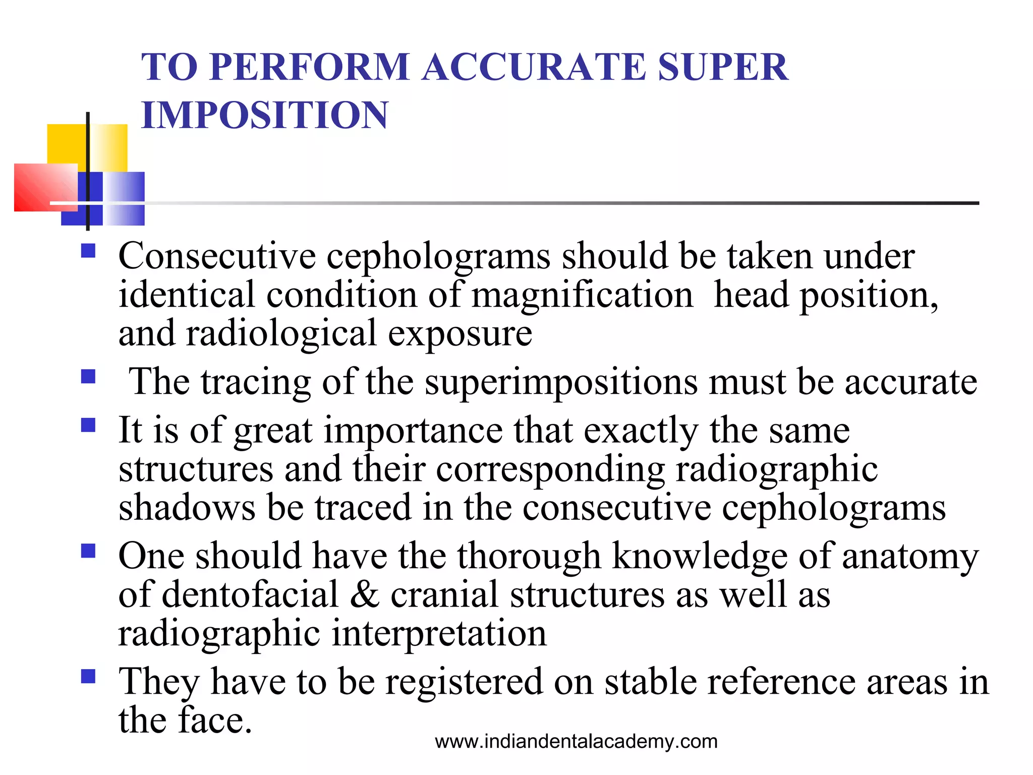
SUPERIMPOSITION
TECHNIQUES
INDIAN DENTAL ACADEMY
Leader in continuing dental education
www.indiandentalacademy.com

www.indiandentalacademy.com
Contents








Introduction
Methods of assessing dentofacial changes
Method of superimposing radiographs
Maxillary super-impositions
Mandibular superimpositions
Ricketts superimposition for treatment design
Ricketts four-step method
www.indiandentalacademy.com
Definition
Superimposition is the placement of an image on
top of an already existing image usually to add to
overall image effect and to conceal something eg.
Such as a different figure is superimposed over an
original figure.
 The transformation of one or more figures to
achieve the geometric relationship to another
figure
Wikipedia


www.indiandentalacademy.com
Introduction







In 1931 Broadbent (USA) and Hofrath (Germany)
- cephalometric technique using a cephalostat.
Treatment planning.
Evaluation of treatment results by quantifying the
changes brought about by treatment
Predicting growth related changes

www.indiandentalacademy.com
What is cephalometric
superimposition ?


“A cephalometric superimposition is an
analysis of lateral cephalograms of the same
patient taken at different times”.

www.indiandentalacademy.com
TO PERFORM ACCURATE SUPER
IMPOSITION










Consecutive cepholograms should be taken under
identical condition of magnification head position,
and radiological exposure
The tracing of the superimpositions must be accurate
It is of great importance that exactly the same
structures and their corresponding radiographic
shadows be traced in the consecutive cepholograms
One should have the thorough knowledge of anatomy
of dentofacial & cranial structures as well as
radiographic interpretation
They have to be registered on stable reference areas in
the face.
www.indiandentalacademy.com
CEPHALOMETRIC SUPER IMPOSITIONS
INVOLVE THE EVALUATION OF:







¨
¨
¨
¨

Changes in the overall face
Changes in the maxilla and its dentition
Changes in the mandible and its dentition
Amount and direction of condylar growth
Mandibular rotation

www.indiandentalacademy.com
Method of superimposing
radiographs

Superimposing on a stable plane
or structure

Registration on a stable
landmark

www.indiandentalacademy.com
Validity and reproducibility




Validity: It is the extent to which the value
obtained represents the object of interest.
 Planes and landmarks should be anatomically
valid and should agree with the anatomic
structures they represent
Reproducibility: It is the closeness of successive
measurements of the same object.

www.indiandentalacademy.com
Methods of assessing
Dentofacial changes






Brodie and Broadbent AJODO 1987-Dentofacial
growth patterns are established at a very early age
and thereafter are subject to proportional changes.
Downs and Ricketts AJO 1960 -several angles and
dimensions change with age but in an orderly and
progressive manner
Hellman -suggested that the infant face is
transformed into that of an adult face by increase
in size by changes in proportion and by
adjustment in position.
www.indiandentalacademy.com
Methods of assessing
Dentofacial changes
Color coding suggested by ABO:
 Pretreatment – Black


Progress – Blue



End of treatment – Red



Retention - Green
www.indiandentalacademy.com
Evaluation of overall changes
in the face


Superimposition methods:
 Broadbent triangle
 Sella nasion line
 Basion horizontal
 Basion nasion plane
 De Costers anterior cranial base reference line
 Viazis cranial base triangle
 Frankl’s occipital reference base
www.indiandentalacademy.com
Evaluation of overall changes
in the face


Objectives
 Overall assessment of growth and treatment
changes of the facial structures
 Amount of change in direction of displacement
and growth of maxilla and mandible.
 Changes in soft tissue
 Changes in maxillo mandibular relationship
 Overall displacement of teeth.
www.indiandentalacademy.com
Evaluation of overall changes
in the face
Superimposition at registration point
R with Bolton-nasion planes parallel

Broadbent triangle

Among the first structures
used for superimposition
 On observations of dried
skulls and a comparative
study of cranial base planes
(Bolton-nasion, porionnasion, sella-nasion) in
persons 3 to 18 years of age.
 Bolton point maybe obscured
by Mastoid
www.indiandentalacademy.com

Evaluation of overall changes
in the face
Sella nasion plane:




.

SN is a frequently used
reference line that has been
reported to be relatively
stable.
Both points S and N are
located in the mid sagittal
plane and are displaced a
minimal degree by
movement of the head.

www.indiandentalacademy.com
Steiner -SN with registration
point
 At sella to evaluate sagittal
changes in mandibular
positions and
 At nasion to evaluate the
position of the maxilla
through changes in the
angle SNA

www.indiandentalacademy.com
Evaluation of overall changes
in the face
Sella nasion plane:
 Unlike Steiner, Björk
used sella as
registration point to
assess changes in
position of both jaws.

www.indiandentalacademy.com
Evaluation of overall changes
in the face

Basion horizontal
 Basion is used as the
registration point.
 The SN planes are made
parallel by the help of the
Basion horizontal line and it’s
constant relationship with SN.
 A coordinate grid system is
used to superimpose the
radiographs.

www.indiandentalacademy.com
Evaluation of overall changes
in the face
Basion - Nasion plane:
 Suggested by Ricketts as
Facial Axis
 He considered Ba-N plane as
a line of separation of the
face from the skull and hence
a basic cranial axis for
growth and structural
reference.

www.indiandentalacademy.com


Based on studies of
laminograph sections,
Ricketts suggested that
the cranial base angle,
while constant on
average, exhibits a
change of 5° in either
direction over a 3-year
period

www.indiandentalacademy.com
Evaluation of overall changes
in the face
De Coster lineAjodo1952
The line represents the axis of skull base.
 He advocated tracing the inner contour of the frontal
bone through the cerebral aspect of the ethmoid , the
planumsphenoidale and the anterior aspect of the
sella turcica.

www.indiandentalacademy.com
Evaluation of overall changes
in the face
Viazis cranial base
triangleJco;25
 The anterior wall of
sella turcica and the
cribriform plate
(laminar cribrosa)
remain unchanged
after age of 5 years.

www.indiandentalacademy.com


Superimposition on the
anterior wall of sella turcica
and the stable TC (cranial
base) line, with registration
on T point, provides a
practical and reliable
formation in both the
anteroposterior and vertical
planes.

www.indiandentalacademy.com
Evaluation of overall changes
in the face
Viazis cranial base triangle
 First priority - to
registering on T point,
followed by
superimposing on the
inner structure of the
triangle, and finally
superimposing on the TC
line. This "best-fit"
approach meets the
realistic expectations of
any superimposition
technique.
www.indiandentalacademy.com
Frankel’s occipital reference
base





Among the basal structures of the neurocranium,
the occipital bone around the foramen magnum is
the first to ossify (between the third and fourth
year of life).
Minimalpostnatal growth of this structure and
surrounding tissues is seen.
www.indiandentalacademy.com
Frankel’s occipital reference
base


Reference plane –
based on natural head
posture and parallel to
the ground

www.indiandentalacademy.com
Evaluation of overall changes
in the face
Reliability of the various cranial base reference
planes used.
For meaningful interpretations of
superimpositions they have to be registered on
stable reference landmarks.
 Cranial base superimpositions are subject to error
due to the continued growth of the sphenoccipital
synchondrosis (Knott).
 Bone remodelling at sella and Nasion are also
responsible for further errors.
www.indiandentalacademy.com






Nasion position can change in a vertical direction
(Nelson and Knott).
Melsen’s study’s on human autopsy material has
shown that the position of sella may change in a
downward or a downward and backward direction.
She also showed that the position of Basion
changed due to remodelling of the clivus.
The Bolton point could be difficult to locate in
children due to the shadow of the mastoid process
www.indiandentalacademy.com
Growth changes in position of Nasion and sella

www.indiandentalacademy.com
Reference structures for
overall face superimpositions.


Nelson’s and Melsen’s - stable structures in the
anterior cranial base.
 Anterior wall of sella turcica
 The contour of the cribriform plate of the
ethmoid
 Trabecular system of the ethmoid air cells
 The median border of the orbital roof
 Planum sphenoidale
www.indiandentalacademy.com
SUPERIMPOSITION OF THE
MAXILLA
Evaluates

Movement of maxillary teeth

Rotation of maxilla

Two method for superimposing the maxillary
structutres are recommended- the structural
method and a modified best fit method.
Structural Method : - Suggested by Bjork & Skieller
in 1976. is recommended if the details of the
zygomatic process of the maxilla are clearly
identified in both cephalograms.
Modified best fit method : - If the details of
zygomatic process of maxilla are not identified in
both cephalograms.
www.indiandentalacademy.com
Structural method of superimposition of maxilla

Pre treatment
Tracing

Post treatment
Tracing

Super imposition of
pre & post treatment tracing

On each cephologram, trace the contours of palate, maxillary 1st
molar, central incisors, zygomatic process of maxilla, floor of the
www.indiandentalacademy.com
orbit N-S line and the construction line.
Modified best fit method

Pre treatment
Tracing





Post treatment
Tracing

Super imposition of
pre & post treatment tracing

On each cephalogram trace the outline of the palate 1st
permanent molars and central incisors.
Following structures are in a best fit alignment.
 Contour of the oral part of the palate
 Contour of the nasal floor
www.indiandentalacademy.com
Evaluation of changes in maxilla
and its dentition in relation to the
maxilla
Superimposition along the
palatal plane registered at
ANS
Broadbent, Moore, Salzman,
Ricketts, McNamara
 Compromised by
remodelling of the palatal
shelves and ANS
Bjork and Skeiler


www.indiandentalacademy.com
Evaluation of changes in maxilla
and its dentition in relation to the
maxilla
Superimposition on
the nasal floors with
films registered at the
anterior surface of the
maxilla
Downs and Brodie.


www.indiandentalacademy.com
Evaluation of changes in maxilla
and its dentition in relation to the
maxilla
Superimposition
along the palatal plane
registered at the
pterygomaxillary
fissure
Moore


www.indiandentalacademy.com
Evaluation of changes in maxilla
and its dentition in relation to the
maxilla
Superimposition on
the outline of the
infratemporal fossa
and the posterior
margin of the hard
palate.
Reidel


www.indiandentalacademy.com
Evaluation of changes in maxilla
and its dentition in relation to the
maxilla
Superimpostion
registered at the
common Ptm
coordinate
maintaining the basion
horizontal
relationship.
Coben


www.indiandentalacademy.com
Evaluation of changes in maxilla
and its dentition in relation to the
maxilla
superimposition on the
best fit of the internal
palatal structures.
McNamara


www.indiandentalacademy.com
Evaluation of changes in maxilla
and its dentition in relation to the
maxilla




The structural
superimposition on the
anterior surface of the
zygomatic process of
the maxilla
Bjork and Skeiler

www.indiandentalacademy.com
Evaluation of changes in maxilla
and its dentition in relation to the
maxilla


Neilsen on a study of various maxillary
superimposition techniques concluded that:
 The best fit method significantly under estimates the
vertical displacement of skeletal and dental
landmarks
 With the structural method ANS showed twice as
much vertical displacement as PNS

www.indiandentalacademy.com




Structural method did not show any significant
differences
The structural method is a valid method of
assessing maxillary growth and treatment
changes

www.indiandentalacademy.com
Mandibular superimposition
Evaluates
 Movement of the mandibular teeth
 Mandibular rotation
 Amount & direction of condylar growth

www.indiandentalacademy.com
Stable areas according to
Bjork and Skieller (1983)
 Anterior contour of the chin
 The inner contour of the
cortical plates at the inferior
border of the symphysis
 Contours of the mandibular
canal
 Lower contour of the
mineralized molar tooth germ

www.indiandentalacademy.com
Step – by – step approach for
mandibular superimpositions

Pre treatment
Tracing

Progress treatment
tracing

Super imposition of
pre & progress treatment
tracing

On each cephologram trace the following
 The symphysis with inner cortical bone
 The inferior and posterior contour of the mandible
 The anterior contour of the ramus
 The most labially positioned lower incisor, and
 The first molar.
www.indiandentalacademy.com
Methods to assess growth Vs
treatment changes




Differentiate between changes produced due to
growth and changes produced due to treatment.
Assess treatment changes against the background of
natural growth of the individual

www.indiandentalacademy.com
RICKETTS SUPER
IMPOSITION TECHNIQUE
It is one of the most important superimposition
technique used.

Ricketts super imposition is used to differentiate the
changes in normal growth and those due to treatment
mechanics.
This techniques involves five super imposition areas to
evaluate

The chin

The maxilla

The teeth in the mandible

The teeth in the maxilla

The facial profile www.indiandentalacademy.com

Rickett ’s Eleven factor
summary analysis




Eleven factors of the basic facial and skeletal
structures are recorded from the cephalometric
tracing to describe the chin, maxilla, teeth and soft
tissue profile.
Five areas of superimposition within which are a
total of seven areas of evaluation are used to
evaluate in amount and direction, change in
normal growth and change due to treatment.
www.indiandentalacademy.com
Rickett’s Eleven factor
summary analysis

www.indiandentalacademy.com
Rickett’s Eleven factor
summary analysis
MEAN

CHANGE

FACIAL AXIS

90+/-3

No change

FACIAL DEPTH

87+/-3

+10 for 3 yrs

MAND PLANE

26+/-4

-1o for 3 yrs

FACIAL TAPER

68+/-3

No change

LFH

47+/-4

No change

MAND ARC

26=/-4

Mand arc
closes1/2yr
Angle inc 1/2yr

CON AT A

2mm+/-2mm

-1mm / 3 yrs

L1 TO APO

1+/-2

No change

L1 INCL

22+/-4

No change

U6 TO PTV

Age+3+2

1mm / year

L LIP TO E LINE

-2+/-2

Less with age

www.indiandentalacademy.com
Rickett’s Eleven factor
summary analysis
SUPERIMPOSITION AREA 1 – EVALUATION
AREA 1
• FACIAL AXIS OPENS 1o FOR
DOLICOFACIAL PATTERN
•FACIAL AXIS CLOSES 1O FOR
BRACHYFACIAL PATTERNS
•FACIAL AXIS OPENS 1O FOR 5mm
CONVEXITY REDUCTION
•FACIAL AXIS OPENS 1O FOR 3mm
MOLAR CORRECTION
•FACIAL AXIS OPENS 1O OR 4mm
OVERBITE CORRECTION
•FACIAL AXIS OPENS 1 TO 1.5O FOR
CROSS BITE CORRECTION AND
RECOVERS ONE HALF

BASION-NASION PLANE at CC
www.indiandentalacademy.com
Rickett’s Eleven factor
summary analysis
SUPERIMPOSITION AREA 2 – EVALUATION
AREA 2
• THE BASION-NASION-POINT A
ANGLE OF 66O DOES NOT CHANGE
WITH GROWTH
•SO ANY CHANGES PRODUCED
MUST BE DUE TO TREATMENT
•HEADGEAR - -8mm
•CLASS II ELASTICS - -3mm
•ACTIVATOR - -2mm
•TORQUE- - 1 TO 2mm
•CLASS III ELASTICS 2 TO 3mm
•FACIAL MASK - 2 TO 4mm
BASION NASION PLANE AT
NASION
www.indiandentalacademy.com
Rickett’s Eleven factor
summary analysis
SUPERIMPOSITION AREA 3 – EVALUATION AREAS 3
AND 4
• LOWER DENTURE REMAINS
CONSTANT WITH THE A Pog LINE
•Without treatment, the lower molar will erupt
directly upward to the new occlusal plane.
•The LOWER INCISAL angle is 22° at +1mm
to the APo plane and + 1 mm to occlusal
plane, but the angle increases 2° with each mm
of forward compromise.

CORPUS AXIS AT PM
www.indiandentalacademy.com






OCCLUSAL PLANE TO
CORPUS AXIS DOES NOT
CHANGE
LOWER MOLAR ERUPTS IN A
DIRECTION PERPENDICULAR
TO THE FH PLANE
OCCLUSAL PLANE ERUPTS
0.8mm UPWARDS FROM THE
CORPUS AXIS.

www.indiandentalacademy.com
Rickett’s Eleven factor
summary analysis
SUPERIMPOSITION AREA 4 – EVALUATION AREAS 5
AND 6
• THE UPPER MOLAR AND INCISOR
FOLLOW THEIR POLAR AXIS WITH
GROWTH
•CHANGES IN POSITION OF THE
MOLAR OR INCISOR IS DUETO
TREATMENT.
•UPPER DENTAL ARCH ERUPTS
DOWNWARD AND FORWARD 0.2 TO
0.3mm PER YEAR

PALATE AT ANS
www.indiandentalacademy.com
Rickett’s Eleven factor
summary analysis
SUPERIMPOSITION AREA 5 – EVALUATION AREA 7
•

ESTHETIC PLANE AT INTERSECTION
OF OCCLUSAL PLANES

www.indiandentalacademy.com
Rickett’s four position analysis
Takes into consideration two superimposition areas
to evaluate skeletal change and two
superimposition areas to evaluate dental change
against growth.

www.indiandentalacademy.com
Rickett’s four position analysis




Position 1
Mandible or chin
Central axis 90+/-3

www.indiandentalacademy.com
Rickett’s four position analysis




Position 2
Maxilla
Backward moment
of .A of 3 help
reduce convexity

www.indiandentalacademy.com
Rickett’s four position analysis








Position 3
Upper teeth
Tracing are
superimposed on
palatal plane and
registered at ans
1 intrusion&
retrusion 7mm
Molar 2mm forward
www.indiandentalacademy.com
Rickett’s four position analysis
Position 4
Lower teeth
Tracings are
superimposed on
corpus axis and
registered at pm
Incisor crown
7mm,root 3mm
molar2mm
www.indiandentalacademy.com
Pitchfork analysis







Johnston in 1985
Used to evaluate
growth/displacement of
the max and mand
To register moments of
max and mand
Data recorded in the
form of a pitch fork

www.indiandentalacademy.com
Pitchfork analysis
Growth and displacement of
max and mand are
measured relation to cranial
base(registration at sella)
The changes of max and mand
molars and incisors are
measured relative to basal
bone
All measurement are executed
parallel to MFOP and are
given signs appropriate to
the nature of their
contribution to the molar and
overjet corrections or
changes.
www.indiandentalacademy.com
Pitchfork analysis




Superimposition on
D
Measurement of
molar and incisal
changes

www.indiandentalacademy.com
Pitchfork analysis


Measurement of
amount of molar
correction

www.indiandentalacademy.com
Pitchfork analysis


Measurement of
overjet correction

www.indiandentalacademy.com
CONCLUSION







Rickett’s Cephalometric superimposition are a
valuable tool in treatment planning.
These analysis help in orthodontic diagnosis by
enabling the study of skeletal, dental and soft tissue
structures of the cranio facial region.
These analysis also classify the skeletal and dental
abnormalities and establish the facial type.

www.indiandentalacademy.com
References:








Orthodontic cephalometry Text book-Athanasios
E Athanasiou
Radiographic cephalometry second ed.,-Alexander
Jacobson(Text book)
Bioprogressive therapy-Robert M.Ricketts(Text
book)
A Four- step method to distinguish orthodontic
changes from natural growth:J.C.O,Vol 9,1975
pgs208-228
www.indiandentalacademy.com
THANK YOU

www.indiandentalacademy.com

Superimposition techniques

  • 1.
    SUPERIMPOSITION TECHNIQUES INDIAN DENTAL ACADEMY Leaderin continuing dental education www.indiandentalacademy.com www.indiandentalacademy.com
  • 2.
    Contents        Introduction Methods of assessingdentofacial changes Method of superimposing radiographs Maxillary super-impositions Mandibular superimpositions Ricketts superimposition for treatment design Ricketts four-step method www.indiandentalacademy.com
  • 3.
    Definition Superimposition is theplacement of an image on top of an already existing image usually to add to overall image effect and to conceal something eg. Such as a different figure is superimposed over an original figure.  The transformation of one or more figures to achieve the geometric relationship to another figure Wikipedia  www.indiandentalacademy.com
  • 4.
    Introduction     In 1931 Broadbent(USA) and Hofrath (Germany) - cephalometric technique using a cephalostat. Treatment planning. Evaluation of treatment results by quantifying the changes brought about by treatment Predicting growth related changes www.indiandentalacademy.com
  • 5.
    What is cephalometric superimposition?  “A cephalometric superimposition is an analysis of lateral cephalograms of the same patient taken at different times”. www.indiandentalacademy.com
  • 6.
    TO PERFORM ACCURATESUPER IMPOSITION      Consecutive cepholograms should be taken under identical condition of magnification head position, and radiological exposure The tracing of the superimpositions must be accurate It is of great importance that exactly the same structures and their corresponding radiographic shadows be traced in the consecutive cepholograms One should have the thorough knowledge of anatomy of dentofacial & cranial structures as well as radiographic interpretation They have to be registered on stable reference areas in the face. www.indiandentalacademy.com
  • 7.
    CEPHALOMETRIC SUPER IMPOSITIONS INVOLVETHE EVALUATION OF:      ¨ ¨ ¨ ¨ Changes in the overall face Changes in the maxilla and its dentition Changes in the mandible and its dentition Amount and direction of condylar growth Mandibular rotation www.indiandentalacademy.com
  • 8.
    Method of superimposing radiographs Superimposingon a stable plane or structure Registration on a stable landmark www.indiandentalacademy.com
  • 9.
    Validity and reproducibility   Validity:It is the extent to which the value obtained represents the object of interest.  Planes and landmarks should be anatomically valid and should agree with the anatomic structures they represent Reproducibility: It is the closeness of successive measurements of the same object. www.indiandentalacademy.com
  • 10.
    Methods of assessing Dentofacialchanges    Brodie and Broadbent AJODO 1987-Dentofacial growth patterns are established at a very early age and thereafter are subject to proportional changes. Downs and Ricketts AJO 1960 -several angles and dimensions change with age but in an orderly and progressive manner Hellman -suggested that the infant face is transformed into that of an adult face by increase in size by changes in proportion and by adjustment in position. www.indiandentalacademy.com
  • 11.
    Methods of assessing Dentofacialchanges Color coding suggested by ABO:  Pretreatment – Black  Progress – Blue  End of treatment – Red  Retention - Green www.indiandentalacademy.com
  • 12.
    Evaluation of overallchanges in the face  Superimposition methods:  Broadbent triangle  Sella nasion line  Basion horizontal  Basion nasion plane  De Costers anterior cranial base reference line  Viazis cranial base triangle  Frankl’s occipital reference base www.indiandentalacademy.com
  • 13.
    Evaluation of overallchanges in the face  Objectives  Overall assessment of growth and treatment changes of the facial structures  Amount of change in direction of displacement and growth of maxilla and mandible.  Changes in soft tissue  Changes in maxillo mandibular relationship  Overall displacement of teeth. www.indiandentalacademy.com
  • 14.
    Evaluation of overallchanges in the face Superimposition at registration point R with Bolton-nasion planes parallel Broadbent triangle Among the first structures used for superimposition  On observations of dried skulls and a comparative study of cranial base planes (Bolton-nasion, porionnasion, sella-nasion) in persons 3 to 18 years of age.  Bolton point maybe obscured by Mastoid www.indiandentalacademy.com 
  • 15.
    Evaluation of overallchanges in the face Sella nasion plane:   . SN is a frequently used reference line that has been reported to be relatively stable. Both points S and N are located in the mid sagittal plane and are displaced a minimal degree by movement of the head. www.indiandentalacademy.com
  • 16.
    Steiner -SN withregistration point  At sella to evaluate sagittal changes in mandibular positions and  At nasion to evaluate the position of the maxilla through changes in the angle SNA www.indiandentalacademy.com
  • 17.
    Evaluation of overallchanges in the face Sella nasion plane:  Unlike Steiner, Björk used sella as registration point to assess changes in position of both jaws. www.indiandentalacademy.com
  • 18.
    Evaluation of overallchanges in the face Basion horizontal  Basion is used as the registration point.  The SN planes are made parallel by the help of the Basion horizontal line and it’s constant relationship with SN.  A coordinate grid system is used to superimpose the radiographs. www.indiandentalacademy.com
  • 19.
    Evaluation of overallchanges in the face Basion - Nasion plane:  Suggested by Ricketts as Facial Axis  He considered Ba-N plane as a line of separation of the face from the skull and hence a basic cranial axis for growth and structural reference. www.indiandentalacademy.com
  • 20.
     Based on studiesof laminograph sections, Ricketts suggested that the cranial base angle, while constant on average, exhibits a change of 5° in either direction over a 3-year period www.indiandentalacademy.com
  • 21.
    Evaluation of overallchanges in the face De Coster lineAjodo1952 The line represents the axis of skull base.  He advocated tracing the inner contour of the frontal bone through the cerebral aspect of the ethmoid , the planumsphenoidale and the anterior aspect of the sella turcica. www.indiandentalacademy.com
  • 22.
    Evaluation of overallchanges in the face Viazis cranial base triangleJco;25  The anterior wall of sella turcica and the cribriform plate (laminar cribrosa) remain unchanged after age of 5 years. www.indiandentalacademy.com
  • 23.
     Superimposition on the anteriorwall of sella turcica and the stable TC (cranial base) line, with registration on T point, provides a practical and reliable formation in both the anteroposterior and vertical planes. www.indiandentalacademy.com
  • 24.
    Evaluation of overallchanges in the face Viazis cranial base triangle  First priority - to registering on T point, followed by superimposing on the inner structure of the triangle, and finally superimposing on the TC line. This "best-fit" approach meets the realistic expectations of any superimposition technique. www.indiandentalacademy.com
  • 25.
    Frankel’s occipital reference base   Amongthe basal structures of the neurocranium, the occipital bone around the foramen magnum is the first to ossify (between the third and fourth year of life). Minimalpostnatal growth of this structure and surrounding tissues is seen. www.indiandentalacademy.com
  • 26.
    Frankel’s occipital reference base  Referenceplane – based on natural head posture and parallel to the ground www.indiandentalacademy.com
  • 27.
    Evaluation of overallchanges in the face Reliability of the various cranial base reference planes used. For meaningful interpretations of superimpositions they have to be registered on stable reference landmarks.  Cranial base superimpositions are subject to error due to the continued growth of the sphenoccipital synchondrosis (Knott).  Bone remodelling at sella and Nasion are also responsible for further errors. www.indiandentalacademy.com
  • 28.
       Nasion position canchange in a vertical direction (Nelson and Knott). Melsen’s study’s on human autopsy material has shown that the position of sella may change in a downward or a downward and backward direction. She also showed that the position of Basion changed due to remodelling of the clivus. The Bolton point could be difficult to locate in children due to the shadow of the mastoid process www.indiandentalacademy.com
  • 29.
    Growth changes inposition of Nasion and sella www.indiandentalacademy.com
  • 30.
    Reference structures for overallface superimpositions.  Nelson’s and Melsen’s - stable structures in the anterior cranial base.  Anterior wall of sella turcica  The contour of the cribriform plate of the ethmoid  Trabecular system of the ethmoid air cells  The median border of the orbital roof  Planum sphenoidale www.indiandentalacademy.com
  • 31.
  • 32.
    SUPERIMPOSITION OF THE MAXILLA Evaluates  Movementof maxillary teeth  Rotation of maxilla  Two method for superimposing the maxillary structutres are recommended- the structural method and a modified best fit method. Structural Method : - Suggested by Bjork & Skieller in 1976. is recommended if the details of the zygomatic process of the maxilla are clearly identified in both cephalograms. Modified best fit method : - If the details of zygomatic process of maxilla are not identified in both cephalograms. www.indiandentalacademy.com
  • 33.
    Structural method ofsuperimposition of maxilla Pre treatment Tracing Post treatment Tracing Super imposition of pre & post treatment tracing On each cephologram, trace the contours of palate, maxillary 1st molar, central incisors, zygomatic process of maxilla, floor of the www.indiandentalacademy.com orbit N-S line and the construction line.
  • 34.
    Modified best fitmethod Pre treatment Tracing   Post treatment Tracing Super imposition of pre & post treatment tracing On each cephalogram trace the outline of the palate 1st permanent molars and central incisors. Following structures are in a best fit alignment.  Contour of the oral part of the palate  Contour of the nasal floor www.indiandentalacademy.com
  • 35.
    Evaluation of changesin maxilla and its dentition in relation to the maxilla Superimposition along the palatal plane registered at ANS Broadbent, Moore, Salzman, Ricketts, McNamara  Compromised by remodelling of the palatal shelves and ANS Bjork and Skeiler  www.indiandentalacademy.com
  • 36.
    Evaluation of changesin maxilla and its dentition in relation to the maxilla Superimposition on the nasal floors with films registered at the anterior surface of the maxilla Downs and Brodie.  www.indiandentalacademy.com
  • 37.
    Evaluation of changesin maxilla and its dentition in relation to the maxilla Superimposition along the palatal plane registered at the pterygomaxillary fissure Moore  www.indiandentalacademy.com
  • 38.
    Evaluation of changesin maxilla and its dentition in relation to the maxilla Superimposition on the outline of the infratemporal fossa and the posterior margin of the hard palate. Reidel  www.indiandentalacademy.com
  • 39.
    Evaluation of changesin maxilla and its dentition in relation to the maxilla Superimpostion registered at the common Ptm coordinate maintaining the basion horizontal relationship. Coben  www.indiandentalacademy.com
  • 40.
    Evaluation of changesin maxilla and its dentition in relation to the maxilla superimposition on the best fit of the internal palatal structures. McNamara  www.indiandentalacademy.com
  • 41.
    Evaluation of changesin maxilla and its dentition in relation to the maxilla   The structural superimposition on the anterior surface of the zygomatic process of the maxilla Bjork and Skeiler www.indiandentalacademy.com
  • 42.
    Evaluation of changesin maxilla and its dentition in relation to the maxilla  Neilsen on a study of various maxillary superimposition techniques concluded that:  The best fit method significantly under estimates the vertical displacement of skeletal and dental landmarks  With the structural method ANS showed twice as much vertical displacement as PNS www.indiandentalacademy.com
  • 43.
      Structural method didnot show any significant differences The structural method is a valid method of assessing maxillary growth and treatment changes www.indiandentalacademy.com
  • 44.
    Mandibular superimposition Evaluates  Movementof the mandibular teeth  Mandibular rotation  Amount & direction of condylar growth www.indiandentalacademy.com
  • 45.
    Stable areas accordingto Bjork and Skieller (1983)  Anterior contour of the chin  The inner contour of the cortical plates at the inferior border of the symphysis  Contours of the mandibular canal  Lower contour of the mineralized molar tooth germ www.indiandentalacademy.com
  • 46.
    Step – by– step approach for mandibular superimpositions Pre treatment Tracing Progress treatment tracing Super imposition of pre & progress treatment tracing On each cephologram trace the following  The symphysis with inner cortical bone  The inferior and posterior contour of the mandible  The anterior contour of the ramus  The most labially positioned lower incisor, and  The first molar. www.indiandentalacademy.com
  • 47.
    Methods to assessgrowth Vs treatment changes   Differentiate between changes produced due to growth and changes produced due to treatment. Assess treatment changes against the background of natural growth of the individual www.indiandentalacademy.com
  • 48.
    RICKETTS SUPER IMPOSITION TECHNIQUE Itis one of the most important superimposition technique used.  Ricketts super imposition is used to differentiate the changes in normal growth and those due to treatment mechanics. This techniques involves five super imposition areas to evaluate  The chin  The maxilla  The teeth in the mandible  The teeth in the maxilla  The facial profile www.indiandentalacademy.com 
  • 49.
    Rickett ’s Elevenfactor summary analysis   Eleven factors of the basic facial and skeletal structures are recorded from the cephalometric tracing to describe the chin, maxilla, teeth and soft tissue profile. Five areas of superimposition within which are a total of seven areas of evaluation are used to evaluate in amount and direction, change in normal growth and change due to treatment. www.indiandentalacademy.com
  • 50.
    Rickett’s Eleven factor summaryanalysis www.indiandentalacademy.com
  • 51.
    Rickett’s Eleven factor summaryanalysis MEAN CHANGE FACIAL AXIS 90+/-3 No change FACIAL DEPTH 87+/-3 +10 for 3 yrs MAND PLANE 26+/-4 -1o for 3 yrs FACIAL TAPER 68+/-3 No change LFH 47+/-4 No change MAND ARC 26=/-4 Mand arc closes1/2yr Angle inc 1/2yr CON AT A 2mm+/-2mm -1mm / 3 yrs L1 TO APO 1+/-2 No change L1 INCL 22+/-4 No change U6 TO PTV Age+3+2 1mm / year L LIP TO E LINE -2+/-2 Less with age www.indiandentalacademy.com
  • 52.
    Rickett’s Eleven factor summaryanalysis SUPERIMPOSITION AREA 1 – EVALUATION AREA 1 • FACIAL AXIS OPENS 1o FOR DOLICOFACIAL PATTERN •FACIAL AXIS CLOSES 1O FOR BRACHYFACIAL PATTERNS •FACIAL AXIS OPENS 1O FOR 5mm CONVEXITY REDUCTION •FACIAL AXIS OPENS 1O FOR 3mm MOLAR CORRECTION •FACIAL AXIS OPENS 1O OR 4mm OVERBITE CORRECTION •FACIAL AXIS OPENS 1 TO 1.5O FOR CROSS BITE CORRECTION AND RECOVERS ONE HALF BASION-NASION PLANE at CC www.indiandentalacademy.com
  • 53.
    Rickett’s Eleven factor summaryanalysis SUPERIMPOSITION AREA 2 – EVALUATION AREA 2 • THE BASION-NASION-POINT A ANGLE OF 66O DOES NOT CHANGE WITH GROWTH •SO ANY CHANGES PRODUCED MUST BE DUE TO TREATMENT •HEADGEAR - -8mm •CLASS II ELASTICS - -3mm •ACTIVATOR - -2mm •TORQUE- - 1 TO 2mm •CLASS III ELASTICS 2 TO 3mm •FACIAL MASK - 2 TO 4mm BASION NASION PLANE AT NASION www.indiandentalacademy.com
  • 54.
    Rickett’s Eleven factor summaryanalysis SUPERIMPOSITION AREA 3 – EVALUATION AREAS 3 AND 4 • LOWER DENTURE REMAINS CONSTANT WITH THE A Pog LINE •Without treatment, the lower molar will erupt directly upward to the new occlusal plane. •The LOWER INCISAL angle is 22° at +1mm to the APo plane and + 1 mm to occlusal plane, but the angle increases 2° with each mm of forward compromise. CORPUS AXIS AT PM www.indiandentalacademy.com
  • 55.
       OCCLUSAL PLANE TO CORPUSAXIS DOES NOT CHANGE LOWER MOLAR ERUPTS IN A DIRECTION PERPENDICULAR TO THE FH PLANE OCCLUSAL PLANE ERUPTS 0.8mm UPWARDS FROM THE CORPUS AXIS. www.indiandentalacademy.com
  • 56.
    Rickett’s Eleven factor summaryanalysis SUPERIMPOSITION AREA 4 – EVALUATION AREAS 5 AND 6 • THE UPPER MOLAR AND INCISOR FOLLOW THEIR POLAR AXIS WITH GROWTH •CHANGES IN POSITION OF THE MOLAR OR INCISOR IS DUETO TREATMENT. •UPPER DENTAL ARCH ERUPTS DOWNWARD AND FORWARD 0.2 TO 0.3mm PER YEAR PALATE AT ANS www.indiandentalacademy.com
  • 57.
    Rickett’s Eleven factor summaryanalysis SUPERIMPOSITION AREA 5 – EVALUATION AREA 7 • ESTHETIC PLANE AT INTERSECTION OF OCCLUSAL PLANES www.indiandentalacademy.com
  • 58.
    Rickett’s four positionanalysis Takes into consideration two superimposition areas to evaluate skeletal change and two superimposition areas to evaluate dental change against growth. www.indiandentalacademy.com
  • 59.
    Rickett’s four positionanalysis    Position 1 Mandible or chin Central axis 90+/-3 www.indiandentalacademy.com
  • 60.
    Rickett’s four positionanalysis    Position 2 Maxilla Backward moment of .A of 3 help reduce convexity www.indiandentalacademy.com
  • 61.
    Rickett’s four positionanalysis      Position 3 Upper teeth Tracing are superimposed on palatal plane and registered at ans 1 intrusion& retrusion 7mm Molar 2mm forward www.indiandentalacademy.com
  • 62.
    Rickett’s four positionanalysis Position 4 Lower teeth Tracings are superimposed on corpus axis and registered at pm Incisor crown 7mm,root 3mm molar2mm www.indiandentalacademy.com
  • 63.
    Pitchfork analysis     Johnston in1985 Used to evaluate growth/displacement of the max and mand To register moments of max and mand Data recorded in the form of a pitch fork www.indiandentalacademy.com
  • 64.
    Pitchfork analysis Growth anddisplacement of max and mand are measured relation to cranial base(registration at sella) The changes of max and mand molars and incisors are measured relative to basal bone All measurement are executed parallel to MFOP and are given signs appropriate to the nature of their contribution to the molar and overjet corrections or changes. www.indiandentalacademy.com
  • 65.
    Pitchfork analysis   Superimposition on D Measurementof molar and incisal changes www.indiandentalacademy.com
  • 66.
    Pitchfork analysis  Measurement of amountof molar correction www.indiandentalacademy.com
  • 67.
    Pitchfork analysis  Measurement of overjetcorrection www.indiandentalacademy.com
  • 68.
    CONCLUSION    Rickett’s Cephalometric superimpositionare a valuable tool in treatment planning. These analysis help in orthodontic diagnosis by enabling the study of skeletal, dental and soft tissue structures of the cranio facial region. These analysis also classify the skeletal and dental abnormalities and establish the facial type. www.indiandentalacademy.com
  • 69.
    References:     Orthodontic cephalometry Textbook-Athanasios E Athanasiou Radiographic cephalometry second ed.,-Alexander Jacobson(Text book) Bioprogressive therapy-Robert M.Ricketts(Text book) A Four- step method to distinguish orthodontic changes from natural growth:J.C.O,Vol 9,1975 pgs208-228 www.indiandentalacademy.com
  • 70.