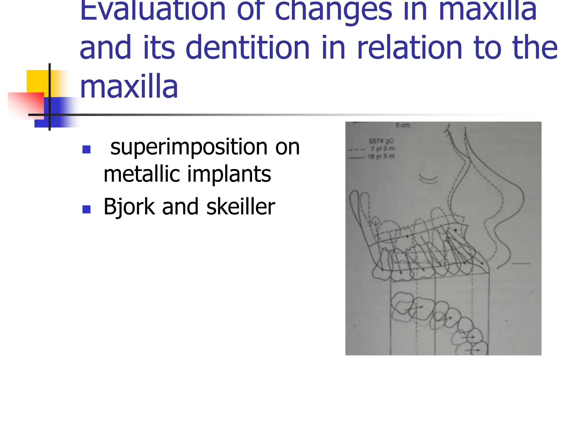
SUPERIMPOSITION
TECHNIQUES
Introduction
 In 1931 Broadbent in USA and Hofrath in Germany
simultaneously presented a standardised
cephalometric technique using a high powered X-ray
source and a head holder called a cephalostat.
 Orthodontic diagnosis by enabling the study of skeletal,
dental and soft tissue structures of the craniofacial region
 Treatment planning.
 Evaluation of treatment results by quantifying the changes
brought about by treatment
 Predicting growth related changes
What is cephalometric
superimposition ?
 A cephalometric superimposition is an
analysis of lateral cephalograms of the same
patient taken at different times.
 Uses:
 Evaluate a patients growth pattern at different
ages
 To evaluate changes in basal and dentoalveolar
relationships after treatment
 To quantify growth and treatment changes in
dentoalveolar and basal relationships.
Method of superimposing
radiographs
Superimposing on a stable plane
or structure
Registration on a stable
landmark
Validity and reproducibility
 Validity: it is the extent to which the
value obtained represents the object of
interest.
 Planes and landmarks should be
anatomically valid and should agree with
the anatomic structures they represent
 Reproducibility: it is the closeness of
successive measurements of the same
object.
Methods of assessing
Dentofacial changes
 Study of changes in dentofacial dimensions using
cephalometric superimposition have shown varying
results
 Brodie and broadbent have shown that dentofacial
growth patterns are established at a very early age
and thereafter are subject to proportional changes.
 Downs and Rickkets pointed out that several angles
and dimensions change with age but in an orderly
and progressive manner
 Hellman suggested that the infant face is transformed
into that of an adult face by increase in size by
changes in proportion and by adjustment in position.
Methods of assessing
Dentofacial changes
 Areas studied to assess changes due to
growth or treatment or both include:
 Changes in the overall face
 Changes in the maxilla and it’s dentition
 Changes in the mandible and its dentition
 Amount and direction of condylar growth
 Mandibular rotation
Methods of assessing
Dentofacial changes
 Color coding of consecutive
cephalograms suggested by ABO:
 Pretreatment – black
 Progress – blue
 End of treatment – red
 Retention - green
Evaluation of overall changes
in the face
 Superimposition methods:
 Broadbent triangle
 Sella nasion line
 Basion horizontal
 Basion nasion plane
 De costers anterior cranial base reference line
 Viazis cranial base triangle
 Frankl’s occipital reference base
Evaluation of overall changes
in the face
 Objectives
 Overall assessment of growth and treatment
changes of the facial structures
 Amount of change in direction of displacement
and growth of maxilla and mandible.
 Changes in soft tissue
 Changes in maxillo mandibular relationship
 Overall displacement of teeth.
Evaluation of overall changes
in the face
 Broadbent triangle
 Among the first structures used for
superimposition
 Broadbent based this method on
observations of dried skulls and a
comparative study of cranial base
planes (Bolton-nasion, porion-nasion,
sella-nasion) in persons 3 to 18 years
of age.
 Bolton point maybe obscured by
Mastoid
Superimposition at registration point R with Bolton-nasion planes parallel
Evaluation of overall changes
in the face
 Sella nasion plane:
 SN is a frequently used reference line
that has been reported to be relatively
stable.
 Both points S and N are located in the
midsagittal plane and are displaced a
minimal degree by movement of the
head.
 Steiner used SN with registration point
at sella to evaluate sagittal changes in
mandibular positions and at nasion to
evaluate the position of the maxilla
through changes in the angle SNA.

Evaluation of overall changes
in the face
 Sella nasion plane:
 Unlike Steiner, Björk used sella as
registration point to assess changes
in position of both jaws.
 Later, Björk reported that errors of
biologic origins of S and N may
weaken the SN reference for
estimation of facial changes. He
stated that an upward or downward
displacement of nasion may occur
with growth at the frontonasal
suture. Likewise, a posterior
displacement of sella may be
induced by the remodeling of
dorsum sellae connected with the
increased size of the pituitary
gland.
Evaluation of overall changes
in the face
 Basion horizontal
 Coben presented the Basion
horizontal concept.
 Basion is used as the
registration point.
 The SN planes are made parallel
by the help of the Basion
horizontal line and it’s constant
relationship with SN.
 A coordinate grid system is used
to superimpose the radiographs.
Evaluation of overall changes
in the face
 Basion - Nasion plane:
 Suggested by Ricketts
 He considered Ba-N plane as a line of
separation of the face from the skull
and hence a basic cranial axis for
growth and structural reference.
 Based on studies of laminograph
sections, Ricketts suggested that the
cranial base angle, while constant on
average, exhibits a change of 5° in
either direction over a 3-year period
Evaluation of overall changes
in the face
 Basion - Nasion plane:
 one may doubt the reliability of this
axis because growth at nasion is
subjected to individual variations.
Moreover, the position of basion is
influenced by remodeling processes on
the clivus surface and on the anterior
border of the foramen magnum, and by
changes in the position of the pars
basilaris ossis occipitalis associated
with growth in the spheno-occipital
synchondrosis.
Evaluation of overall changes
in the face
 De Coster line
 He advocated tracing
the inner contour of
the frontal bone
through the cerebral
aspect of the ethmoid ,
the planum
sphenoidale and the
anterior aspect of the
sella turcica.
Evaluation of overall changes
in the face
 Viazis cranial base triangle
 The anterior wall of sella
turcica and the cribriform plate
(laminar cribrosa) remain
unchanged after age 5.
 Superimposition on the anterior
wall of sella turcica and the
stable TC (cranial base) line,
with registration on T point,
provides a practical and
reliable formation in both the
anteroposterior and vertical
planes.
Evaluation of overall changes
in the face
 Viazis cranial base triangle
 Superimposition on the anterior
wall of sella turcica and the stable
TC (cranial base) line, with
registration on T point, provides a
practical and reliable formation in
both the anteroposterior and
vertical planes.
 First priority should be given to
registering on T point, followed by
superimposing on the inner
structure of the triangle, and finally
superimposing on the TC line. This
"best-fit" approach meets the
realistic expectations of any
superimposition technique.
Evaluation of overall changes
in the face – Viazis Triangle
Frankl’s occipital reference
base
 Among the basal structures of the neurocranium, the occipital bone
around the foramen magnum is the first to ossify (between the third
and fourth year of life).
 Phylogenetically and ontogenetically as the midbrain is highly
conserved minimalpostnatal growth of this structure and surrounding
tissues is seen.
 It permits the study of craniofacial growth in relation to an individual
bone and its immediate structures
 Landmarks – O’ and
O”
 Reference plane –
based on natural
head posture and
parallel to the
ground
Frankl’s occipital reference
base
Frankl’s occipital reference
base
 Reliability of the various cranial base reference planes used.
 For meaningful interpretations of superimpositions they have to be
registered on stable reference landmarks.
 Cranial base superimpositions are subject to error due to the continued
growth of the sphenoccipital synchondrosis (Knott).
 Bone remodelling at sella and Nasion are also responsible for further
errors.
 Nasion position can change in a vertical direction (Nelson and Knott).
 Melsen’s study’s on human autopsy material has shown that the
position of sella may change in a downward or a downward and
backward direction. She also showed that the position of Basion
changed due to remodelling of the clivus.
 The Bolton point could be difficult to locate in children due to the
shadow of the mastoid process
Evaluation of overall changes
in the face
Growth changes in position of Nasion and sella
Reference structures for
overall face superimpositions.
 Nelson’s cephalometric study and Melsen’s
histological study on human autopsy
materials have reveled a few stable structures
in the anterior cranial base for use in
superimposition.
 Anterior wall of sella turcica
 The contour of the cribriform plate of the ethmoid
 Trabecular system of the ethmoid air cells
 The median border of the orbital roof
 Planum sphenoidale
Method of superimposition of
radiographs to assess overall
changes
Pretreatment tracing Progress tracing
Method of superimposition of
radiographs to assess overall
changes
Superimposition using ‘Best fit method’
Evaluation of changes in maxilla
and its dentition in relation to the
maxilla
 Superimposition along
the palatal plane
registered at ANS
 Broadbent, Moore,
Salzman, Ricketts,
McNamara
 Compromised by
remodelling of the
palatal shelves and
ANS- Bjork and Skeiler
Evaluation of changes in maxilla
and its dentition in relation to the
maxilla
 Superimposition on
the nasal floors with
films registered at
the anterior surface
of the maxilla
 Downs and Brodie.
Evaluation of changes in maxilla
and its dentition in relation to the
maxilla
 Superimposition
along the palatal
plane registered at
the pterygomaxillary
fissure
 Moore
Evaluation of changes in maxilla
and its dentition in relation to the
maxilla
 Superimposition on
the outline of the
infratemporal fossa
and the posterior
margin of the hard
palate.
 Reidel
Evaluation of changes in maxilla
and its dentition in relation to the
maxilla
 superimpostion
registered at the
common Ptm
cordinate
maintaining the
basion horizontal
relationship.
 Coben
Evaluation of changes in maxilla
and its dentition in relation to the
maxilla
 superimposition on
the best fit of the
internal palatal
structures.
 McNamara
Evaluation of changes in maxilla
and its dentition in relation to the
maxilla
 superimposition on
metallic implants
 Bjork and skeiller
Evaluation of changes in maxilla
and its dentition in relation to the
maxilla
 the structural
superimposition on
the anterior surface
of the zygomatic
process of the
maxilla
 Bjork and Skeiler
Evaluation of changes in maxilla
and its dentition in relation to the
maxilla
 Neilsen on a study of various maxillary
superimposition techniques concluded that:
 The best fit method significantly under estimates
the vertical displacement of skeletal and dental
landmarks
 With the implant method and the structural
method ANS showed twice as much vertical
displacement as PNS
 Structural method and implant methd did not
show any significant differences
 The structural method is a valid method of
assessing maxillary growth and treatment changes
Structural method of
superimposing maxillary
structures
Pretreatment tracing
Structural method of
superimposing maxillary
structures
superimposition
Mandibular superimposition
 Stable areas for
superimposition:
 Anterior contour of the chin
 The inner contour of the
cortical plates at the inferior
border of the symphysis
 Contours of the mandibular
canal
 Lower contour of the
mineralized molar tooth
germ
Mandibular superimposition
technique
Mandibular superimposition
technique
Methods to assess growth vs
treatment changes
 Though the techniques described till now will assess
the amount of growth changes in a given duration of
time or the overall changes of treatment and growth
during a given treatment period they do not however
differentiate between changes produced due to
growth and changes produced due to treatment.
 The following cephalometric analyses help us to
assess treatment changes against the background of
natural growth of the individual
Methods to assess growth vs
treatment changes
 Rickett’s eleven factor summary analysis
 Four position analysis
 The analysis is based on Rickkets short
term growth forecasting, data which was
obtained on patients – both male and
female of different ages and growth
patterns undergoing orthodontic treatment.
Rickett’s Eleven factor
summary analysis
 Eleven factors of the basic facial and skeletal
structures are recorded from the
cephalometric tracing to describe the chin,
maxilla, teeth and soft tissue profile.
 Five areas of superimposition within which
are a total of seven areas of evaluation are
used to evaluate in amount and direction,
change in normal growth and change due to
treatment.
Rickett’s Eleven factor
summary analysis
Rickett’s Eleven factor
summary analysis
CHIN IN SPACE MEAN CHANGE
FACIAL AXIS 90+/-3 No change
FACIAL DEPTH 87+/-3 +10 for 3 yrs
MAND PLANE 26+/-4 -1o for 3 yrs
FACIAL TAPER 68+/-3 No change
CONVEXITY
CON AT A 2+/-2 -1mm / 3 yrs
TEETH
L1 TO APO 1+/-2 No change
L1 INCL 22+/-4 No change
U6 TO PTV Age+3+2 1mm / year
PROFILE
L LIP TO E LINE -2+/-2 Less with age
Rickett’s Eleven factor
summary analysis
SUPERIMPOSITION AREA 1 – EVALUATION AREA 1
• FACIAL AXIS OPENS 1o FOR
DOLICOFACIAL PATTERN
•FACIAL AXIS CLOSES 1O FOR
BRACHYFACIAL PATTERNS
•FACIAL AXIS OPENS 1O FOR 5mm
CONVEXITY REDUCTION
•FACIAL AXIS OPENS 1O FOR 3mm
MOLAR CORRECTION
•FACIAL AXIS OPENS 1O OR 4mm
OVERBITE CORRECTION
•FACIAL AXIS OPENS 1 TO 1.5O FOR
CROSS BITE CORRECTION AND
RECOVERS ONE HALF
BASION-NASION PLANE at CC
Rickett’s Eleven factor
summary analysis
SUPERIMPOSITION AREA 2 – EVALUATION AREA 2
• THE BASION-NASION-POINT A
ANGLE OF 66O DOES NOT CHANGE
WITH GROWTH
•SO ANY CHANGES PRODUCED MUST
BE DUE TO TREATMENT
•HEADGEAR - -8mm
•CLASS II ELASTICS - -3mm
•ACTIVATOR - -2mm
•TORQUE- - 1 TO 2mm
•CLASS III ELASTICS 2 TO 3mm
•FACIAL MASK - 2 TO 4mm
BASION NASION PLANE AT NASION
Rickett’s Eleven factor
summary analysis
SUPERIMPOSITION AREA 3 – EVALUATION AREAS 3 AND 4
• LOWER DENTURE REMAINS CONSTANT
WITH THE A Pog LINE
•Without treatment, the lower molar will
erupt directly upward to the new occlusal
plane.
•The LOWER INCISAL angle is 22° at
+1mm to the APo plane and + 1 mm to
occlusal plane, but the angle increases 2°
with each mm of forward compromise.
•OCCLUSAL PLANE TO CORPUS AXIS DOES
NOT CHANGE
•LOWER MOLAR ERUPTS IN A DIRECTION
PERPENDICULAR TO THE FH PLANE
•OCCLUSAL PLANE ERUPTS 0.8mm
UPWARDS FROM THE CORPUS AXIS.
CORPUS AXIS AT PM
Rickett’s Eleven factor
summary analysis
SUPERIMPOSITION AREA 4 – EVALUATION AREAS 5 AND 6
• THE UPPER MOLAR AND INCISOR
FOLLOW THEIR POLAR AXIS WITH
GROWTH
•CHANGES IN POSITION OF THE
MOLAR OR INCISOR IS DUETO
TREATMENT.
•UPPER DENTAL ARCH ERUPTS
DOWNWARD AND FORWARD 0.2 TO
0.3mm PER YEAR
PALATE AT ANS
Rickett’s Eleven factor
summary analysis
SUPERIMPOSITION AREA 5 – EVALUATION AREA 7
•
ESTHETIC PLANE AT INTERSECTION OF
OCCLUSAL PLANES
Rickett’s four position analysis
 Takes into consideration two
superimposition areas to evaluate
orthopedic change and two
superimposition areas to evaluate
orthodontic change against growth.
Rickett’s four position analysis
 Position 1
Rickett’s four position analysis
 Position 2
Rickett’s four position analysis
 Position 3
Rickett’s four position analysis
 Position 4
Superimposition methods to assess
dentoalveolar and skeletal changes in
Class II treatment.
 Pancherz analysis
 Modified Pancherz analysis
 Johnstons Pitchfork analysis
Pancherz analysis
Pancherz analysis
 is/OLp minus ii/OLp— Overjet.
 ms/OLp minus mi/OLp— Molar
relation (a positive value indicates a
distal relation; a negative value
indicates a normal relation).
 Skeletal measuring points
 ss/OLp— Position of the maxillary
base.
 pg/OLp— Position of the mandibular
base.
 co/OLp— Position of the condylar
head.
 pg/OLp + co/OLp— Mandibular
length.
Pancherz analysis
 Dental measuring points
 is/OLp— Position of the maxillary
central incisor.
 ii/OLp— Position of the mandibular
central incisor.
 ms/OLp— Position of the maxillary
permanent first molar.
 mi/OLp— Position of the mandibular
permanent first molar
Pancherz analysis
 11. is/OLp (d) minus ss/OLp
(d)— Change in position of the
maxillary central incisor within
the maxilla.
 12. ii/OLp (d) minus pg/OLp
(d)— Change in position of the
mandibular central incisor within
the mandible.
 13. ms/OLp (d) minus ss/OLp
(d)— Change in position of the
maxillary permanent first molar
within the maxilla.
 14. mi/OLp(d) minus
pg/OLp(d)— Change in position
of the mandibular permanent
first molar within the mandible.
Modified Pancherz Analysis
The original analysis The modified Analysis
Modified Pancherz Analysis
LINEAR
MEASUREMENTS
Co-pg
Co-go
Go-pg
Modified Pancherz Analysis
ANGULAR MEASUREMENTS
FMN-T-ba
FMN-T-ar
Cl_ml
Ar-goi-me
Nl-T-FMN
Ml-T-FMN
Nl-ml
Modified Pancherz Analysis
VERTICAL DENTAL
T-FMN/is
T-FMN/ii
T-FMN/ms
T-FMN/mi
Pitchfork analysis
 Johnston in 1985
 Used to describethe
treatment effects of
different treatment
strategies used to
correct Class II
patients
 Data recorded in the
form of a pitch fork
Pitchfork analysis
 Reference planes – SE
perpendicular to MFOP
 D perpendicular to
MFOP
 Measurement of Max
change
 Measurement of ABCH
 Calculation of Mand
Pitchfork analysis
 Superimposition on
D
 Measurement of
molar and incisal
changes
Pitchfork analysis
 Measurement of
amount of molar
correction
Pitchfork analysis
 Measurement of
overjet correction
ORTHODONTICS SUPERIMPOSITION_TECHNIQUES.ppt

ORTHODONTICS SUPERIMPOSITION_TECHNIQUES.ppt

  • 1.
  • 2.
    Introduction  In 1931Broadbent in USA and Hofrath in Germany simultaneously presented a standardised cephalometric technique using a high powered X-ray source and a head holder called a cephalostat.  Orthodontic diagnosis by enabling the study of skeletal, dental and soft tissue structures of the craniofacial region  Treatment planning.  Evaluation of treatment results by quantifying the changes brought about by treatment  Predicting growth related changes
  • 3.
    What is cephalometric superimposition?  A cephalometric superimposition is an analysis of lateral cephalograms of the same patient taken at different times.  Uses:  Evaluate a patients growth pattern at different ages  To evaluate changes in basal and dentoalveolar relationships after treatment  To quantify growth and treatment changes in dentoalveolar and basal relationships.
  • 4.
    Method of superimposing radiographs Superimposingon a stable plane or structure Registration on a stable landmark
  • 5.
    Validity and reproducibility Validity: it is the extent to which the value obtained represents the object of interest.  Planes and landmarks should be anatomically valid and should agree with the anatomic structures they represent  Reproducibility: it is the closeness of successive measurements of the same object.
  • 6.
    Methods of assessing Dentofacialchanges  Study of changes in dentofacial dimensions using cephalometric superimposition have shown varying results  Brodie and broadbent have shown that dentofacial growth patterns are established at a very early age and thereafter are subject to proportional changes.  Downs and Rickkets pointed out that several angles and dimensions change with age but in an orderly and progressive manner  Hellman suggested that the infant face is transformed into that of an adult face by increase in size by changes in proportion and by adjustment in position.
  • 7.
    Methods of assessing Dentofacialchanges  Areas studied to assess changes due to growth or treatment or both include:  Changes in the overall face  Changes in the maxilla and it’s dentition  Changes in the mandible and its dentition  Amount and direction of condylar growth  Mandibular rotation
  • 8.
    Methods of assessing Dentofacialchanges  Color coding of consecutive cephalograms suggested by ABO:  Pretreatment – black  Progress – blue  End of treatment – red  Retention - green
  • 9.
    Evaluation of overallchanges in the face  Superimposition methods:  Broadbent triangle  Sella nasion line  Basion horizontal  Basion nasion plane  De costers anterior cranial base reference line  Viazis cranial base triangle  Frankl’s occipital reference base
  • 10.
    Evaluation of overallchanges in the face  Objectives  Overall assessment of growth and treatment changes of the facial structures  Amount of change in direction of displacement and growth of maxilla and mandible.  Changes in soft tissue  Changes in maxillo mandibular relationship  Overall displacement of teeth.
  • 11.
    Evaluation of overallchanges in the face  Broadbent triangle  Among the first structures used for superimposition  Broadbent based this method on observations of dried skulls and a comparative study of cranial base planes (Bolton-nasion, porion-nasion, sella-nasion) in persons 3 to 18 years of age.  Bolton point maybe obscured by Mastoid Superimposition at registration point R with Bolton-nasion planes parallel
  • 12.
    Evaluation of overallchanges in the face  Sella nasion plane:  SN is a frequently used reference line that has been reported to be relatively stable.  Both points S and N are located in the midsagittal plane and are displaced a minimal degree by movement of the head.  Steiner used SN with registration point at sella to evaluate sagittal changes in mandibular positions and at nasion to evaluate the position of the maxilla through changes in the angle SNA. 
  • 13.
    Evaluation of overallchanges in the face  Sella nasion plane:  Unlike Steiner, Björk used sella as registration point to assess changes in position of both jaws.  Later, Björk reported that errors of biologic origins of S and N may weaken the SN reference for estimation of facial changes. He stated that an upward or downward displacement of nasion may occur with growth at the frontonasal suture. Likewise, a posterior displacement of sella may be induced by the remodeling of dorsum sellae connected with the increased size of the pituitary gland.
  • 14.
    Evaluation of overallchanges in the face  Basion horizontal  Coben presented the Basion horizontal concept.  Basion is used as the registration point.  The SN planes are made parallel by the help of the Basion horizontal line and it’s constant relationship with SN.  A coordinate grid system is used to superimpose the radiographs.
  • 15.
    Evaluation of overallchanges in the face  Basion - Nasion plane:  Suggested by Ricketts  He considered Ba-N plane as a line of separation of the face from the skull and hence a basic cranial axis for growth and structural reference.  Based on studies of laminograph sections, Ricketts suggested that the cranial base angle, while constant on average, exhibits a change of 5° in either direction over a 3-year period
  • 16.
    Evaluation of overallchanges in the face  Basion - Nasion plane:  one may doubt the reliability of this axis because growth at nasion is subjected to individual variations. Moreover, the position of basion is influenced by remodeling processes on the clivus surface and on the anterior border of the foramen magnum, and by changes in the position of the pars basilaris ossis occipitalis associated with growth in the spheno-occipital synchondrosis.
  • 17.
    Evaluation of overallchanges in the face  De Coster line  He advocated tracing the inner contour of the frontal bone through the cerebral aspect of the ethmoid , the planum sphenoidale and the anterior aspect of the sella turcica.
  • 18.
    Evaluation of overallchanges in the face  Viazis cranial base triangle  The anterior wall of sella turcica and the cribriform plate (laminar cribrosa) remain unchanged after age 5.  Superimposition on the anterior wall of sella turcica and the stable TC (cranial base) line, with registration on T point, provides a practical and reliable formation in both the anteroposterior and vertical planes.
  • 19.
    Evaluation of overallchanges in the face  Viazis cranial base triangle  Superimposition on the anterior wall of sella turcica and the stable TC (cranial base) line, with registration on T point, provides a practical and reliable formation in both the anteroposterior and vertical planes.  First priority should be given to registering on T point, followed by superimposing on the inner structure of the triangle, and finally superimposing on the TC line. This "best-fit" approach meets the realistic expectations of any superimposition technique.
  • 20.
    Evaluation of overallchanges in the face – Viazis Triangle
  • 21.
    Frankl’s occipital reference base Among the basal structures of the neurocranium, the occipital bone around the foramen magnum is the first to ossify (between the third and fourth year of life).  Phylogenetically and ontogenetically as the midbrain is highly conserved minimalpostnatal growth of this structure and surrounding tissues is seen.  It permits the study of craniofacial growth in relation to an individual bone and its immediate structures
  • 23.
     Landmarks –O’ and O”  Reference plane – based on natural head posture and parallel to the ground Frankl’s occipital reference base
  • 24.
  • 25.
     Reliability ofthe various cranial base reference planes used.  For meaningful interpretations of superimpositions they have to be registered on stable reference landmarks.  Cranial base superimpositions are subject to error due to the continued growth of the sphenoccipital synchondrosis (Knott).  Bone remodelling at sella and Nasion are also responsible for further errors.  Nasion position can change in a vertical direction (Nelson and Knott).  Melsen’s study’s on human autopsy material has shown that the position of sella may change in a downward or a downward and backward direction. She also showed that the position of Basion changed due to remodelling of the clivus.  The Bolton point could be difficult to locate in children due to the shadow of the mastoid process Evaluation of overall changes in the face
  • 26.
    Growth changes inposition of Nasion and sella
  • 27.
    Reference structures for overallface superimpositions.  Nelson’s cephalometric study and Melsen’s histological study on human autopsy materials have reveled a few stable structures in the anterior cranial base for use in superimposition.  Anterior wall of sella turcica  The contour of the cribriform plate of the ethmoid  Trabecular system of the ethmoid air cells  The median border of the orbital roof  Planum sphenoidale
  • 29.
    Method of superimpositionof radiographs to assess overall changes Pretreatment tracing Progress tracing
  • 30.
    Method of superimpositionof radiographs to assess overall changes Superimposition using ‘Best fit method’
  • 31.
    Evaluation of changesin maxilla and its dentition in relation to the maxilla  Superimposition along the palatal plane registered at ANS  Broadbent, Moore, Salzman, Ricketts, McNamara  Compromised by remodelling of the palatal shelves and ANS- Bjork and Skeiler
  • 32.
    Evaluation of changesin maxilla and its dentition in relation to the maxilla  Superimposition on the nasal floors with films registered at the anterior surface of the maxilla  Downs and Brodie.
  • 33.
    Evaluation of changesin maxilla and its dentition in relation to the maxilla  Superimposition along the palatal plane registered at the pterygomaxillary fissure  Moore
  • 34.
    Evaluation of changesin maxilla and its dentition in relation to the maxilla  Superimposition on the outline of the infratemporal fossa and the posterior margin of the hard palate.  Reidel
  • 35.
    Evaluation of changesin maxilla and its dentition in relation to the maxilla  superimpostion registered at the common Ptm cordinate maintaining the basion horizontal relationship.  Coben
  • 36.
    Evaluation of changesin maxilla and its dentition in relation to the maxilla  superimposition on the best fit of the internal palatal structures.  McNamara
  • 37.
    Evaluation of changesin maxilla and its dentition in relation to the maxilla  superimposition on metallic implants  Bjork and skeiller
  • 38.
    Evaluation of changesin maxilla and its dentition in relation to the maxilla  the structural superimposition on the anterior surface of the zygomatic process of the maxilla  Bjork and Skeiler
  • 39.
    Evaluation of changesin maxilla and its dentition in relation to the maxilla  Neilsen on a study of various maxillary superimposition techniques concluded that:  The best fit method significantly under estimates the vertical displacement of skeletal and dental landmarks  With the implant method and the structural method ANS showed twice as much vertical displacement as PNS  Structural method and implant methd did not show any significant differences  The structural method is a valid method of assessing maxillary growth and treatment changes
  • 40.
    Structural method of superimposingmaxillary structures Pretreatment tracing
  • 41.
    Structural method of superimposingmaxillary structures superimposition
  • 42.
    Mandibular superimposition  Stableareas for superimposition:  Anterior contour of the chin  The inner contour of the cortical plates at the inferior border of the symphysis  Contours of the mandibular canal  Lower contour of the mineralized molar tooth germ
  • 43.
  • 44.
  • 45.
    Methods to assessgrowth vs treatment changes  Though the techniques described till now will assess the amount of growth changes in a given duration of time or the overall changes of treatment and growth during a given treatment period they do not however differentiate between changes produced due to growth and changes produced due to treatment.  The following cephalometric analyses help us to assess treatment changes against the background of natural growth of the individual
  • 46.
    Methods to assessgrowth vs treatment changes  Rickett’s eleven factor summary analysis  Four position analysis  The analysis is based on Rickkets short term growth forecasting, data which was obtained on patients – both male and female of different ages and growth patterns undergoing orthodontic treatment.
  • 47.
    Rickett’s Eleven factor summaryanalysis  Eleven factors of the basic facial and skeletal structures are recorded from the cephalometric tracing to describe the chin, maxilla, teeth and soft tissue profile.  Five areas of superimposition within which are a total of seven areas of evaluation are used to evaluate in amount and direction, change in normal growth and change due to treatment.
  • 48.
  • 49.
    Rickett’s Eleven factor summaryanalysis CHIN IN SPACE MEAN CHANGE FACIAL AXIS 90+/-3 No change FACIAL DEPTH 87+/-3 +10 for 3 yrs MAND PLANE 26+/-4 -1o for 3 yrs FACIAL TAPER 68+/-3 No change CONVEXITY CON AT A 2+/-2 -1mm / 3 yrs TEETH L1 TO APO 1+/-2 No change L1 INCL 22+/-4 No change U6 TO PTV Age+3+2 1mm / year PROFILE L LIP TO E LINE -2+/-2 Less with age
  • 50.
    Rickett’s Eleven factor summaryanalysis SUPERIMPOSITION AREA 1 – EVALUATION AREA 1 • FACIAL AXIS OPENS 1o FOR DOLICOFACIAL PATTERN •FACIAL AXIS CLOSES 1O FOR BRACHYFACIAL PATTERNS •FACIAL AXIS OPENS 1O FOR 5mm CONVEXITY REDUCTION •FACIAL AXIS OPENS 1O FOR 3mm MOLAR CORRECTION •FACIAL AXIS OPENS 1O OR 4mm OVERBITE CORRECTION •FACIAL AXIS OPENS 1 TO 1.5O FOR CROSS BITE CORRECTION AND RECOVERS ONE HALF BASION-NASION PLANE at CC
  • 51.
    Rickett’s Eleven factor summaryanalysis SUPERIMPOSITION AREA 2 – EVALUATION AREA 2 • THE BASION-NASION-POINT A ANGLE OF 66O DOES NOT CHANGE WITH GROWTH •SO ANY CHANGES PRODUCED MUST BE DUE TO TREATMENT •HEADGEAR - -8mm •CLASS II ELASTICS - -3mm •ACTIVATOR - -2mm •TORQUE- - 1 TO 2mm •CLASS III ELASTICS 2 TO 3mm •FACIAL MASK - 2 TO 4mm BASION NASION PLANE AT NASION
  • 52.
    Rickett’s Eleven factor summaryanalysis SUPERIMPOSITION AREA 3 – EVALUATION AREAS 3 AND 4 • LOWER DENTURE REMAINS CONSTANT WITH THE A Pog LINE •Without treatment, the lower molar will erupt directly upward to the new occlusal plane. •The LOWER INCISAL angle is 22° at +1mm to the APo plane and + 1 mm to occlusal plane, but the angle increases 2° with each mm of forward compromise. •OCCLUSAL PLANE TO CORPUS AXIS DOES NOT CHANGE •LOWER MOLAR ERUPTS IN A DIRECTION PERPENDICULAR TO THE FH PLANE •OCCLUSAL PLANE ERUPTS 0.8mm UPWARDS FROM THE CORPUS AXIS. CORPUS AXIS AT PM
  • 53.
    Rickett’s Eleven factor summaryanalysis SUPERIMPOSITION AREA 4 – EVALUATION AREAS 5 AND 6 • THE UPPER MOLAR AND INCISOR FOLLOW THEIR POLAR AXIS WITH GROWTH •CHANGES IN POSITION OF THE MOLAR OR INCISOR IS DUETO TREATMENT. •UPPER DENTAL ARCH ERUPTS DOWNWARD AND FORWARD 0.2 TO 0.3mm PER YEAR PALATE AT ANS
  • 54.
    Rickett’s Eleven factor summaryanalysis SUPERIMPOSITION AREA 5 – EVALUATION AREA 7 • ESTHETIC PLANE AT INTERSECTION OF OCCLUSAL PLANES
  • 55.
    Rickett’s four positionanalysis  Takes into consideration two superimposition areas to evaluate orthopedic change and two superimposition areas to evaluate orthodontic change against growth.
  • 56.
    Rickett’s four positionanalysis  Position 1
  • 57.
    Rickett’s four positionanalysis  Position 2
  • 58.
    Rickett’s four positionanalysis  Position 3
  • 59.
    Rickett’s four positionanalysis  Position 4
  • 60.
    Superimposition methods toassess dentoalveolar and skeletal changes in Class II treatment.  Pancherz analysis  Modified Pancherz analysis  Johnstons Pitchfork analysis
  • 61.
  • 62.
    Pancherz analysis  is/OLpminus ii/OLp— Overjet.  ms/OLp minus mi/OLp— Molar relation (a positive value indicates a distal relation; a negative value indicates a normal relation).  Skeletal measuring points  ss/OLp— Position of the maxillary base.  pg/OLp— Position of the mandibular base.  co/OLp— Position of the condylar head.  pg/OLp + co/OLp— Mandibular length.
  • 63.
    Pancherz analysis  Dentalmeasuring points  is/OLp— Position of the maxillary central incisor.  ii/OLp— Position of the mandibular central incisor.  ms/OLp— Position of the maxillary permanent first molar.  mi/OLp— Position of the mandibular permanent first molar
  • 64.
    Pancherz analysis  11.is/OLp (d) minus ss/OLp (d)— Change in position of the maxillary central incisor within the maxilla.  12. ii/OLp (d) minus pg/OLp (d)— Change in position of the mandibular central incisor within the mandible.  13. ms/OLp (d) minus ss/OLp (d)— Change in position of the maxillary permanent first molar within the maxilla.  14. mi/OLp(d) minus pg/OLp(d)— Change in position of the mandibular permanent first molar within the mandible.
  • 65.
    Modified Pancherz Analysis Theoriginal analysis The modified Analysis
  • 66.
  • 67.
  • 68.
    Modified Pancherz Analysis ANGULARMEASUREMENTS FMN-T-ba FMN-T-ar Cl_ml Ar-goi-me Nl-T-FMN Ml-T-FMN Nl-ml
  • 69.
    Modified Pancherz Analysis VERTICALDENTAL T-FMN/is T-FMN/ii T-FMN/ms T-FMN/mi
  • 70.
    Pitchfork analysis  Johnstonin 1985  Used to describethe treatment effects of different treatment strategies used to correct Class II patients  Data recorded in the form of a pitch fork
  • 71.
    Pitchfork analysis  Referenceplanes – SE perpendicular to MFOP  D perpendicular to MFOP  Measurement of Max change  Measurement of ABCH  Calculation of Mand
  • 72.
    Pitchfork analysis  Superimpositionon D  Measurement of molar and incisal changes
  • 73.
    Pitchfork analysis  Measurementof amount of molar correction
  • 74.