SlideShare a Scribd company logo
1 of 85
A Consensus Meeting  for The Definitions of Subcortical lesions, Lacunes, Microbleeds, and Subcortical White Matter Change Jei Kim, MD
M/71, 2011. 1. 28
M/71, 2011. 1. 28
M/71, 2011. 1. 28
Subcortical lesions 1. Perivascular spaces (etatcrible) 2. Lacunes 3. Subcortical white matter changes 4. Microbleeds
Perivascular spaces (etatcrible)
1. Perivascular spaces (etatcrible) 1) Histopathologic definition (ref. 1) : the dilatation of perivascular spaces around   cerebral arterioles in the brain of elderly patients   (Virchow-Robin space) 2) MRI definition (ref. 1) : the punctiform dilatations of the perivascular  spaces often seen by brain MRI in the white   matter and in the basal ganglia
1. Perivascular spaces (etatcrible) 2) MRI definition : the punctiform dilatations of the perivascular spaces   often seen by brain MRI in the white matter and   in the basal ganglia : On T2WI – high intensity,  same as the intensity of CSF  : On FLAIR – dark (low), same as the intensity of CSF : On T1WI – dark (low), same as the intensity of CSF http://www.radiologyassistant.nl/en/4556dea65db62
M/80, 2011. 10. 13
Lacunes
2. Lacunes 1) Histopathologic definition : a small, cystic cavity of the brain substance that   usually results from an ischemic infarction in the   territory of a penetrating arteriole (ref. 1)
2. Lacunes 2) Vascular pathology of lacunes (ref. 13)
2. Lacunes 3) Perforating arteries  (1) Anterior perforating arteries (2) Posterior perforating arteries (3) Arterial supply of the brainstem
(1) Anterior perforating arteries
(1) Anterior perforating arteries A. Perforating branches arising from ACA    and the recurrent artery of Heubner
(1) Anterior perforating arteries B. Perforating branches arising from MCA a. Perforating branches arising from MCA
(1) Anterior perforating arteries B. Perforating branches arising from MCA b. Percentage of perforating arteries arising from MCA trunk and its branches
(1) Anterior perforating arteries B. Perforating branches arising from MCA c. Origin of perforating arteries arising     from MCA trunk and its branches
(1) Anterior perforating arteries B. Perforating branches arising from MCA d. Number of perforating arteries arising     from different distances from the origin of MCA
(1) Anterior perforating arteries B. Perforating branches arising from MCA e. Branching characteristics of 508 perforating arteries     arising from common stems of MCA
(2) Posterior perforating arteries
(3) Arterial supply of the brainstem A. Perforating arteries of the midbrain
(3) Arterial supply of the brainstem B. Perforating arteries of the pons
(3) Arterial supply of the brainstem C. Perforating arteries of the midbrain
Schematic diagram of origin of deep perforating branches from a parent artery
2. Lacunes 4) Pathogenic implications of microcirculation (1) Perforating arteries 1) Stenosis or complete occlusion by atherosclerosis 2) Stenosis or occlusion of ostium of a branch point 3) Atherosclerotic narrowing of a parent artery 4) Proximal thrombus or embolus in atherosclerotic artery (2) Cortical branches
4) Pathogenic implications of microcirculation (1) Perforating arteries A. Stenosis or complete occlusion by atherosclerosis
4) Pathogenic implications of microcirculation (1) Perforating arteries B. Stenosis or occlusion of ostium of a branch point
4) Pathogenic implications of microcirculation (1) Perforating arteries C. Atherosclerotic narrowing of a parent artery
4) Pathogenic implications of microcirculation (1) Perforating arteries D. Proximal thrombus or embolus in atherosclerotic artery
4) Pathogenic implications of microcirculation (2) Cortical branches
2. Lacunes 5) MRI definition (ref. 2) : small hyperintense lesions on T2WI (ref. 2) : corresponding distinctive low intensity area on T1WI : Maximum size of lacune (ref. 4)   - with a diameter of 5-10 mm : On CT (ref. 4)   - areas of more or less complete focal tissue destruction   - clearly defined borders with marked central  hypodensity on CT : On MRI (ref. 4)   - low intensity on T1WI, proton-density and FLAIR scans   - high intensity on T2WI     -> isointense to CSF
2. Lacunes 5) MRI definition (ref. 17)
2. Lacunes 1) Histopathologic definition : a small, cystic cavity of the brain substance that   usually results from an ischemic infarction in the   territory of a penetrating arteriole (ref. 1) : defined as cavitatedmicroinfarcts or encephalomalacic  lesions, 2 mm or smaller in greatest dimension, not   identifiable with certainty on gross inspection of the   brain or non-cavitatedmicroinfarcts, focal gliotic areas   without a cystic cavity (ref. 3)
M/80, 2011. 10. 13
2. Lacunes 6) Grading of lacunes (ref. 2) Absent (2) Mild – 1-3 (3) Moderate – 4-10 (4) Severe - >10 7) Locations of lacunes (ref. 2) Cortico-subcortical (2) Basal ganglia (3) Thalamus (4) Brain stem (5) Cerebellum
Subcortical white matter change
3. Subcortical white matter change 1) Definition of Binswanger’s disease (1894) : pronounced atrophy of the white matter, either confined    to one or more gyri of the brain or in several sections    of the hemisphere  : in the most severe cases the entire white matter    of a cerebral lobe appears to have completely wasted away : a severe atheromatosis of the arteries of the brain is always    present in these cases : extensive atrophic degeneration or fatty degeneration    of the small arterial and venous vessels : partial thickening of the inner and middle vascular    membranes : the lumen is correspondingly narrowed
3. Subcortical white matter change 2) Definition of leukoaraiosis (Hachinski et al., 1987) : loss of density of the periventricular white matter    observed by CT of the brain : the white matter changes commonly observed in the    elderly by MRI of the brain
3. Subcortical white matter change 3) Mechanisms hypothesized to be involved     in the pathogenesis of white matter change (ref. 14)
3. Subcortical white matter change 4) Small vessel changes related to white matter changes     (ref. 14)
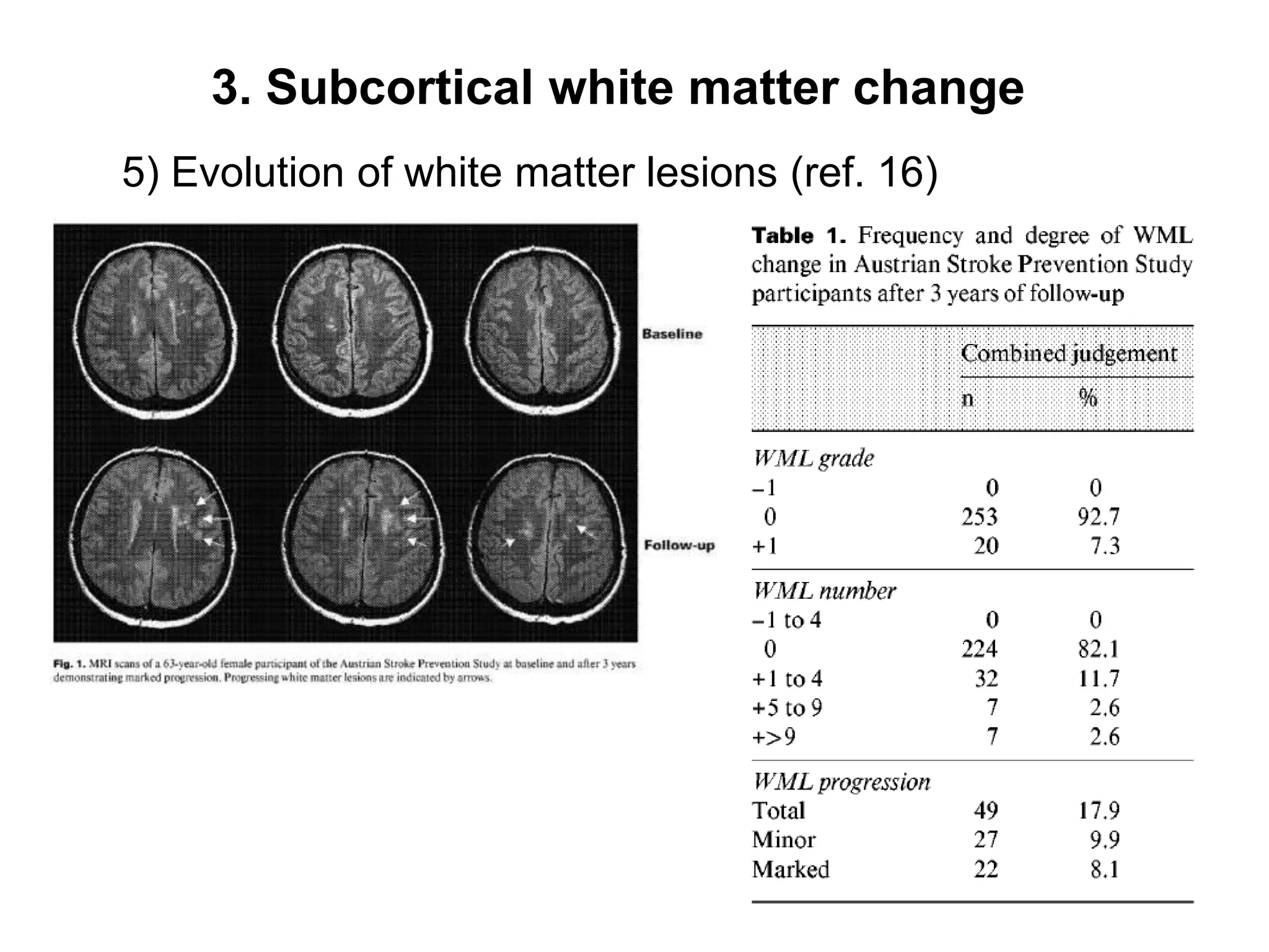
3. Subcortical white matter change 5) Evolution of white matter lesions (ref. 16)
3. Subcortical white matter change 6) Definition of ‘Periventricular’ and ‘Deep white matter’     change (ref.5) (1) Periventricular - Start directly at the ventricular border
3. Subcortical white matter change 6) Definition of ‘Periventricular’ and ‘Deep white matter’     change (ref.5) (2) Both periventricular and deep white matter ,[object Object],  into the adjacent white matter
3. Subcortical white matter change 6) Definition of ‘Periventricular’ and ‘Deep white matter’     change (ref.5) (3) Selective deep white matter lesion - usually characterized by a rim of normal-appearing tissue which separates them from the periventricular region
3. Subcortical white matter change 6) Definition of ‘Periventricular’ and ‘Deep white matter’     change (ref.5) (4) Basal ganglia hypodensities on CT or hyperintensity on MRI (M/82)
3. Subcortical white matter change 6) Definition of ‘Periventricular’ and ‘Deep white matter’     change (ref.24)
3. Subcortical white matter change 6) Definition of ‘Periventricular’ and ‘Deep white matter’     change – (1) (ref.5) Periventricularhyperintensity      0 = absence      1 = “caps” or pencil-thin lining      2 = smooth “halo”      3 = irregular PVH extending into the deep white matter (2) Deep white matter hyperintense signal      0 = absence      1 = punctuate foci      2 = beginning confluence of foci      3 = large confluent areas
3. Subcortical white matter change 7) Definition of ‘Periventricular’ and ‘Deep white matter’ –(3)     (ref. 6) (1) White matter lesions       0 = no lesions  (including symmetrical, well-defined caps or bands)       1 = Focal lesions       2 = Beginning confluence of lesions       3 = Diffuse involvement of the entire region,  with or without involvement of U fibers  (2) Basal ganglia lesions       0 = No lesions       1 = 1 focal lesion (≥ 5 mm)       2 = > 1 focal lesion       3 = Confluent lesions
3. Subcortical white matter change 7) Definition of ‘Periventricular’ and ‘Deep white matter’ –(3)     (ref. 6) 1. Score of 1
3. Subcortical white matter change 7) Definition of ‘Periventricular’ and ‘Deep white matter’ –(3)     (ref. 6) 2. Score of 2
3. Subcortical white matter change 7) Definition of ‘Periventricular’ and ‘Deep white matter’ –(3)     (ref. 6) 3. Score of 3
3. Subcortical white matter change (1) White matter lesions 1 = Focal lesions
3. Subcortical white matter change (1)  White matter lesions    2 = Beginning confluence of lesions
3. Subcortical white matter change (1) White matter lesions    3 = Diffuse involvement of the entire region,  with or without involvement of U fibers  (M/75)
3. Subcortical white matter change (1) White matter lesions   3 = Diffuse involvement of the entire region,  with or without involvement of U fibers  (M/60)
3. Subcortical white matter change (2) Basal ganglia lesions    1 = 1 focal lesion (≥ 5 mm)
3. Subcortical white matter change (2) Basal ganglia lesions    2 = > 1 focal lesion
3. Subcortical white matter change (2) Basal ganglia lesions 3 = Confluent lesions
Microbleeds
4. Microbleeds 1) Histopathologic and MRI definition : paramagnetic material which produces local susceptibility   gradients and thereby causes a faster decay of transverse   magnetization on gradient-echo acquisition (ref. 18) : remnants of even minor blood leakage through   damaged vessel walls
4. Microbleeds 1) Histopathologic definition : Postmortem gradient-echo-T2*-weighted MRI and  histopathologic finding (ref. 19)
4. Microbleeds 2) Severity of amyloidangiopathy (ref. 27) Figure 1. Grading of CAA severity in single brain samples (ref. 27)  0: No cerebral vessels showed immunopositivity for beta amyloid 1+: Amyloid is restricted to a rim around smooth  muscle fibers in the media of occasional  normal vessels 2+: The media is thicker than normal and  circumferentially replaced by amyloid in a few vessels 	 3+: Widespread medial thickening and  circumferential amyloid deposition  with a small halo of immunoreactivity in the surrounding parenchyma  : A focus of wall leakage as evidenced  by fresh hemorrhage or hemosiderin-laden  macrophages, or occlusion, or recanalization
4. Microbleeds 3) MRI definition of microbleed (ref. 2, 19, 20, 21) (1) Homogeneous round signal loss lesion with a diameter of up to 5 mm (or <10 mm)      on gradient echo image Distinct from       a. Vascular flow voids on subarachnoid space      b. Leptomeningealhemasiderosis      c. Non-hemorrhagic subcortical mineralization
[object Object],(ref. 26)
[object Object],(M/80)
[object Object],1. Superficial cortical hemosidersosis (ref. 25)
[object Object],2. Subarachnoid hemosidersosis (ref. 25)
[object Object],3. Schematic drawing illustrating subarachnoid hemosiderosis    and superficial coricalhemosidersosi (ref. 25)
4. Microbleeds 3) MRI definition of microbleed (ref. 20) ,[object Object]
 In CADASIL Pt.
 In H/T Pt.,[object Object]
4. Microbleeds 4) Degree of severity of microbleeds (ref. 2) (1) Absent (2) Mild – total number of MBs, 1-5 (3) Moderate – total number of MBs, 6-15 (4) Severe – total number of MBs, >15
4. Microbleeds 5) The locations of the microbleeds and lacunes (ref. 2) (1) Cortico-subcortical (2) Basal ganglia (3) Thalamus (4) Brain stem (5) Cerebellum
Evaluation of the vessel stenosis
Definition of coronary artery stenosis (ref. 8) 1) Significant stenosis - >50% 2) No significant stenosis - <50%
The degrees of stenoocclusive disease (ref. 7) 1) Normal – 0% - 29% diameter stenosis 2) Mildly stenotic – 30% - 49% 3) Moderately stenotic – 50% - 79% 4) Severely stenotic – 80%- 99% 5) Occluded
Regional location of stenosis (ref. 9) : Schematic representation of 11 arterial segments studied    by transcranial Doppler and duplex ultrasound MCA – 1 and 2 ACA – 3 and 4 PCA – 5 and 6 Siphon ICA – 7 and 8 Extracranial ICA - 9 and 10 Vertebrobasilar artery – 11
Measuement of vessel stenosis (ref. 10) 1. Equation for measuring intracranial arterial stenosis : Percent stenosis = [(1-(Dstenosis/Dnormal))] x 100 ,[object Object],            most severe degree of stenosis ,[object Object],[object Object]
Measuement of vessel stenosis (ref. 10) 2. Criteria for normal proximal artery (3) Third choice A. If the entire intracranial artery was diseased -> the most distal, parallel, non-tortous normal      segment of the feeding artery  B. If the entire middle cerebral artery was diseased -> measured at the most distal, parallel segement      of the supraaclinoid carotid artery C. If the entire intracranial vertebral artery was diseased  -> measured at the most distal, parallel,      non-tortous normal segment of the extracranial      vertebral artery
Measuement of vessel stenosis (ref. 10) 2. Criteria for normal proximal artery 2) For the ICA (1) First choice : The precavernous, cavernous, and postcavernous    stenoses of ICA -> measured at the widest, non-tortous, normal      portion of the petrous carotid artery that had      parallel margins
Measuement of vessel stenosis (ref. 10) 2. Criteria for normal proximal artery 2) For the ICA (2) Second choice - If the entire petrous carotid was diseased -> the most distal, parallel part of the extracranial      internal carotid artery was substituted - If tandem intracranial lesions were present -> percent stenosis of both sites was measured      and the more severe stenosis was selected  - When a “gap sign” was present -- the lumen of the vessel could not be visualized     at the site of severe stenosis -- could not be measured -- defined as 99% luminal stenosis
Measuement of vessel stenosis (ref. 10) 1. Equation for measuring intracranial arterial stenosis
Measuement of vessel stenosis 2. Equation for measuring extracranial arterial stenosis 1) Severity of intracranial stenosis (ref. 11, 12) (1) Mild - <30% (2) Moderate – 30% - 69% (3) Severe – 70% - 99%   - in case of segmental signal void     -> the stenosis was graded as severe (>70%) (4) Occluded
Measuement of vessel stenosis 2. Equation for measuring extracranial arterial stenosis 2) Measurement of the carotid artery stenosis (ref. 12) (1) NASCET    : (1-md/C)x100% (2) ECST     : (1-md/B)x100% (3) CC    : (1-md/A)x100%

More Related Content

Similar to Subcortical lesion-classification-presentation-2011-10-10

Cerebral Infarcts . pptx
Cerebral Infarcts          .         pptxCerebral Infarcts          .         pptx
Cerebral Infarcts . pptxDr Abna J
 
Stroke and stroke mimics
Stroke and stroke mimicsStroke and stroke mimics
Stroke and stroke mimicsNathaliazuos
 
Imaging in cerebral ischemia
Imaging in cerebral ischemiaImaging in cerebral ischemia
Imaging in cerebral ischemiaMilan Silwal
 
Diagnostic Imaging of Spinal Infection & Inflammation
Diagnostic Imaging of Spinal Infection & InflammationDiagnostic Imaging of Spinal Infection & Inflammation
Diagnostic Imaging of Spinal Infection & InflammationMohamed M.A. Zaitoun
 
art%3A10.1007%2Fs10237-015-0729-2 (5)
art%3A10.1007%2Fs10237-015-0729-2 (5)art%3A10.1007%2Fs10237-015-0729-2 (5)
art%3A10.1007%2Fs10237-015-0729-2 (5)Desmond Dillon-Murphy
 
131 the evolution of coronary atherosclerosis
131 the evolution of coronary atherosclerosis131 the evolution of coronary atherosclerosis
131 the evolution of coronary atherosclerosisSHAPE Society
 
Important radiological classification of fracture and AVN
Important radiological classification of fracture and AVNImportant radiological classification of fracture and AVN
Important radiological classification of fracture and AVNDr pradeep Kumar
 
Introduction to musculoskeletal radiology
Introduction to musculoskeletal radiologyIntroduction to musculoskeletal radiology
Introduction to musculoskeletal radiologySubhanjan Das
 
Patterns of Enhancement in the Brain
Patterns of Enhancement in the BrainPatterns of Enhancement in the Brain
Patterns of Enhancement in the BrainMohamed M.A. Zaitoun
 
Presentation1.pptx, radiological imaging of cerebral ischemia.
Presentation1.pptx, radiological imaging of cerebral ischemia.Presentation1.pptx, radiological imaging of cerebral ischemia.
Presentation1.pptx, radiological imaging of cerebral ischemia.Abdellah Nazeer
 
Left ventricular function evaluation
Left ventricular function evaluationLeft ventricular function evaluation
Left ventricular function evaluationMostafa Sayed
 
Normal & abnormal radiology of brain part ii
Normal & abnormal radiology of brain part iiNormal & abnormal radiology of brain part ii
Normal & abnormal radiology of brain part iiMohammed Fathy
 
Intracoronaryopticalcoherencetomography 130909083234-
Intracoronaryopticalcoherencetomography 130909083234-Intracoronaryopticalcoherencetomography 130909083234-
Intracoronaryopticalcoherencetomography 130909083234-Mashiul Alam
 
MRI evaluation of developing brain and its role in congenital brain malformat...
MRI evaluation of developing brain and its role in congenital brain malformat...MRI evaluation of developing brain and its role in congenital brain malformat...
MRI evaluation of developing brain and its role in congenital brain malformat...Shaurya Agarwal
 

Similar to Subcortical lesion-classification-presentation-2011-10-10 (20)

Cerebral Infarcts . pptx
Cerebral Infarcts          .         pptxCerebral Infarcts          .         pptx
Cerebral Infarcts . pptx
 
Stroke and stroke mimics
Stroke and stroke mimicsStroke and stroke mimics
Stroke and stroke mimics
 
Imaging in cerebral ischemia
Imaging in cerebral ischemiaImaging in cerebral ischemia
Imaging in cerebral ischemia
 
Diagnostic Imaging of Spinal Infection & Inflammation
Diagnostic Imaging of Spinal Infection & InflammationDiagnostic Imaging of Spinal Infection & Inflammation
Diagnostic Imaging of Spinal Infection & Inflammation
 
art%3A10.1007%2Fs10237-015-0729-2 (5)
art%3A10.1007%2Fs10237-015-0729-2 (5)art%3A10.1007%2Fs10237-015-0729-2 (5)
art%3A10.1007%2Fs10237-015-0729-2 (5)
 
131 the evolution of coronary atherosclerosis
131 the evolution of coronary atherosclerosis131 the evolution of coronary atherosclerosis
131 the evolution of coronary atherosclerosis
 
Dr stone
Dr stoneDr stone
Dr stone
 
131 the evolution of coronary atherosclerosis
131 the evolution of coronary atherosclerosis131 the evolution of coronary atherosclerosis
131 the evolution of coronary atherosclerosis
 
ECR2013_C-1604
ECR2013_C-1604ECR2013_C-1604
ECR2013_C-1604
 
Important radiological classification of fracture and AVN
Important radiological classification of fracture and AVNImportant radiological classification of fracture and AVN
Important radiological classification of fracture and AVN
 
Introduction to musculoskeletal radiology
Introduction to musculoskeletal radiologyIntroduction to musculoskeletal radiology
Introduction to musculoskeletal radiology
 
Patterns of Enhancement in the Brain
Patterns of Enhancement in the BrainPatterns of Enhancement in the Brain
Patterns of Enhancement in the Brain
 
Presentation1.pptx, radiological imaging of cerebral ischemia.
Presentation1.pptx, radiological imaging of cerebral ischemia.Presentation1.pptx, radiological imaging of cerebral ischemia.
Presentation1.pptx, radiological imaging of cerebral ischemia.
 
Left ventricular function evaluation
Left ventricular function evaluationLeft ventricular function evaluation
Left ventricular function evaluation
 
Normal & abnormal radiology of brain part ii
Normal & abnormal radiology of brain part iiNormal & abnormal radiology of brain part ii
Normal & abnormal radiology of brain part ii
 
Intracoronaryopticalcoherencetomography 130909083234-
Intracoronaryopticalcoherencetomography 130909083234-Intracoronaryopticalcoherencetomography 130909083234-
Intracoronaryopticalcoherencetomography 130909083234-
 
Case record...Spinal multiple sclerosis
Case record...Spinal multiple sclerosisCase record...Spinal multiple sclerosis
Case record...Spinal multiple sclerosis
 
Degenerative spine
Degenerative  spineDegenerative  spine
Degenerative spine
 
MRI evaluation of developing brain and its role in congenital brain malformat...
MRI evaluation of developing brain and its role in congenital brain malformat...MRI evaluation of developing brain and its role in congenital brain malformat...
MRI evaluation of developing brain and its role in congenital brain malformat...
 
Frank Gijsen
Frank GijsenFrank Gijsen
Frank Gijsen
 

Recently uploaded

Nutritional Needs Presentation - HLTH 104
Nutritional Needs Presentation - HLTH 104Nutritional Needs Presentation - HLTH 104
Nutritional Needs Presentation - HLTH 104misteraugie
 
CARE OF CHILD IN INCUBATOR..........pptx
CARE OF CHILD IN INCUBATOR..........pptxCARE OF CHILD IN INCUBATOR..........pptx
CARE OF CHILD IN INCUBATOR..........pptxGaneshChakor2
 
Measures of Central Tendency: Mean, Median and Mode
Measures of Central Tendency: Mean, Median and ModeMeasures of Central Tendency: Mean, Median and Mode
Measures of Central Tendency: Mean, Median and ModeThiyagu K
 
Mastering the Unannounced Regulatory Inspection
Mastering the Unannounced Regulatory InspectionMastering the Unannounced Regulatory Inspection
Mastering the Unannounced Regulatory InspectionSafetyChain Software
 
1029 - Danh muc Sach Giao Khoa 10 . pdf
1029 -  Danh muc Sach Giao Khoa 10 . pdf1029 -  Danh muc Sach Giao Khoa 10 . pdf
1029 - Danh muc Sach Giao Khoa 10 . pdfQucHHunhnh
 
Interactive Powerpoint_How to Master effective communication
Interactive Powerpoint_How to Master effective communicationInteractive Powerpoint_How to Master effective communication
Interactive Powerpoint_How to Master effective communicationnomboosow
 
Presentation by Andreas Schleicher Tackling the School Absenteeism Crisis 30 ...
Presentation by Andreas Schleicher Tackling the School Absenteeism Crisis 30 ...Presentation by Andreas Schleicher Tackling the School Absenteeism Crisis 30 ...
Presentation by Andreas Schleicher Tackling the School Absenteeism Crisis 30 ...EduSkills OECD
 
Introduction to ArtificiaI Intelligence in Higher Education
Introduction to ArtificiaI Intelligence in Higher EducationIntroduction to ArtificiaI Intelligence in Higher Education
Introduction to ArtificiaI Intelligence in Higher Educationpboyjonauth
 
Contemporary philippine arts from the regions_PPT_Module_12 [Autosaved] (1).pptx
Contemporary philippine arts from the regions_PPT_Module_12 [Autosaved] (1).pptxContemporary philippine arts from the regions_PPT_Module_12 [Autosaved] (1).pptx
Contemporary philippine arts from the regions_PPT_Module_12 [Autosaved] (1).pptxRoyAbrique
 
Call Girls in Dwarka Mor Delhi Contact Us 9654467111
Call Girls in Dwarka Mor Delhi Contact Us 9654467111Call Girls in Dwarka Mor Delhi Contact Us 9654467111
Call Girls in Dwarka Mor Delhi Contact Us 9654467111Sapana Sha
 
Q4-W6-Restating Informational Text Grade 3
Q4-W6-Restating Informational Text Grade 3Q4-W6-Restating Informational Text Grade 3
Q4-W6-Restating Informational Text Grade 3JemimahLaneBuaron
 
A Critique of the Proposed National Education Policy Reform
A Critique of the Proposed National Education Policy ReformA Critique of the Proposed National Education Policy Reform
A Critique of the Proposed National Education Policy ReformChameera Dedduwage
 
Paris 2024 Olympic Geographies - an activity
Paris 2024 Olympic Geographies - an activityParis 2024 Olympic Geographies - an activity
Paris 2024 Olympic Geographies - an activityGeoBlogs
 
Separation of Lanthanides/ Lanthanides and Actinides
Separation of Lanthanides/ Lanthanides and ActinidesSeparation of Lanthanides/ Lanthanides and Actinides
Separation of Lanthanides/ Lanthanides and ActinidesFatimaKhan178732
 
Advanced Views - Calendar View in Odoo 17
Advanced Views - Calendar View in Odoo 17Advanced Views - Calendar View in Odoo 17
Advanced Views - Calendar View in Odoo 17Celine George
 
Sanyam Choudhary Chemistry practical.pdf
Sanyam Choudhary Chemistry practical.pdfSanyam Choudhary Chemistry practical.pdf
Sanyam Choudhary Chemistry practical.pdfsanyamsingh5019
 
Arihant handbook biology for class 11 .pdf
Arihant handbook biology for class 11 .pdfArihant handbook biology for class 11 .pdf
Arihant handbook biology for class 11 .pdfchloefrazer622
 

Recently uploaded (20)

Nutritional Needs Presentation - HLTH 104
Nutritional Needs Presentation - HLTH 104Nutritional Needs Presentation - HLTH 104
Nutritional Needs Presentation - HLTH 104
 
TataKelola dan KamSiber Kecerdasan Buatan v022.pdf
TataKelola dan KamSiber Kecerdasan Buatan v022.pdfTataKelola dan KamSiber Kecerdasan Buatan v022.pdf
TataKelola dan KamSiber Kecerdasan Buatan v022.pdf
 
CARE OF CHILD IN INCUBATOR..........pptx
CARE OF CHILD IN INCUBATOR..........pptxCARE OF CHILD IN INCUBATOR..........pptx
CARE OF CHILD IN INCUBATOR..........pptx
 
Measures of Central Tendency: Mean, Median and Mode
Measures of Central Tendency: Mean, Median and ModeMeasures of Central Tendency: Mean, Median and Mode
Measures of Central Tendency: Mean, Median and Mode
 
Mastering the Unannounced Regulatory Inspection
Mastering the Unannounced Regulatory InspectionMastering the Unannounced Regulatory Inspection
Mastering the Unannounced Regulatory Inspection
 
1029 - Danh muc Sach Giao Khoa 10 . pdf
1029 -  Danh muc Sach Giao Khoa 10 . pdf1029 -  Danh muc Sach Giao Khoa 10 . pdf
1029 - Danh muc Sach Giao Khoa 10 . pdf
 
Mattingly "AI & Prompt Design: Structured Data, Assistants, & RAG"
Mattingly "AI & Prompt Design: Structured Data, Assistants, & RAG"Mattingly "AI & Prompt Design: Structured Data, Assistants, & RAG"
Mattingly "AI & Prompt Design: Structured Data, Assistants, & RAG"
 
Interactive Powerpoint_How to Master effective communication
Interactive Powerpoint_How to Master effective communicationInteractive Powerpoint_How to Master effective communication
Interactive Powerpoint_How to Master effective communication
 
Código Creativo y Arte de Software | Unidad 1
Código Creativo y Arte de Software | Unidad 1Código Creativo y Arte de Software | Unidad 1
Código Creativo y Arte de Software | Unidad 1
 
Presentation by Andreas Schleicher Tackling the School Absenteeism Crisis 30 ...
Presentation by Andreas Schleicher Tackling the School Absenteeism Crisis 30 ...Presentation by Andreas Schleicher Tackling the School Absenteeism Crisis 30 ...
Presentation by Andreas Schleicher Tackling the School Absenteeism Crisis 30 ...
 
Introduction to ArtificiaI Intelligence in Higher Education
Introduction to ArtificiaI Intelligence in Higher EducationIntroduction to ArtificiaI Intelligence in Higher Education
Introduction to ArtificiaI Intelligence in Higher Education
 
Contemporary philippine arts from the regions_PPT_Module_12 [Autosaved] (1).pptx
Contemporary philippine arts from the regions_PPT_Module_12 [Autosaved] (1).pptxContemporary philippine arts from the regions_PPT_Module_12 [Autosaved] (1).pptx
Contemporary philippine arts from the regions_PPT_Module_12 [Autosaved] (1).pptx
 
Call Girls in Dwarka Mor Delhi Contact Us 9654467111
Call Girls in Dwarka Mor Delhi Contact Us 9654467111Call Girls in Dwarka Mor Delhi Contact Us 9654467111
Call Girls in Dwarka Mor Delhi Contact Us 9654467111
 
Q4-W6-Restating Informational Text Grade 3
Q4-W6-Restating Informational Text Grade 3Q4-W6-Restating Informational Text Grade 3
Q4-W6-Restating Informational Text Grade 3
 
A Critique of the Proposed National Education Policy Reform
A Critique of the Proposed National Education Policy ReformA Critique of the Proposed National Education Policy Reform
A Critique of the Proposed National Education Policy Reform
 
Paris 2024 Olympic Geographies - an activity
Paris 2024 Olympic Geographies - an activityParis 2024 Olympic Geographies - an activity
Paris 2024 Olympic Geographies - an activity
 
Separation of Lanthanides/ Lanthanides and Actinides
Separation of Lanthanides/ Lanthanides and ActinidesSeparation of Lanthanides/ Lanthanides and Actinides
Separation of Lanthanides/ Lanthanides and Actinides
 
Advanced Views - Calendar View in Odoo 17
Advanced Views - Calendar View in Odoo 17Advanced Views - Calendar View in Odoo 17
Advanced Views - Calendar View in Odoo 17
 
Sanyam Choudhary Chemistry practical.pdf
Sanyam Choudhary Chemistry practical.pdfSanyam Choudhary Chemistry practical.pdf
Sanyam Choudhary Chemistry practical.pdf
 
Arihant handbook biology for class 11 .pdf
Arihant handbook biology for class 11 .pdfArihant handbook biology for class 11 .pdf
Arihant handbook biology for class 11 .pdf
 

Subcortical lesion-classification-presentation-2011-10-10

  • 1. A Consensus Meeting for The Definitions of Subcortical lesions, Lacunes, Microbleeds, and Subcortical White Matter Change Jei Kim, MD
  • 5. Subcortical lesions 1. Perivascular spaces (etatcrible) 2. Lacunes 3. Subcortical white matter changes 4. Microbleeds
  • 7. 1. Perivascular spaces (etatcrible) 1) Histopathologic definition (ref. 1) : the dilatation of perivascular spaces around cerebral arterioles in the brain of elderly patients (Virchow-Robin space) 2) MRI definition (ref. 1) : the punctiform dilatations of the perivascular spaces often seen by brain MRI in the white matter and in the basal ganglia
  • 8. 1. Perivascular spaces (etatcrible) 2) MRI definition : the punctiform dilatations of the perivascular spaces often seen by brain MRI in the white matter and in the basal ganglia : On T2WI – high intensity, same as the intensity of CSF : On FLAIR – dark (low), same as the intensity of CSF : On T1WI – dark (low), same as the intensity of CSF http://www.radiologyassistant.nl/en/4556dea65db62
  • 11. 2. Lacunes 1) Histopathologic definition : a small, cystic cavity of the brain substance that usually results from an ischemic infarction in the territory of a penetrating arteriole (ref. 1)
  • 12. 2. Lacunes 2) Vascular pathology of lacunes (ref. 13)
  • 13. 2. Lacunes 3) Perforating arteries (1) Anterior perforating arteries (2) Posterior perforating arteries (3) Arterial supply of the brainstem
  • 15. (1) Anterior perforating arteries A. Perforating branches arising from ACA and the recurrent artery of Heubner
  • 16. (1) Anterior perforating arteries B. Perforating branches arising from MCA a. Perforating branches arising from MCA
  • 17. (1) Anterior perforating arteries B. Perforating branches arising from MCA b. Percentage of perforating arteries arising from MCA trunk and its branches
  • 18. (1) Anterior perforating arteries B. Perforating branches arising from MCA c. Origin of perforating arteries arising from MCA trunk and its branches
  • 19. (1) Anterior perforating arteries B. Perforating branches arising from MCA d. Number of perforating arteries arising from different distances from the origin of MCA
  • 20. (1) Anterior perforating arteries B. Perforating branches arising from MCA e. Branching characteristics of 508 perforating arteries arising from common stems of MCA
  • 22. (3) Arterial supply of the brainstem A. Perforating arteries of the midbrain
  • 23. (3) Arterial supply of the brainstem B. Perforating arteries of the pons
  • 24. (3) Arterial supply of the brainstem C. Perforating arteries of the midbrain
  • 25. Schematic diagram of origin of deep perforating branches from a parent artery
  • 26. 2. Lacunes 4) Pathogenic implications of microcirculation (1) Perforating arteries 1) Stenosis or complete occlusion by atherosclerosis 2) Stenosis or occlusion of ostium of a branch point 3) Atherosclerotic narrowing of a parent artery 4) Proximal thrombus or embolus in atherosclerotic artery (2) Cortical branches
  • 27. 4) Pathogenic implications of microcirculation (1) Perforating arteries A. Stenosis or complete occlusion by atherosclerosis
  • 28. 4) Pathogenic implications of microcirculation (1) Perforating arteries B. Stenosis or occlusion of ostium of a branch point
  • 29. 4) Pathogenic implications of microcirculation (1) Perforating arteries C. Atherosclerotic narrowing of a parent artery
  • 30. 4) Pathogenic implications of microcirculation (1) Perforating arteries D. Proximal thrombus or embolus in atherosclerotic artery
  • 31. 4) Pathogenic implications of microcirculation (2) Cortical branches
  • 32. 2. Lacunes 5) MRI definition (ref. 2) : small hyperintense lesions on T2WI (ref. 2) : corresponding distinctive low intensity area on T1WI : Maximum size of lacune (ref. 4) - with a diameter of 5-10 mm : On CT (ref. 4) - areas of more or less complete focal tissue destruction - clearly defined borders with marked central hypodensity on CT : On MRI (ref. 4) - low intensity on T1WI, proton-density and FLAIR scans - high intensity on T2WI -> isointense to CSF
  • 33. 2. Lacunes 5) MRI definition (ref. 17)
  • 34. 2. Lacunes 1) Histopathologic definition : a small, cystic cavity of the brain substance that usually results from an ischemic infarction in the territory of a penetrating arteriole (ref. 1) : defined as cavitatedmicroinfarcts or encephalomalacic lesions, 2 mm or smaller in greatest dimension, not identifiable with certainty on gross inspection of the brain or non-cavitatedmicroinfarcts, focal gliotic areas without a cystic cavity (ref. 3)
  • 36. 2. Lacunes 6) Grading of lacunes (ref. 2) Absent (2) Mild – 1-3 (3) Moderate – 4-10 (4) Severe - >10 7) Locations of lacunes (ref. 2) Cortico-subcortical (2) Basal ganglia (3) Thalamus (4) Brain stem (5) Cerebellum
  • 38. 3. Subcortical white matter change 1) Definition of Binswanger’s disease (1894) : pronounced atrophy of the white matter, either confined to one or more gyri of the brain or in several sections of the hemisphere : in the most severe cases the entire white matter of a cerebral lobe appears to have completely wasted away : a severe atheromatosis of the arteries of the brain is always present in these cases : extensive atrophic degeneration or fatty degeneration of the small arterial and venous vessels : partial thickening of the inner and middle vascular membranes : the lumen is correspondingly narrowed
  • 39. 3. Subcortical white matter change 2) Definition of leukoaraiosis (Hachinski et al., 1987) : loss of density of the periventricular white matter observed by CT of the brain : the white matter changes commonly observed in the elderly by MRI of the brain
  • 40. 3. Subcortical white matter change 3) Mechanisms hypothesized to be involved in the pathogenesis of white matter change (ref. 14)
  • 41. 3. Subcortical white matter change 4) Small vessel changes related to white matter changes (ref. 14)
  • 42. 3. Subcortical white matter change 5) Evolution of white matter lesions (ref. 16)
  • 43. 3. Subcortical white matter change 6) Definition of ‘Periventricular’ and ‘Deep white matter’ change (ref.5) (1) Periventricular - Start directly at the ventricular border
  • 44.
  • 45. 3. Subcortical white matter change 6) Definition of ‘Periventricular’ and ‘Deep white matter’ change (ref.5) (3) Selective deep white matter lesion - usually characterized by a rim of normal-appearing tissue which separates them from the periventricular region
  • 46. 3. Subcortical white matter change 6) Definition of ‘Periventricular’ and ‘Deep white matter’ change (ref.5) (4) Basal ganglia hypodensities on CT or hyperintensity on MRI (M/82)
  • 47. 3. Subcortical white matter change 6) Definition of ‘Periventricular’ and ‘Deep white matter’ change (ref.24)
  • 48. 3. Subcortical white matter change 6) Definition of ‘Periventricular’ and ‘Deep white matter’ change – (1) (ref.5) Periventricularhyperintensity 0 = absence 1 = “caps” or pencil-thin lining 2 = smooth “halo” 3 = irregular PVH extending into the deep white matter (2) Deep white matter hyperintense signal 0 = absence 1 = punctuate foci 2 = beginning confluence of foci 3 = large confluent areas
  • 49. 3. Subcortical white matter change 7) Definition of ‘Periventricular’ and ‘Deep white matter’ –(3) (ref. 6) (1) White matter lesions 0 = no lesions (including symmetrical, well-defined caps or bands) 1 = Focal lesions 2 = Beginning confluence of lesions 3 = Diffuse involvement of the entire region, with or without involvement of U fibers (2) Basal ganglia lesions 0 = No lesions 1 = 1 focal lesion (≥ 5 mm) 2 = > 1 focal lesion 3 = Confluent lesions
  • 50. 3. Subcortical white matter change 7) Definition of ‘Periventricular’ and ‘Deep white matter’ –(3) (ref. 6) 1. Score of 1
  • 51. 3. Subcortical white matter change 7) Definition of ‘Periventricular’ and ‘Deep white matter’ –(3) (ref. 6) 2. Score of 2
  • 52. 3. Subcortical white matter change 7) Definition of ‘Periventricular’ and ‘Deep white matter’ –(3) (ref. 6) 3. Score of 3
  • 53. 3. Subcortical white matter change (1) White matter lesions 1 = Focal lesions
  • 54. 3. Subcortical white matter change (1) White matter lesions 2 = Beginning confluence of lesions
  • 55. 3. Subcortical white matter change (1) White matter lesions 3 = Diffuse involvement of the entire region, with or without involvement of U fibers (M/75)
  • 56. 3. Subcortical white matter change (1) White matter lesions 3 = Diffuse involvement of the entire region, with or without involvement of U fibers (M/60)
  • 57. 3. Subcortical white matter change (2) Basal ganglia lesions 1 = 1 focal lesion (≥ 5 mm)
  • 58. 3. Subcortical white matter change (2) Basal ganglia lesions 2 = > 1 focal lesion
  • 59. 3. Subcortical white matter change (2) Basal ganglia lesions 3 = Confluent lesions
  • 61. 4. Microbleeds 1) Histopathologic and MRI definition : paramagnetic material which produces local susceptibility gradients and thereby causes a faster decay of transverse magnetization on gradient-echo acquisition (ref. 18) : remnants of even minor blood leakage through damaged vessel walls
  • 62. 4. Microbleeds 1) Histopathologic definition : Postmortem gradient-echo-T2*-weighted MRI and histopathologic finding (ref. 19)
  • 63. 4. Microbleeds 2) Severity of amyloidangiopathy (ref. 27) Figure 1. Grading of CAA severity in single brain samples (ref. 27) 0: No cerebral vessels showed immunopositivity for beta amyloid 1+: Amyloid is restricted to a rim around smooth muscle fibers in the media of occasional normal vessels 2+: The media is thicker than normal and circumferentially replaced by amyloid in a few vessels 3+: Widespread medial thickening and circumferential amyloid deposition with a small halo of immunoreactivity in the surrounding parenchyma : A focus of wall leakage as evidenced by fresh hemorrhage or hemosiderin-laden macrophages, or occlusion, or recanalization
  • 64. 4. Microbleeds 3) MRI definition of microbleed (ref. 2, 19, 20, 21) (1) Homogeneous round signal loss lesion with a diameter of up to 5 mm (or <10 mm) on gradient echo image Distinct from a. Vascular flow voids on subarachnoid space b. Leptomeningealhemasiderosis c. Non-hemorrhagic subcortical mineralization
  • 65.
  • 66.
  • 67.
  • 68.
  • 69.
  • 70.
  • 72.
  • 73. 4. Microbleeds 4) Degree of severity of microbleeds (ref. 2) (1) Absent (2) Mild – total number of MBs, 1-5 (3) Moderate – total number of MBs, 6-15 (4) Severe – total number of MBs, >15
  • 74. 4. Microbleeds 5) The locations of the microbleeds and lacunes (ref. 2) (1) Cortico-subcortical (2) Basal ganglia (3) Thalamus (4) Brain stem (5) Cerebellum
  • 75. Evaluation of the vessel stenosis
  • 76. Definition of coronary artery stenosis (ref. 8) 1) Significant stenosis - >50% 2) No significant stenosis - <50%
  • 77. The degrees of stenoocclusive disease (ref. 7) 1) Normal – 0% - 29% diameter stenosis 2) Mildly stenotic – 30% - 49% 3) Moderately stenotic – 50% - 79% 4) Severely stenotic – 80%- 99% 5) Occluded
  • 78. Regional location of stenosis (ref. 9) : Schematic representation of 11 arterial segments studied by transcranial Doppler and duplex ultrasound MCA – 1 and 2 ACA – 3 and 4 PCA – 5 and 6 Siphon ICA – 7 and 8 Extracranial ICA - 9 and 10 Vertebrobasilar artery – 11
  • 79.
  • 80. Measuement of vessel stenosis (ref. 10) 2. Criteria for normal proximal artery (3) Third choice A. If the entire intracranial artery was diseased -> the most distal, parallel, non-tortous normal segment of the feeding artery B. If the entire middle cerebral artery was diseased -> measured at the most distal, parallel segement of the supraaclinoid carotid artery C. If the entire intracranial vertebral artery was diseased -> measured at the most distal, parallel, non-tortous normal segment of the extracranial vertebral artery
  • 81. Measuement of vessel stenosis (ref. 10) 2. Criteria for normal proximal artery 2) For the ICA (1) First choice : The precavernous, cavernous, and postcavernous stenoses of ICA -> measured at the widest, non-tortous, normal portion of the petrous carotid artery that had parallel margins
  • 82. Measuement of vessel stenosis (ref. 10) 2. Criteria for normal proximal artery 2) For the ICA (2) Second choice - If the entire petrous carotid was diseased -> the most distal, parallel part of the extracranial internal carotid artery was substituted - If tandem intracranial lesions were present -> percent stenosis of both sites was measured and the more severe stenosis was selected - When a “gap sign” was present -- the lumen of the vessel could not be visualized at the site of severe stenosis -- could not be measured -- defined as 99% luminal stenosis
  • 83. Measuement of vessel stenosis (ref. 10) 1. Equation for measuring intracranial arterial stenosis
  • 84. Measuement of vessel stenosis 2. Equation for measuring extracranial arterial stenosis 1) Severity of intracranial stenosis (ref. 11, 12) (1) Mild - <30% (2) Moderate – 30% - 69% (3) Severe – 70% - 99% - in case of segmental signal void -> the stenosis was graded as severe (>70%) (4) Occluded
  • 85. Measuement of vessel stenosis 2. Equation for measuring extracranial arterial stenosis 2) Measurement of the carotid artery stenosis (ref. 12) (1) NASCET : (1-md/C)x100% (2) ECST : (1-md/B)x100% (3) CC : (1-md/A)x100%