Micron X: Optimized Imaging for Large Animal Eye Research
•
2 likes•1,645 views

The document describes a laser-induced choroidal neovascularization model in pigs for studying retinal diseases. It discusses the Micron X imaging system for obtaining high-quality images of the retina and fluorescein angiograms in large animals like pigs. The model involves using laser shots to induce CNV lesions and then treating them with drugs like aflibercept to evaluate effectiveness. Quantitative analysis of the lesions is done using OCT, fluorescein angiography, and flat mounts, showing that aflibercept significantly reduces CNV development. The pig model is said to be more clinically relevant and translatable than rodent models for testing new therapies.

Recommended
Recommended
More Related Content
What's hot
What's hot (20)
Similar to Micron X: Optimized Imaging for Large Animal Eye Research
Similar to Micron X: Optimized Imaging for Large Animal Eye Research (20)
More from InsideScientific
More from InsideScientific (20)
Recently uploaded
Recently uploaded (20)
Micron X: Optimized Imaging for Large Animal Eye Research
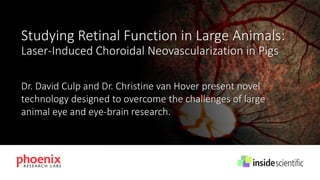
- 1. Studying Retinal Function in Large Animals: Laser-Induced Choroidal Neovascularization in Pigs Dr. David Culp and Dr. Christine van Hover present novel technology designed to overcome the challenges of large animal eye and eye-brain research.
- 2. InsideScientific is an online educational environment designed for life science researchers. Our goal is to aid in the sharing and distribution of scientific information regarding innovative technologies, protocols, research tools and laboratory services. JOIN FOR FREE AT WWW.INSIDESCIENTIFIC.COM
- 3. Christine van Hover, PhD Chief Scientist, Phoenix Research Labs W. David Culp, PhD Director of Research, Powered Research, LLC Studying Retinal Function in Large Animals: Laser-Induced Choroidal Neovascularization in Pigs
- 4. Copyright 2017 Phoenix Research Labs and InsideScientific. All Rights Reserved. Christine van Hover, PhD Chief Scientist, Phoenix Research Labs Micron X: Optimized Image System for Large Animal Eye Research
- 5. Micron X Imaging Large animal camera
- 6. And: Anterior segment, Ganzfeld ERG, Laser-induced CNV image examine measure Geography Micron IV Structure OCT2 Function ERG Phoenix Research Labs
- 7. The Micron X is optimized for large animals ✓ Wide angle 100° field-of-view ✓ Resolution of 8 um ✓ Bright field and fluorescein angiography Images courtesy of Powered Research
- 8. Why large animals? • Critical step between rodents and clinical trials • Similar eye size to humans • Allergy testing
- 9. Four Step Process 1. Sedate and dilate the animal 2. Position and apply gel 3. Contact the Micron X lens onto the eye. Adjust light level, focus, and gain 4. Take images and video as the Micron X scans the retina
- 10. Imaging Versatility in a Range of Species Cat retina Pig retina Dog retina
- 11. Albino rabbit retina Imaging Versatility Crisp images of both light albino retinas and darkly pigmented retinas Pigmented macaque primate retina
- 12. Stunning Fluorescein Angiography Ability Bright field macaque primate retina Fluorescein angiography Images courtesy of Ora Clinical
- 13. Fluorescein Angiography Video Ability Video of fluorescein infiltration in a macaque primate Images courtesy of Ora Clinical Artery Vein
- 14. Fluorescein Angiography Video Ability Slowed down video of fluorescein infiltration in a macaque primate Images courtesy of Ora Clinical
- 15. Time lapse in a macaque primate: Clearly see the arteries, capillaries, and then veins filling with fluorescein Fluorescein Angiography Time Lapse Images courtesy of Ora Clinical
- 16. Bright field pig retina with laser shots Fluorescein angiography of pig retina showing CNV induction Images courtesy of Powered Research CNV Induction in Pigs
- 17. Eylea® treatment significantly decreases CNV development Pig retina with laser shots and no treatment shows CNV development Saline Eylea® Treating CNV Induction in Pigs with Eylea® Images courtesy of Powered Research
- 18. Micron X: A unique retinal imaging system optimized for eye research using large animals. Micron X delivers bright field images of pinpoint resolution and high contrast. Angiograms using fluorescein can also be obtained utilizing the Phoenix Research Labs Micron X. Learn More >>
- 19. Copyright 2017 D. Culp, Phoenix Research Labs and InsideScientific. All Rights Reserved. W. David Culp, PhD Director of Research, Powered Research, LLC Evaluation of Aflibercept in a Laser-Induced Model of Choroidal Neovascularization in Swine
- 20. • Preclinical contract research organization based in Research Triangle Park, North Carolina. • Provide high quality and cost effective, non-GLP studies. • More than 80% of our business is in the ophthalmic space. Learn More >>
- 21. Preclinical Models of Choroidal Neovascularization (CNV) are Needed to Assess New Therapies… Limitations of current models: • Pharmacokinetically (e.g. rodent models) • Pathophysiologically (e.g. VEGF induced models) • Economically (e.g. non-human primates)
- 22. Swine Model of Laser-Induced Choroidal Neovascularization (CNV) • Weanling Swine, Sus scrofa domesticus • Female and male • 6-12 week, 12 – 20 kilograms • Approximately 6 lesions per swine eye, equidistant from the optic nerve were created using an 810 nm diode laser and an indirect ophthalmoscope
- 23. Prior to laser Post laser 2 weeks 3 weeks 4 weeks BSS Aflibercept Swine Model of Laser-Induced CNV Clinical images
- 25. Prior to laser Post laser 2 weeks 4 weeks Swine Model of Laser-Induced CNV OCT images
- 26. 0 2 3 4 0 50 100 150 200 250 Adjacent-lesion retinal thickness Weeks after injection mean+/-SDuM BSS Aflibercept 0 2 3 4 0 50 100 150 200 250 Central-lesion retinal thickness Weeks after injection mean+/-SDuM BSS Aflibe 150 200 250 Central-lesion retinal thickness -SDuM BSS Aflibercept Swine Model of Laser-Induced CNV OCT measurements
- 27. Day 7 BSS Day 7 Eylea® Micron X, Phoenix Research Labs
- 28. Day 14 BSS Day 14 Eylea® Micron X, Phoenix Research Labs
- 29. CTLF: Corrected Total Lesion Fluorescence Integrated Density: Area of lesion X Mean fluorescence of background readings
- 30. Swine Model of Laser-Induced CNV FA measurements
- 31. BSS Aflibercept W eek 2 W eek 4 0 100000 200000 300000 400000 Weeks after injection µm 2 IsolectinIB4Signal BSS Aflibercept Swine Model of Laser-Induced CNV Flat mount measurements Blue : DAPI (nucleus); Green : AF-488 Phalloidin (F-actin); Red : AF-594 Isolectin IB4 (blood vessels and microglia)
- 32. Aflibercept 200 X magnification; hematoxylin and eosin staining Swine Model of Laser-Induced CNV: Histopathology Day 28 BSS
- 33. ✓ The larger size of the swine eye is more relevant to human eyes. ✓ The model provides quantitative assessment by evaluating neovascularization by fluorescein angiography and flat mount analysis. ✓ The model is easy to reproduce and is economical. ✓ Characterization of this sensitive and translatable model allows effective evaluation and proof of concept of new treatments or delivery. Conclusions
- 34. Acknowledgments • Dr. Brian Gilger, Professor of Ophthalmology, North Carolina State University • Kristie Powell, Powered Research • Dr. Grazia Spiga, Powered Research • Justin Prater, Powered Research info@poweredresearch.com
- 35. Christine van Hover, PhD Chief Scientist, Phoenix Research Labs Send Email >> W. David Culp, PhD Director of Research, Powered Research, LLC Send Email >> Thank You: For additional information on the products and applications presented during this webinar please visit www.phoenixreslabs.com