A survey on nuclear to-cytoplasmic ratio analysis using image segmentation
Wheeler_Benjamin_MyPURA
1. Benjamin Wheeler 1
A Vesicle Based Biosensor for Real Time Identification of Mild Traumatic Brain Injury.
Sponsor: Dr. Peter Searson, Materials Science and Engineering
Objective: The overall objective of this project is to design and fabricate a wearable biosensor
composed of lipid vesicles suspended in a polymeric biofilm in order to identify subjects at risk for mild
traumatic brain injury following head trauma. This sensor will be designed to allow for attachment or
insertion into the protective headwear of groups like professional athletes and military personnel. The
vesicles will be fabricated in such a way that they will
contain both a fluorescent dye and quencher. Under
normal conditions, the vesicles will hold both
molecules in proximity such that there will be no
fluorescence. However, when there is a mechanical
disturbance or event that disrupts the vesicles, the two
molecules will leak and separate enough to allow for
the fluorescence to be observed. Preliminary results
measuring vesicle leakage in an electromagnetic
shaker indicate the feasibility of this design.
Significance: Mild Traumatic Brain injury (mTBI) is
defined as a state when there is alteration of minimal duration and severity in the patient’s baseline
neurological or mental status after an injury. Annually, there is an estimate of 3.8 million concussions
occurring in the United States. Unfortunately, 50% of the injuries go unreported, making it a serious
problem for the health care field.1 mTBI is the leading cause of mortality in the active/working
population under 35 in the United States,2 and a major source of these injuries is both collegiate and
professional sports. For instance, at least 60% of those playing soccer on the collegiate level developed
symptoms compatible with a concussion during a season. Additionally, it has been reported that as high
as 18% of soldiers returning from Iraq and Afghanistan experience mild traumatic brain injury.3 In both
of these sectors, there is particular interest in the development of a functional real-time identification and
diagnostic device to aid in the development of functional return-to-action guidelines. The current
diagnosis is straightforward through computed topography (CT) and magnetic resonance imaging (MRI);
however, these clinical methods are costly, time consuming, and not readily translatable to devices for on-site
use.
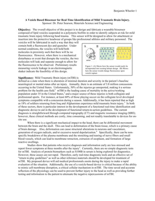
A
B
Figure 1: (A) Shows how the sensor would easily be
incorporated into existing helmet design. (B) Shows
how the sensor would change fluorescence upon
vesicle rupture
When there is a significant mechanical impact to the head, there can be differential movement
between the brain and the skull. This can lead to deformation of the brain tissue, which is a primary cause
of brain damage. Also, deformation can cause structural alterations to neurons and vasculature,
generation of oxygen radicals, and/or excessive neural depolarization.4 Specifically, there can be non-specific
breakdown of the plasma membrane and the stretching and tearing of nerve fibers and small
blood vessels, which can lead to membrane retraction, extrusion of axoplasm, and formation of large
reactive swellings.
Studies show that patients who receive diagnosis and information early are less stressed and
report fewer symptoms at three months after the injury5. Currently, there are no simple diagnostic tests
for mTBI. Analysis of protein biomarkers such as S100B in serum is being explored for diagnostics.
However, these assays are not rapid. Therefore, early real-time diagnostic tools and an effective set of
“return to play guidelines” as well as other reference materials should be developed for treatment of
mTBI. My proposed device will aid medical professionals onsite during the injury to make a rapid
evaluation of the situation. Additionally, the use of a vesicle-based device is critical because of the ability
to mimic cell membrane disruption, a critical cellular mechanism in mTBI. The time saved and accurate
reflection of the physiology can be used to prevent further injury to the head as well as providing further
testing and information to the patient to attenuate the negative repercussions of mTBI.
2. Benjamin Wheeler 2
Originality: Currently in the field the only studies done to characterize the effects of force on the brain at
a cellular level have been done in vivo using animal models. These tests usually consist of either fluid
perfused through the tissue to create a pressure differential or a weight dropped from a height that impacts
the tissue with a predetermined force. Typically in these studies, a pressure of 2 to 5 atmospheres is
applied to the tissue to simulate the impact.6 These tests, while critical in understanding the mechanisms
of mTBI, cause significant harm to the animals involved and do not readily translate to a testable in vitro
model. Compared to the existing systems, the proposed system is unique in its ability to very finely
control the magnitude of force applied. Additionally, this design will allow for testing in multiple
platforms including various cell culture dishes, microfluidic devices, and cuvettes filled with vesicles in
various solutions. This will allow for easy transition to a functional field diagnostic tool.
Project Design: The objective of this project is achieved in two aims. The first aim is to characterize the
relationship between vesicle leakage and the applied mechanical force, and the second aim is to create a
wearable sensor composed of an optimized vesicle-hydrogel matrix, which can be incorporated into
protective headwear.
Aim 1: The initial aim of the project will be to
characterize the relationship between vesicle
leakage and the applied mechanical force. The
goal of this is to create leakage versus impact
calibration curves that will be used in the design
of the sensor and for calibration in the field. The
vesicles will be made using the standard method
of desiccation, re-suspension, and extrusion. In
preliminary work, vesicles have been formed
from a physiologically relevant
composition of 1-palmitoyl-2-oleoyl-sn-glycero-
3-phosphocholine (POPC),
polyethylene glycol (PEG2k), and
cholesterol. Encapsulated within these
vesicles is a solution of the dye/quencher
pair, 8-aminonapthalene-1,3,6 trisulfonic
acid (ANTS)/p-xylene-bis-pyridinium
bromide (DPX). This setup is shown in
figure 2.
To characterize the relationship
A
B
Figure 2: (A) Mechanical disturbance of the
vesicles leads to a change in the fluorescence of the
solution. (B) Shows the vesicle in greater detail
illustrating how the close proximity of dye and
A
B
Figure 3: (A) In preliminary work I have
assembled an electromagnetic shaker with a
sample stage, coupled to the cooling vacuum and
the connection to the amplifier leading out of
view. (B) Amplifier, transformer, and function
generator shown in functional arrangement. (C)
Close up of the custom stage with cuvette holder
attached.
C
between vesicle disruption and force, I
have obtained an electromagnetic shaker
(labworks et-140). The shaker has been
assembled with accompanying amplifier,
cooling fan, and function generator, as
shown in figure 3. In this configuration,
the shaker applies a displacement on the order of centimeters with variable amplitude and frequency, and
is used to model the impact force. To accompany this setup I have custom designed and fabricated a
sample stage to mount the cuvettes holding the vesicle samples to the shaker. This design can be easily
modified to allow attachment of a range of sample types, including: cuvettes, 96 well plates, and custom
made microfluidic devices. This setup accommodates a high degree of control, and allows an exact
application of impact force in agreement with previous studies and data from the literature. I will be
working with Dr. Joe Katz in mechanical engineering to relate the output of the electromagnetic shaker to
impact force.
Aim 2: The second aim of the project is to use the results previously mentioned to design and fabricate a
wearable sensor that can be easily incorporated into existing protective headwear. The sensor envisioned
3. Benjamin Wheeler 3
is a hydrogel strip composed of vesicles suspended in a polymeric matrix encased in a protective outer
layer of flexible plastic to allow for easy interpretation of fluorescence change as well as efficient
transduction of force from the impact to the sensor. It makes sense to fabricate this device using vesicles
in a gel matrix versus the aqueous suspension used to initially characterize the force -leakage relationship
for two reasons. First, and foremost, the gel will ensure even distribution of the vesicles throughout the
sensor, which will allow the sensor to be equally sensitive to impact force in any direction. Second, the
structural support provided by the gel will increase the longevity and resilience of the sensor. To create
the most effective gel for these purposes, gel composition as well as matrix and vesicle density will have
to be optimized. As a starting point, a gel composed of 5mg/mL collagen will be tested. This will allow
for an ideal balance between porosity and stiffness. To create a matrix that accurately mirrors the
mechanical properties of native tissue, the concentration of collagen as well as the introduction of other
polymers can be varied. A key aspect of this sensor is the real-time decision making it allows. To do so,
it must be easily distinguishable between a positive and negative result. To accommodate this design
parameter, the vesicle concentration will also be varied to produce an obvious response when the impact
exceeds a predetermined threshold. Beyond the fluorescence protocol previously described, other dyes
that have colorimetric function will be investigated to provide the optimal means of producing real-time
results.
Timetable: Week 1-6: Production of force-leakage calibration curves
Week 6 – 10: Optimization of the gel and vesicle composition
Week 10-14: Design and fabrication of the actual sensor and quality testing
Summary of Expected Results: Preliminary results show the force-leakage relationship can be readily
characterized and predicted based upon vesicle composition. As shown in figure 4 below, preliminary
results indicate there is at first a linear correlation between duration of force and vesicle leakage. Once
the time and steady state responses are characterized, it is expected that the magnitude of the force and
vesicle leakage can be characterized in a similar manor. It is expected at first the fluorescence will exhibit
a linear correlation with the force applied and eventually reach a plateau, as shown in the time dependent
case below at the 100-minute time point. This effect can be visualized in figure 5. Both cuvettes contain
800 μL of 100 nm diameter vesicles composed entirely of POPC. In the picture, the cuvettes are exposed
to light at the excitation wavelength of ANTS/DPX. The cuvette on the left contains undisturbed
Figure 5: Demonstration of
fluorescence readout of 100
nm POPC vesicles in
solution before and after
mechanical Figure 4: Leakage versus time for disturbance
POPC vesicles under continuous force
vesicles, while the cuvette on the right was exposed to mechanical displacement for 100 minutes. The
emission of yellow light indicates vesicle leakage, as expected from impact to the vesicles. The
combination of these results will allow for easy optimization, design, and fabrication of the sensor.
4. Benjamin Wheeler 4
Background: I have worked in Dr. Searson’s lab since May 2013, committed to understanding the
galvanotaxis of 612 glioblastoma cells under the super vision of PhD candidate Yu-Ja Huang. During my
time with the Searson Group I have learned many critical skills through the major aspects of the project
such as microfabrication, cell culture, normal light and fluorescence miscroscopy, and various biological
assays. This had led to me developing skills in and not limited to the following: photolithography,
hydrogel formation, device assembly, media preparation, stem cell culture, confocal microscopy, live and
dead cell fixing and staining, real time PCR, and cell tracking.
In addition to the hands-on laboratory work, I am also ahead on degree requirements for my junior
standing as a BME. I have already completed essential math classes such as Calculus III, Differential
Equations, and Probability and Statistics. Additionally, I have modeling experience including MATLAB
and completion of the class Models and Simulations along with preliminary course work in Systems
Bioengineering. I also have background with biology and bioengineering, as I have completed organic
chemistry, molecules and cells, and tissue engineering while currently working on Biomaterials I and
Cellular Engineering.
Should some parts of this project have complexity beyond the theoretical and practical preparation I have
obtained, I will have several experienced researches as mentors. I will be working closely with PhD
candidates Chloe Kim and Yu-Ja Huang who combined have over 8 years of research experience. Dr.
Peter Searson, the Reynolds Professor of Materials Science and Engineering and the head of the Institute
for NanoBioTechnology, will be overseeing the project. I believe the combination of these rich and
expansive has given me the ability to successfully complete this project.
Presentation and Evaluation: The results of this project will be presented in poster form, including
results of experiments carried out and reported weekly to Dr. Searson and Yu-Ja Huang at weekly
working group meetings. Project and experimental designs will be created, evaluated, and decided upon
at these meetings as well.
5. Benjamin Wheeler 5
References:
1 Hodge, Samuel D., Jr. "A Heads Up on Traumatic Brains Injuries in Sports." Journal of Health Care Law & Policy,
1 Mar. 2014. Web. 2 Oct. 2014.
2 Kovesdi, E et al. "
Update on protein biomarkers in traumatic brain injury with emphasis on clinical use in adults
2 Kovesdi, E et al. "
Update on protein biomarkers in traumatic brain injury with emphasis on clinical use in adults
and pediatrics." National Center for Biotechnology Information. U.S. National Library of Medicine, Jan. 2010. Web.
1 Oct. 2014.
3
Hoge, Charles W. "Mild Traumatic Brain Injury in U.S. Soldiers Returning from Iraq — NEJM." New England
Journal of Medicine. N.p., 31 Jan. 2008. Web. 1 Oct. 2014.
4
Dixon, C. Edward, PhD, Wiliam C. Taft, PhD, and Ronald L. Hayes, PhD. "Mechanisms of Mild Traumatic Brain
Injury. : The Journal of Head Trauma Rehabilitation." Mechanisms of Mild Traumatic Brain Injury. : The Journal of
Head Trauma Rehabilitation. Journal of Head Trauma Rehabilitation, Sept. 1993. Web. 10 Oct. 2014.
5 Ponsford, J., C. Willmott, A. Rothwell, P. Cameron, A. Kelly, R. Nelms, and C. Curran. "Impact of early
intervention on outcome following mild head injury in adults." National Center for Biotechnology Information. U.S.
National Library of Medicine, 21 Feb. 0006. Web. 09 Oct. 2014.
6 DeAngelis, M. M. "Traumatic Brain Injury Causes a Decrease in M2 Muscarinic Cholinergic Receptor Binding in
the Rat Brain." National Center for Biotechnology Information. U.S. National Library of Medicine, Aug. 1994.
Web. 10 Oct. 2014.