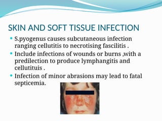
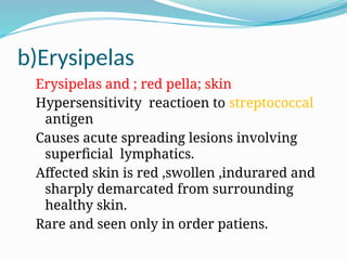
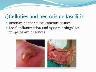
The document discusses Streptococcus pyogenes, highlighting its classification, morphology, cultural characteristics, pathogenicity, and diagnosis. It outlines the infections caused by this bacterium, including pyogenic and non-pyogenic diseases, along with their symptoms and treatments. Laboratory diagnosis methods are also presented, emphasizing the importance of specific tests for identifying infections and the recommended antibiotic therapies.

![INTRODUTION
Streptococci [or] Gram positive cocci arranged in
chains or pairs ,they are normal flora of humans and
animals .
Some of them are human pathogens.
The most important one are streptococcus pyogenes
causing pyogenic infection ,which are spread as
opposed to staphylococcal lesions,which are
typically , are localized.
Cocci in Billroth [1874] ,who called them
streptococci.](https://image.slidesharecdn.com/pooja-240813043449-513d042f/85/Streptococcus-pyogenes-Introduction-classification-3-320.jpg)
![MORPHOLOGY
The individual cocci are spherical ,oval in shape [0.5µm]
The size may be variated from cultural conditions
E,g. Anaerobically growing streptococcus are some what
smaller
They are arranged in chains (i-e) chains being longer in
liquid media than in solid media
Streptococci are non motile, non-spore formers](https://image.slidesharecdn.com/pooja-240813043449-513d042f/85/Streptococcus-pyogenes-Introduction-classification-5-320.jpg)
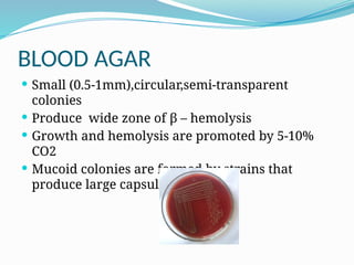
![CULTURAL CHARACTERS
It is an aerobe & facultative anaerobe, growing
best at temperature of 37°C [range 22 - 42°C].
It is exacting in nutritive requirements ,growth
occurring only in media containing fermented
carbohydrads [or ] enriched whith blood
[or]serum.
In blood agar at 24 hrs the organisms product
small [0.5-1µm ],circular ,semi transparent,
clearheamolysis with low convex disc.
The growth & hemolysis only promoted by
10%CO 2](https://image.slidesharecdn.com/pooja-240813043449-513d042f/85/Streptococcus-pyogenes-Introduction-classification-6-320.jpg)
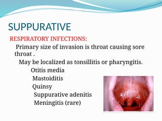
![a) Impetigo [pyoderma]
Pyo-purulent and derma-skin
Caused by higher numeberd M types S. pyrogen.
Superficial discrete crused spot of less than one
inch in diameter seen in children .
Lasts for 1-2 weeks and heals spontaneously
wihtout any scars .](https://image.slidesharecdn.com/pooja-240813043449-513d042f/85/Streptococcus-pyogenes-Introduction-classification-12-320.jpg)
![NON SUPPERATIVE
After a latent period of 1-4 weeks
Followed by rheumatic fever and acute
glomerulonephritis
A) Rheumatic fever
complication of S.pyogenus pharyngitis due to
specific 1M protein tyoes,
characterized by aschoff nodules [sub
cutaneous nodule] degeneration of haert valves
Mimics epidemiologic character of
streptococcalpharygitis.](https://image.slidesharecdn.com/pooja-240813043449-513d042f/85/Streptococcus-pyogenes-Introduction-classification-15-320.jpg)
![LAB DIAGNOSIS
Throat swab culture ;detectin of group a antigen .
Specific nucleic acid based test.
Elevation of anti hyalurinidase antibodies [strong
evidence]
Specimen;
Throat swab , pus swab or exudates are collected .
Microscopy ;
Gram –staining of pus can be examinated
Presence of gram positive cocci in chain can be
indication.](https://image.slidesharecdn.com/pooja-240813043449-513d042f/85/Streptococcus-pyogenes-Introduction-classification-17-320.jpg)