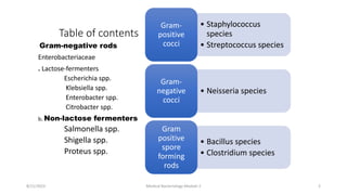
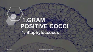
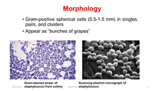
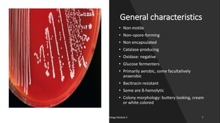
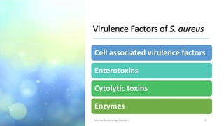
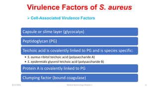
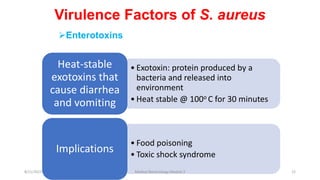
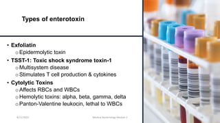
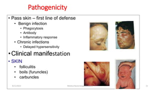
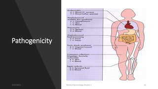
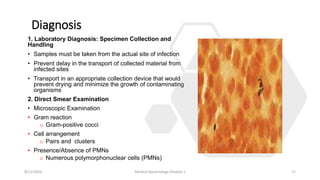
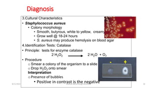
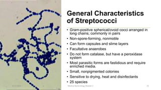
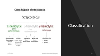
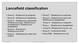
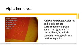
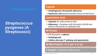
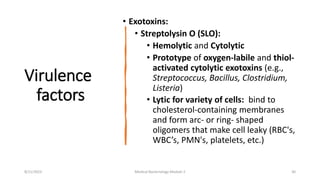
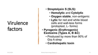
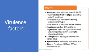
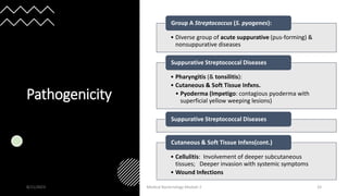
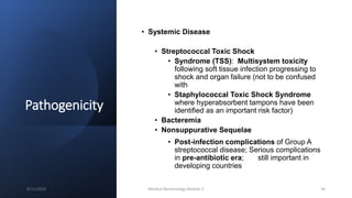
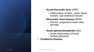
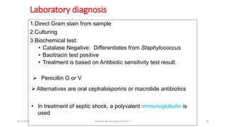
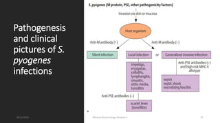
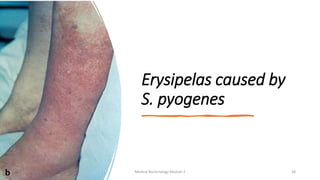
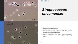
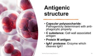
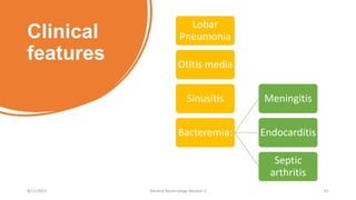
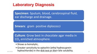
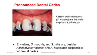
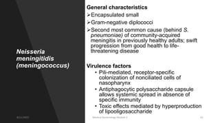
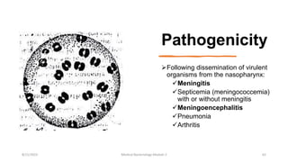
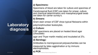
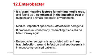
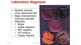
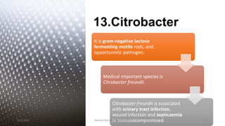
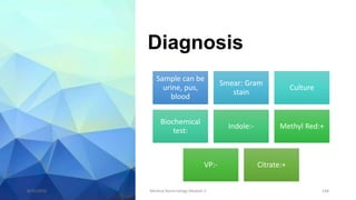
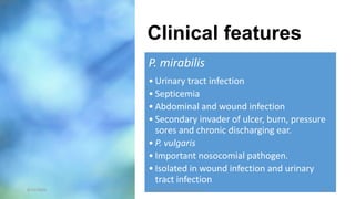
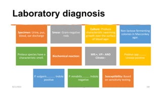
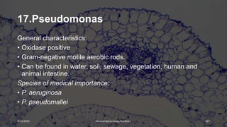
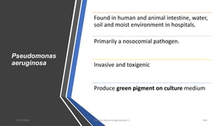
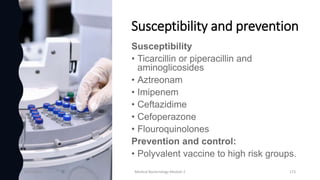
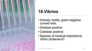
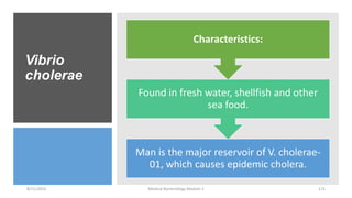
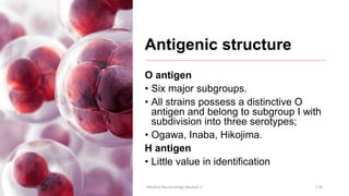
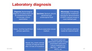
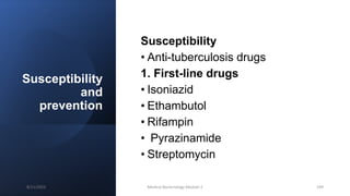
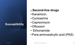
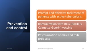
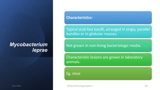
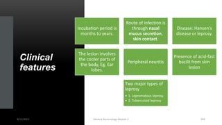
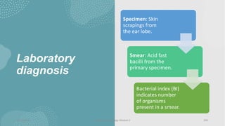
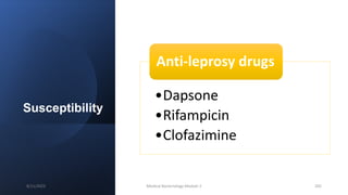
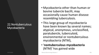
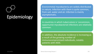
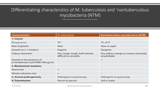
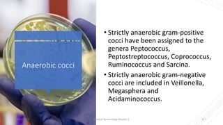
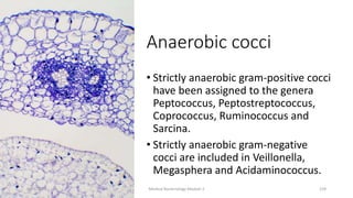
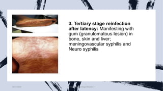
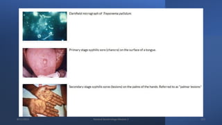
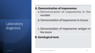
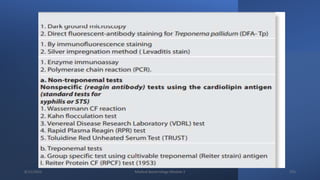
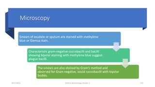
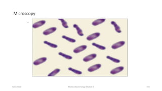
Medical
Bacteriology
[Bacteria causing medically important diseases]
8/11/2023 Medical Bacteriology Module 2 1
Table of contents
Gram-negative rods
Enterobacteriaceae
. Lactose-fermenters
Escherichia spp.
Klebsiella spp.
Enterobacter spp.
Citrobacter spp.
b. Non-lactose fermenters
Salmonella spp.
Shigella spp.
Proteus spp.
• Staphylococcus
species
• Streptococcus species
Gram-
positive
cocci
• Neisseria species
Gram-
negative
cocci
• Bacillus species
• Clostridium species
Gram
positive
spore
forming
rods
8/11/2023 Medical Bacteriology Module 2 2
• Mycoplasma
• Ureaplasma
• Rickettsiaceae,
Bartonellaceae and
Coxiella
• EHRLICHIA,
• ANAPLASMA AND
• NEORICKETTSIA
• Bartonella and
• Grahamella
Gram negative
rods
Oxidase
Positive
Pseudomonas
spp.
Vibrio
Campylobacter
Helicobacter
Borelia species
Leptospira
8/11/2023 Medical Bacteriology Module 2 3
Chlamydia and
Chlamydophila
Yersinia,
Pasteurella,
Francisella
8/11/2023 Medical Bacteriology Module 2 4
1.GRAM
POSITIVE COCCI
1. Staphyloccocus
8/11/2023 Medical Bacteriology Module 2 5
• Gram-positive spherical cells (0.5-1.5 mm) in singles,
pairs, and clusters
• Appear as “bunches of grapes”
Gram-stained smear of
staphylococci from colony
Scanning electron micrograph of
staphylococci
Morphology
8/11/2023 Medical Bacteriology Module 2 6
General characteristics
• Non motile
• Non–spore-forming
• Non encapsulated
• Catalase-producing
• Oxidase: negative
• Glucose fermenters
• Primarily aerobic, some facultatively
anaerobic
• Bacitracin resistant
• Some are ß-hemolytic
• Colony morphology: buttery looking, cream
or white colored
8/11/2023 Medical Bacteriology Module 2 7
Staphylococcus aureus
Primary pathogen of the genus
Habitat:
• Anterior nares (carriers)
• Colonization: axilla, vagina, pharynx
Produce superficial to systemic infections
• Skin
• Bacterial sepsis
• Hospital acquired infections
8/11/2023 Medical Bacteriology Module 2 8
• Mode of transmission
oTraumatic introduction
oDirect contact with infected person
oInanimate objects
• Predisposing conditions
oChronic infections
oIndwelling devices
oSkin injuries
oImmune response defects
• Infection will elaborate
inflammatory response with
accumulating pus
• Pus: mix of active and
inactive neutrophils, bacterial
cells and extravascular fluid
Staphylococcus aureus con’t
8/11/2023 Medical Bacteriology Module 2 9
Virulence Factors of S. aureus
Cell associated virulence factors
Enterotoxins
Cytolytic toxins
Enzymes
8/11/2023 Medical Bacteriology Module 2 10
Capsule or slime layer (glycocalyx)
Peptidoglycan (PG)
Teichoic acid is covalently linked to PG and is species specific:
• S. aureus ribitol teichoic acid (polysaccharide A)
• S. epidermidis glycerol teichoic acid (polysaccharide B)
Protein A is covalently linked to PG
Clumping factor (bound coagulase)
Cell-Associated Virulence Factors
Virulence Factors of S. aureus
8/11/2023 Medical Bacteriology Module 2 11
• Exotoxin: protein produced by a
bacteria and released into
environment
• Heat stable @ 100o C for 30 minutes
Heat-stable
exotoxins that
cause diarrhea
and vomiting
• Food poisoning
• Toxic shock syndrome
Implications
Virulence Factors of S. aureus
Enterotoxins
8/11/2023 Medical Bacteriology Module 2 12
Types of enterotoxin
• Exfoliatin
o Epidermolytic toxin
• TSST-1: Toxic shock syndrome toxin-1
o Multisystem disease
o Stimulates T cell production & cytokines
• Cytolytic Toxins
oAffects RBCs and WBCs
o Hemolytic toxins: alpha, beta, gamma, delta
o Panton-Valentine leukocin, lethal to WBCs
8/11/2023 Medical Bacteriology Module 2 13
Virulence Factors of S. aureus
• Hydrolyzes hyaluronic acid in connective
tissue allowing spread of infection
Hyaluronidase:
• Fibrinolysin which allows spread of
infection
Staphylokinase:
• Virulence marker
Coagulase:
• Lipase:
oAllows colonization by acting on
lipids present on the surface of
the skin.
• Penicillinase:
oConfers resistance
• DNase:
oDegrades DNA
• Beta-lactamase:
oCuts the beta-lactam wall of
certain antibiotics
Extracellular Enzymes
8/11/2023 Medical Bacteriology Module 2 14
• Pass skin – first line of defense
• Benign infection
• Phagocytosis
• Antibody
• Inflammatory response
• Chronic infections
• Delayed hypersensitivity
•Clinical manifestation
• SKIN
• folliculitis
• boils (furuncles)
• carbuncles
Pathogenicity
8/11/2023 Medical Bacteriology Module 2 15
Pathogenicity
8/11/2023 Medical Bacteriology Module 2 16
Diagnosis
1. Laboratory Diagnosis: Specimen Collection and
Handling
• Samples must be taken from the actual site of infection
• Prevent delay in the transport of collected material from
infected sites
• Transport in an appropriate collection device that would
prevent drying and minimize the growth of contaminating
organisms
2. Direct Smear Examination
• Microscopic Examination
• Gram reaction
o Gram-positive cocci
• Cell arrangement
o Pairs and clusters
• Presence/Absence of PMNs
o Numerous polymorphonuclear cells (PMNs)
8/11/2023 Medical Bacteriology Module 2 17
3.Cultural Characteristics
• Staphylococcus aureus
• Colony morphology
• Smooth, butyrous, white to yellow, creamy
• Grow well @ 18-24 hours
• S. aureus may produce hemolysis on blood agar
4.Identification Tests: Catalase
• Principle: tests for enzyme catalase
2 H2O2 2 H2O + O2
• Procedure
o Smear a colony of the organism to a slide
o Drop H2O2 onto smear
Interpretation
oPresence of bubbles
• Positive in contrast is the negative
Diagnosis
8/11/2023 Medical Bacteriology Module 2 18
Diagnosis con’t
5.Identification test: slide coagulase test
• Differentiates members within the Staphylococci
• Detects clumping factor found in S. aureus
• Procedure
oPlace a drop of sterile water on a slide and emulsify a colony
oAdd a drop of rabbit plasma to the suspension
oObserve
• Agglutination = Positive
• No agglutination= Negative
8/11/2023 Medical Bacteriology Module 2 19
Susceptibility
• Deep/metastatic infections
o semi-synthetic penicillins
o cephalosporins
o erythromycin
o clindamycin
• Endocarditis
o semi-synthetic penicillin +
an aminoglycoside
Note:
The treatment is guided by
the result of the antibiotic
sensitivity test.
8/11/2023 Medical Bacteriology Module 2 20
Medically important Staphylococcus Sp
* CNS: coagulase-negative staphylococci
8/11/2023 Medical Bacteriology Module 2 21
2. Streptococcus species
8/11/2023 Medical Bacteriology Module 2 22
General Characteristics
of Streptococci
• Gram-positive spherical/ovoid cocci arranged in
long chains; commonly in pairs
• Non-spore-forming, nonmotile
• Can form capsules and slime layers
• Facultative anaerobes
• Do not form catalase, but have a peroxidase
system
• Most parasitic forms are fastidious and require
enriched media.
• Small, nonpigmented colonies
• Sensitive to drying, heat and disinfectants
• 25 species
8/11/2023 Medical Bacteriology Module 2 23
Classification
8/11/2023 Medical Bacteriology Module 2 24
Lancefield classification
• Group A - Streptococcus pyogenes
• Group B - Streptococcus agalactiae
• Group C - Streptococcus equisimilis,
Streptococcus equi, Streptococcus
zooepidemicus, Streptococcus
dysgalactiae
• Group D - Enterococci,
Streptococcus bovis
• Group E - Streptococcus milleri and
mutans
• Group F - Streptococcus anginosus
• Group G - Streptococcus canis and
Streptococcus dysgalactiae
• Group H - Streptococcus sanguinis
• Group L - Streptococcus dysgalactiae
• Group M: Streptococcus fryi sp. nov
• Group N - Lactococcus lactis
• Group R&S - Streptococcus suis
8/11/2023 Medical Bacteriology Module 2 25
Alpha hemolysis
• Alpha-hemolysis. Colonies
on blood agar are
surrounded by a green
zone. This “greening” is
caused by H2O2, which
converts hemoglobin into
methemoglobin.
8/11/2023 Medical Bacteriology Module 2 26
Beta hemolysis
• Beta-hemolysis.
Colonies on blood agar
are surrounded by a large,
yellowish hemolytic zone
in which no more intact
erythrocytes are present
and the hemoglobin is
decomposed.
8/11/2023 Medical Bacteriology Module 2 27
Gamma hemolysis
• Gamma hemolysis is a lack of
hemolysis in the area around a
bacterial colony. A blood agar
plate displaying gamma
hemolysis actually appears
brownish. This is a normal
reaction of the blood to the
growth conditions used (37° C
in the presence of carbon
dioxide).
8/11/2023 Medical Bacteriology Module 2 28
Streptococcus
pyogenes (A
Streptococci)
 Virulence factors
Capsule
• Antiphagocytic; Nonspecific adherence
• Hyaluronic acid (polysaccharide) mimics animal tissue
Lipoteichoic Acid
• Cytotoxic for wide variety of cells
• Adherence: Complexes with M protein (LTA-M) and
binds to fibronectin on epithelial cells
M-Protein
• LTA-M protein is adhesin
• Antiphagocytic
• Inhibits alternate C’ pathway and opsonization
M-like Proteins: bind IgM and IgG
F Protein: mediates adherence
8/11/2023 Medical Bacteriology Module 2 29
Virulence
factors
• Exotoxins:
• Streptolysin O (SLO):
• Hemolytic and Cytolytic
• Prototype of oxygen-labile and thiol-
activated cytolytic exotoxins (e.g.,
Streptococcus, Bacillus, Clostridium,
Listeria)
• Lytic for variety of cells: bind to
cholesterol-containing membranes
and form arc- or ring- shaped
oligomers that make cell leaky (RBC's,
WBC’s, PMN's, platelets, etc.)
8/11/2023 Medical Bacteriology Module 2 30
Virulence
factors
• Streptolysin S (SLS):
• Hemolytic and Cytolytic
• Oxygen stable, non-antigenic
• Lytic for red and white blood
cells and wall-less forms
(protoplast, L- forms)
• Pyrogenic (Erythrogenic)
Exotoxins (Types A, B &C)
• Produced by more than 90% of
Grp A strep
• Cardiohepatic toxin
8/11/2023 Medical Bacteriology Module 2 31
Virulence
factors
• Nucleases: Four antigenic types (A,B,C,D)
• Facilitate liquefication of pus generating
growth substrates
• Nucleases A, C have DNase activity
producing runny pus
• Nucleases B, D also have RNase activity
• Streptokinases: Two different forms
• Lyse blood clots: catalyze conversion of
plasminogen to plasmin, leading to
digestion of fibrin
• C5a Peptidase: destroys C’ chemotactic
signals (C5a)
• Hyaluronidase: hydrolyzes hyaluronic acid
• Others: Proteinase, NADase, ATPase,
phosphatase, etc
Enzymes:
8/11/2023 Medical Bacteriology Module 2 32
Pathogenicity
• Diverse group of acute suppurative (pus-forming) &
nonsuppurative diseases
Group A Streptococcus (S. pyogenes):
• Pharyngitis (& tonsilitis):
• Cutaneous & Soft Tissue Infxns.
• Pyoderma (Impetigo: contagious pyoderma with
superficial yellow weeping lesions)
Suppurative Streptococcal Diseases
• Cellulitis: Involvement of deeper subcutaneous
tissues; Deeper invasion with systemic symptoms
• Wound Infections
Cutaneous & Soft Tissue Infxns(cont.)
8/11/2023 Medical Bacteriology Module 2 33
Pathogenicity
• Systemic Disease
• Streptococcal Toxic Shock
• Syndrome (TSS): Multisystem toxicity
following soft tissue infection progressing to
shock and organ failure (not to be confused
with
• Staphylococcal Toxic Shock Syndrome
where hyperabsorbent tampons have been
identified as an important risk factor)
• Bacteremia
• Nonsuppurative Sequelae
• Post-infection complications of Group A
streptococcal disease; Serious complications
in pre-antibiotic era; still important in
developing countries
8/11/2023 Medical Bacteriology Module 2 34
• Acute rheumatic fever (ARF):
• Inflammation of heart, joints, blood
vessels, sub-cutaneous tissues
• Rheumatic heart disease (RHD):
• Chronic, progressive heart valve
damage
• Acute glomerulonephritis (AG):
• Acute inflammation of renal
(kidney) glomeruli
• Foodborne Disease
8/11/2023 Medical Bacteriology Module 2 35
Laboratory diagnosis
1.Direct Gram stain from sample
2.Culturing
3.Biochemical test:
• Catalase Negative: Differentiates from Staphylococcus
• Bacitracin test positive
• Treatment is based on Antibiotic sensitivity test result.
 Penicillin G or V
Alternatives are oral cephalosporins or macrolide antibiotics
• In treatment of septic shock, a polyvalent immunoglobulin is
used
8/11/2023 Medical Bacteriology Module 2 36
Pathogenesis
and clinical
pictures of S.
pyogenes
infections
8/11/2023 Medical Bacteriology Module 2 37
Erysipelas caused by
S. pyogenes
8/11/2023 Medical Bacteriology Module 2 38
Streptococcus
pneumoniae
• Gram positive diplococci.
• Possess a capsule of polysaccharide that permits
typing with specific antisera.
• Found as a normal flora in the upper respiratory
tract.
8/11/2023 Medical Bacteriology Module 2 39
Antigenic
structure
• Capsular polysaccharide:
Pathogenicity determinant with anti-
phagocytic property
• C substance: Cell wall associated
antigen
• Protein M antigen
• IgA1 protease: Enzyme which
cleaves IgA1
8/11/2023 Medical Bacteriology Module 2 40
Clinical
features
Lobar
Pneumonia
Otitis media
Sinusitis
Bacteremia:
Meningitis
Endocarditis
Septic
arthritis
8/11/2023 Medical Bacteriology Module 2 41
Laboratory Diagnosis
Specimen: Sputum, blood, cerebrospinal fluid,
ear discharge and drainage.
Smears: gram positive diplococci
Culture: Grow best in chocolate agar media in
CO2 enriched atmosphere.
• Shows α-hemolytic,
• Greater sensitivity to optochin (ethyl hydrocuprein
hydrochloride) in the disk test or their bile solubility.
8/11/2023 Medical Bacteriology Module 2 42
Antibacterial Susceptibility, & Prevention
Antibacterial Susceptibility
• Amoxicillin
• Chloramphenicol
• Third generation Cephalosporins
Prevention
• Pneumococcal conjugate vaccine: Immunization of individuals with type
specific polysaccharide vaccine that is effective in children under two years
of age.
8/11/2023 Medical Bacteriology Module 2 43
Differentiation of streptococcus
species
Species Catalase Bacitracin Optochin Litmus
milk
reduction
CAMP
(Christie–
Atkins–
Munch-
Peterson)
S. pyogenes - + - - -
S. agalaciae - - - - +
Enterococci - - - + -
Viridans
streptococci
- - - - -
S.
pneumoniae
- - + - -
8/11/2023 Medical Bacteriology Module 2 44
The Most Important Human Pathogen
Streptococci and Enterococci
8/11/2023 Medical Bacteriology Module 2 45
The Most Important Human Pathogen
Streptococci and Enterococci
8/11/2023 Medical Bacteriology Module 2 46
The Most Important
Human Pathogen
Streptococci and
Enterococci
8/11/2023 Medical Bacteriology Module 2 47
Oral Streptococci
 Most of the oral streptococci of the type often known as the
viridans group have no group antigen.
 They usually cause a-hemolysis, some c-hemolysis as well.
 Oral streptococci are responsible for 50–70% of all cases of
bacterial endocarditis
 Predisposing factors include congenital heart defects, acute
rheumatic fever, cardiac surgery, and scarred heart valves
 Laboratory diagnosis of endocarditis involves isolation of
the pathogen from blood cultures
 Drug therapy of endocarditis is carried out with either
penicillin G alone or combined with an aminoglycoside
(mostly gentamicin). Bactericidal activity is the decisive
parameter.
8/11/2023 Medical Bacteriology Module 2 48
Pronounced Dental Caries
• Certain oral streptococci
(S. mutans) are the main
culprits in tooth decay.
 S. mutans, S. sanguis, and S. mitis are, besides
Actinomyces viscosus and A. naeslundii, responsible
for dental caries
8/11/2023 Medical Bacteriology Module 2 49
GRAM NEGATIVE COCCI
3.Neisseria species
8/11/2023 Medical Bacteriology Module 2 50
Neisseria
Introduction
The
Neisseria
species are
gram
negative
cocci.
They occur in
pairs.
Neisseria
gonorrhea
“gonococci”
and Neisseria
meningitidis
“meningococci”
are pathogenic
for human
Die rapidly
outside
the
human
organism
8/11/2023 Medical Bacteriology Module 2 51
General
characteristics
• Aerobic, Nonmotile
• Gram-negative cocci , in pairs
(diplococci) with kidney-shaped.
• Oxidase positive
• Most catalase positive
• Intracellular diplococci
• Ferment carbohydrate producing acid but
not gas
• The main species of medical importance
are:
• N. meningitidis
• N. gonorrhoea
8/11/2023 Medical Bacteriology Module 2 52
N. gonorrhea
& N.
meningitidis
• Gonococci :
1) do not have
capsules
2) They have plasmids
3) They cause genital
infection
4) Ferment glucose
only
• Meningococci:
1) have
polysaccharide
capsules
2) Rarely have
plasmids
3) They cause a
disease called
meningitis
4) Ferment glucose
and maltose
8/11/2023 Medical Bacteriology Module 2 53
Neisseria gonorrhoeae
(gonococcus)
General characteristics
• Readily transmitted by sexual contact
• Gram-negative diplococci
• Susceptible to cool temperatures,
drying and fatty acids
• Produce acid from glucose, but not from
other sugars
8/11/2023 Medical Bacteriology Module 2 54
Virulent factors
1. Pili: Hair-like appendages and enhance attachment to host cells and
evade human defense.
2. Por (Protein I)
Pores on the surface of bacteria through which nutrients enter the cell.
3. Opa (Protein II)
Important for attachment of bacteria to host cells.
4. Protein III: Reduction-modifiable protein. It is associated with por in the
formation of pores in the cell.
5. Lipooligosaccharide(LOS): Responsible to damage epithelial cells.
Toxicity in gonococcal infection is largely due to the endotoxic effects of
LOS
6. IgA1 protease: Splits and inactivates major mucosal IgA(IgA1)
8/11/2023 Medical Bacteriology Module 2 55
Clinical
manifestation
• Route of infection: Sexual contact
Male:
• Gonococcal urethritis
• Gonococcal epididymitis
• Gonococcal epididymo-orchitis (inflammation of
testis)
• Infertility
• Gonococcal suppurative arthritis
Female:
• Gonococcal cervicitis
• Gonococcal salpingitis (infection and
inflammation in fallopian tubes)
• If complicated: Gonococcal tubo-ovarian abscess
8/11/2023 Medical Bacteriology Module 2 56
Females Males
50% risk of infection after single exposure 20% risk of infection after single exposure
Asymptomatic infections frequently not diagnosed Most initially symptomatic (95% acute)
Major reservoir is asymptomatic carriage in females Major reservoir is asymptomatic carriage in females
Genital infection primary site is cervix (cervicitis), but vagina,
urethra, rectum can be colonized
Genital infection generally restricted to urethra (urethritis)
with purulent discharge and dysuria
Ascending infections in 10-20% including salpingitis, tubo-
ovarian abscesses, pelvic inflammatory disease (PID) ,
chronic infections can lead to sterility
Rare complications may include epididymitis, prostatitis,
and periurethral abscesses
Disseminated infections more common, including septicemia,
infection of skin and joints (1-3%)
Disseminated infections are very rare
Can infect infant at delivery (conjunctivitis, opthalmia
neonatorum)
More common in homosexual/bisexual men than in
heterosexual population
8/11/2023 Medical Bacteriology Module 2 57
D) Throat infection occur in abnormal person’s
suffering from Gonococal pharyngitis. It’s common
in homosexual person.
C) Eye infection: occur in new born babies and it
will lead to blindness.
B) Rectal involvement: infection caused by Neisseria
gonorrhoeae is called proctitis and characterized by
purulent discharge from anus.
8/11/2023 Medical Bacteriology Module 2 58
Laboratory
diagnosis
a) Specimens:
Pus and secretions are taken from the urethra,
cervix, rectum, conjunctiva, throat or synovial fluid
for culture and smear.
b) Smears:
Gram stained smears of urethral or endocervical
exudate
reveal many diplococci within pus cells.
c) Culture:
Immediately after collection pus or mucous is
streaked on
enriched selective medium.
d) Serology:
Serum and genital fluid contain IgG and IgA
antibodies by
ELISA
8/11/2023 Medical Bacteriology Module 2 59
Prevention &
Susceptibility
• Penicillin no longer drug of choice due to:
• Continuing rise in the MIC
• Plasmid-encoded beta-lactamase
production
• Chromosomally-mediated resistance
• Uncomplicated infection: ceftriaxone,
cefixime or fluoroquinolone
• Combined with doxycycline or azithromycin
for dual infections with Chlamydia
• Treatment of newborns with opthalmia
neonatorum with ceftriaxone
• Measures to limit epidemic include
education, aggressive detection, and follow-
up screening of sexual partners, use of
condoms or spermicides.
8/11/2023 Medical Bacteriology Module 2 60
Neisseria
meningitidis
(meningococcus)
General characteristics
Encapsulated small
Gram-negative diplococci
Second most common cause (behind S.
pneumoniae) of community-acquired
meningitis in previously healthy adults; swift
progression from good health to life-
threatening disease
Virulence factors
• Pili-mediated, receptor-specific
colonization of nonciliated cells of
nasopharynx
• Antiphagocytic polysaccharide capsule
allows systemic spread in absence of
specific immunity
• Toxic effects mediated by hyperproduction
of lipooligosaccharide
8/11/2023 Medical Bacteriology Module 2 61
Pathogenicity
Following dissemination of virulent
organisms from the nasopharynx:
Meningitis
Septicemia (meningococcemia)
with or without meningitis
Meningoencephalitis
Pneumonia
Arthritis
8/11/2023 Medical Bacteriology Module 2 62
Laboratory
diagnosis
a) Specimens:
Specimens of blood are taken for culture and specimes of
Cerebrospinal fluid (CSF) are taken for smear, culture
and chemical determination and masopharyngeal swab
are taken for carrier survey’s.
b) Smears:
Gram stain smear of CSF show typical Neisseria within
polymorphnuclear leukocytes.
c) Culture:
CSF specimens are placed on heated blood agar
(chocolate)
agar and Thyer martin media) and incubated at 37c.
d) Serology:
Antibodies to meningococcal polysaccharides can be
measured by latex agglutination or by immuno
electrophoresis.
e) PCR
8/11/2023 Medical Bacteriology Module 2 63
Laboratory
diagnosis
a) Specimens:
Specimens of blood are taken for culture and specimes of
Cerebrospinal fluid (CSF) are taken for smear, culture and
chemical determination and masopharyngeal swab are taken
for carrier survey’s.
b) Smears:
Gram stain smear of CSF show typical Neisseria within
polymorphnuclear leukocytes.
c) Culture:
CSF specimens are placed on heated blood agar (chocolate
agar and Thyer martin media) and incubated at 37c.
d) Serology:
Antibodies to meningococcal polysaccharides can be
measured by latex agglutination or by immuno
electrophoresis.
e) PCR
8/11/2023 Medical Bacteriology Module 2 64
Susceptibility
and
Prevention
Susceptibility
Pencillin G is the drug of choice for treating meningococcal disease
and third-generation cephalosporins.
In patient allergic to penicillin, chloramphenicol and ceforamime (or
ceftriaxone) can be used.
Prevention
1) Irradiation of the carrier states (major source).
2) Isolation of the patient.
3) Chemoprophylaxis for contact people.
4) Vaccination.
8/11/2023 Medical Bacteriology Module 2 65
Neisseria gonorrhoeae and
Neisseria meningitidis
N. meningitidis: gram
staining of a preparation of
cerebrospinal fluid sediment.
Clinical diagnosis: acute
purulent meningitis.
N. gonorrheae: gram staining of a
preparation of urethral secretion:
coffee-bean-shaped diplococci,
grouped within a granulocyte.
Clinical diagnosis: gonorrhea
8/11/2023 Medical Bacteriology Module 2 66
GRAM NEGATIVE COCCOBACILLI
4. GENUS: HAEMOPHILUS
This is a group of small gram-negative, non-spore forming, non-motile,
pleomorphic bacteria that require enriched media for growth.
• Growth is enhanced in CO2 enriched atmosphere.
• Present in upper respiratory tract as a normal microbial flora in healthy
people.
• The group is fastidious requiring growth factors for isolation.
The growth factors are X-factor(Hematin) and V-factor (Diphosphopyridine
nucleotide).
8/11/2023 Medical Bacteriology Module 2 67
Cont
GENUS:
HAEMOPHILUS
The main species of
medical importance
are: H. influenza, H.
ducreyii H. aegyptius
Growth factor
required
Haemophilus
species:
X and V factor for H.
influenzae, H.
aegyptius, H.
hemolyticus.
X factor for H.
ducreyii .
V factor for H.
parainfluenzae, H.
parahemolyticus.
The main species of
medical importance
are:
H. influenza, H.
ducreyii and H.
aegyptius
8/11/2023 Medical Bacteriology Module 2 68
Haemophilus
influenzae
Characteristics:
• Gram-negative cocobacilli.
• Fastidious bacteria requiring growth factors for isolation.
• .Found in upper respiratory tract as normal flora in
healthy people.
• Antigenic structure
• Capsular polysaccharide .
• There are six serotypes of H. influenzae, A-F. . Capsular
antigen type b is composed of polyribose ribitol
phosphate.
• H. influenzae type b is the most common cause of disease
in humans.
• It is the main virulence factor which provides anti-
phagocytic property.
• Outer membrane protein
• . Lipo-oligosaccharide
8/11/2023 Medical Bacteriology Module 2 69
Cont
Clinical features: The bacteria causes disease most
commonly in young children.
• Acute pyogenic meningitis, Acute epiglottis ,Pneumonia , Otitis
media , Sinusitis . Cellulitis , Acute pyogenic arthritis
• Laboratory diagnosis:
• Specimen: Cerebrospinal fluid, sputum, blood, pus Smear: Gram-
negative short rods.
• Serology: Quellung reaction (using specific antisera)
Immunofluorescence stain
• Culture: Chocolate agar contain both X and V factor; blood agar
contain only X factor.
• Satellitism test is used to identify H. influenzae in blood agar.
8/11/2023 Medical Bacteriology Module 2 70
Satellitism
test
• Mix a loopful of haemophilus growth in 2ml
of sterile saline.
• Inoculate the bacteria suspension on a plate
of blood agar using a sterile swab.
• Streak a pure culture of S. aureus across the
inoculated plate which provides V-factor for
H. influenzae.
• incubate the plate over night in a CO2 -
enriched environment at 35- 37 Oc.
• . Look for growth and satellite colonies in
next morning. NB: Colonies are largest
nearest to the S. aureus column of growth.
8/11/2023 Medical Bacteriology Module 2 71
H.ducreyii
Slender, gram-negative, ovoid bacilli, slightly larger
than H. influenzae.
It causes chancroid (tender genital ulcer).
• Cultured in special enriched media (20-30% rabbit
blood agar) with colonic morphology of small grey
glistening colonies surrounded by zone of hemolysis.
• It is treated by erythromycin, cotrimoxazole and
third generation cephalosporins
3. H. aegyptius It causes contagious conjunctivitis.
8/11/2023 Medical Bacteriology Module 2 72
5. GENUS:
BORDETELLA
Characteristics: Minute strictly aerobic, non-
motile gram-negative rods.
Bordetella species of medical importance: B.
pertussis.
Antigenic structure:
Pili: Adheres to ciliated epithelial cells of the
respiratory tract. . Filamentous haemagglutinin:
Adheres to the ciliated respiratory tract.
8/11/2023 Medical Bacteriology Module 2 73
BORDETELLA
Pertussis toxin: . Lymphocytosis promoting factor .
Histamine sensitizing factor
. Insulin secretion enhancing factor
. Adenylyl cyclase toxin
. Dermonecrotic toxin
. Hemolysin .
Tracheal cytotoxin: Inhibits DNA synthesis in ciliated
respiratory epithelial cells.
8/11/2023 Medical Bacteriology Module 2 74
BORDETELLA
Clinical features:
Incubation period: 2 weeks
Route of transmission is respiratory from
early cases and possibly carries.
It has three stages: 1. Catarrhal stage 2.
Paroxysmal stage 3. Convalescence stage.
8/11/2023 Medical Bacteriology Module 2 75
BORDETELLA
During catarrhal stage, the patient is highly infectious but not very
ill manifesting with mild coughing and sneezing.
During paroxysmal stage, the patient presents with explosive
repetitive cough with characteristic ‘whoop’ upon inhalation leading
to exhaustion, vomiting, cyanosis and convulsion.
During convalescence stage, the patient presents with prolonged cough
8/11/2023 Medical Bacteriology Module 2 76
BORDETELLA
oLaboratory diagnosis:
• Specimen: Saline nasal wash (Preferred
specimen) Nasopharyngeal swab or cough
droplets on cough plate.
• Smear: Small, non-motile, capsulated,
gram-negative cocobacilli singly or in pair,
and may show bipolar staining.
• Culture: Inoculate the primary specimen on
Bordet-Gengue agar medium and incubate
for 2-6 days at 37 o c in a moist aerobic
atmosphere which produces small, raised,
shiny, mucoid colonies.
• Biochemical reaction:
• No growth on blood agar
• . Oxidase positive .
• Most of them are catalase positive
8/11/2023 Medical Bacteriology Module 2 77
8/11/2023 Medical Bacteriology Module 2 78
GRAM POSITIVE SPORES FORMING
RODS
4. Bacillus and 5. Clostridium
species
8/11/2023 Medical Bacteriology Module 2 79
6.BACILLUS
Characteristics
• Aerobic, non-motile, spore-
forming, gram-positive chain
forming rods.
• Bacillus species are saprophytes
• Important human pathogens
• B. anthracis
• B. cereus
8/11/2023 Medical Bacteriology Module 2 80
Bacillus
anthracis
• Major agent of bioterrorism and biological
warfare
• Major pathogen of domestic herbivores that
come in contact with humans
• Spores can survive in soil for years
Antigenic structure:
• Capsule: Enables the organism to evade
phagocytosis
• Polysaccharide somatic antigen
• Protein somatic antigen
• Anthrax toxin- Protein toxin: Complex of 3
protein factors
• Edema factor + Protective antigen = Edema
toxin
• Lethal factor + Protective antigen = Lehtal toxin
8/11/2023 Medical Bacteriology Module 2 81
Pathogenesis and Clinical feature
• Acquired by the entry of spores through injured skin in
cutaneous anthrax, or mucus membrane in intestinal anthrax, or
inhalation of spores in the lung while handling skin and hides
There are four forms of anthrax
1. Cutaneous anthrax (Malignant pustule): 95 % of anthrax
presentation
• Characterized by a black necrotic lesion with edematous margin
on hands, arms, face or neck with regional lymphadenitis
associated systemic symptoms.
2. Pulmonary anthrax: 5% of anthrax presentation
• Presents with pain, cough with haemorrhagic mediastinitis
8/11/2023 Medical Bacteriology Module 2 82
Pathogenesis and Clinical feature
3. Bacteremic anthrax: presents with
clinical features of sepsis
4. Intestinal anthrax: Presents with
abdominal pain, vomiting, and bloody
diarrhea
Bacteremic and intestinal anthrax are rare
to occur
8/11/2023 Medical Bacteriology Module 2 83
Laboratory
diagnosis
• Specimen: Fluid or pus from skin
lesion, Blood, sputum
• Smear: Non-capsulated gram-
positive rods
• Culture: Grows aerobically in
ordinary media: Non-hemolytic,
large, dense, grey-white irregular
colonies
• Serology: ELISA has been
developed to measure antibodies
to edema toxin and lethal toxin
8/11/2023 Medical Bacteriology Module 2 84
Susceptibility & Prevention
Susceptibility
• Ciprofloxacin
• Penicillin+ gentamicin or streptomycin
Prevention and control:
• Disposal of animal carcasses by deep burial or
burning
• Decontamination (autoclaving) of animal product
• Protective clothing and gloves for handling
potentially infected materials
• Active immunization of domestic
• Immunize high occupation risk persons with anthrax
toxoid
8/11/2023 Medical Bacteriology Module 2 85
Bacillus cereus
• General characteristics:
• Exhibit motility by swarming in
semisolid media
• Produce β lactamase, so not sensitive
to penicillin
8/11/2023 Medical Bacteriology Module 2 86
Clinical features
1. Food poisoning
Pathogenicity
determinant: Exotoxin
a. Emetic type food
poisoning
Characterized by nausea,
vomiting, abdominal
cramps, and self-limited
with in 24 hrs
b. Diarrheal type food
poisoning
Characterized by profuse
diarrhea and abdominal
cramps.
Lab. Diagnosis: Isolation
of B.cereus in stool is not
diagnostic since it is
present in normal stool
specimen
Treatment: Fluid
replacement
Antibiotics not required
8/11/2023 Medical Bacteriology Module 2 87
2. Ocular infection
Ocular disease following
trauma from non-surgical
penetrating objects
Susceptibility: Clindamycin
+ Aminoglycosides
8/11/2023 Medical Bacteriology Module 2 88
7.Clostridium
Characteristics
• Clostridia are anaerobic, spore-forming motile, gram-
positive rods
• Most species are soil saprophytes but a few are
pathogens to human
• They inhabit human and animal intestine, soil, water,
decaying animal and plant matter
• Spores of clostridia are wider than the diameter of
organism and located centrally, subterminally and
terminally
• Species of medical importance:
• C. perfringens
• C. tetani
• C. botulinum
• C. difficile
8/11/2023 Medical Bacteriology Module 2 89
Clostridium
perfringens
Characteristics:
Capsulated, non-motile, short gram-positive
rods in which spores are hardly seen
There are five toxigenic groups : A-E
Human disease is caused by C. perfringens
type A and C
8/11/2023 Medical Bacteriology Module 2 90
Clostridium perfringens
• Clinical diagnosis: gas
gangrene in a gunshot
wound.
• C. perfringens: gram
staining of a preparation
of wound pus. Large,
thick, gram-positive rods.
8/11/2023 Medical Bacteriology Module 2 91
Pathogenicity determinant
1. Enzymes: Digest collagen of
subcutaneous tissue and muscle.
• Collagenase
• Proteinase
• Hyaluronidase
• Dnase
2. Toxins
• PhospholipaseC (α toxin)
• It has lethal, necrotizing and
hemolytic effect on tissue.
• Theta toxin
• It has hemolytic and necrotic effect
on tissue
• Enterotoxin
8/11/2023 Medical Bacteriology Module 2 92
Clinical
manifestation:
1. Clostridial myonecrosis: Gas
gangrene
• IP(Incubation period) =1-3 days
• Colonization of devitalized traumatized
wound by C. perfringens spores, and
organism germination and release of
toxins
• Presentation: Muscle and subcutaneous
tissue necrosis and crepitation
• Foul smelling wound discharge
• Fever, toxaemia, hemolytic anemia, shock
2. Clostridial food poisoning
• It causes secretory diarrhea due to
release of enterotoxin in the intestine
8/11/2023 Medical Bacteriology Module 2 93
Laboratory
diagnosis
Specimen: Infected
tissue, pus, vomitus,
left over food, serum
Smear: Non-motile,
capsulated, thick brick-
shaped gram positive
rods in smears from
tissue; spores are
rarely seen.
1. Blood agar medium
• β-hemolytic colonies
are seen anaerobic
atmosphere.
2. Cooked meat
medium(Chopped meat-
glucose medium)
• Thioglycolate medium
• Nagler reaction:
Lecithinase C activity-
Opacity in the egg-
yolk medium due to
lecithin break down
8/11/2023 Medical Bacteriology Module 2 94
Identification of C. perfringens rests on colony form,
hemolysis pattern, biochemical reaction, and toxin
production and neutralization by specific antisera.
Treatment: Penicillin
• Prompt and extensive wound debridement
• Polyvalent antitoxin
Prevention and control
• Early adequate contaminated wound cleansing and
debridement
8/11/2023 Medical Bacteriology Module 2 95
Clostridium
difficile
General characteristics:
• Not frequently found in the healthy
adult, but is found often in the hospital
environment
• Produce cytotoxins ( A and B)
• Human feces are the expected source of
the organism
Pathogenesis and clinical
features:
• Clinically presents with
pseudomembraneous colitis and
manifests with fever, abdominal cramps,
watery or bloody diarrhea leading to
dehydration, septicemia and shock
8/11/2023 Medical Bacteriology Module 2 96
Lab. Diagnosis :
• Identification of toxin A and B in feces by
latex agglutination test
Susceptibility
• Discontinuation of offending drugs
• Administration of metronidazole or
vancomycin
• Administration of antibiotics like
ampicillin, clindamycin and
cephalosporins results in killing of colonic
normal flora and proliferation of drug
resistant C.difficile and release of
cytoxins
8/11/2023 Medical Bacteriology Module 2 97
Clostridium tetani
General characteristics:
• World wide in distribution in the soil and in
animal feces
• Gram -positive rods with round terminal spores
• There are ten antigenic types of C. tetani but all
produce the same neurotoxin.
• The toxin has two components:
• 1. Tetanospasmin: Neurotoxic property
• 2. Tetanolysin: Hemolytic property
8/11/2023 Medical Bacteriology Module 2 98
Pathogenesis
and Clinical
manifestation
• Infection of devitalized tissue (wound,
burn, injury, umblical stamp, surgical
suture) by spores of C. tetani →
Germination of the spore and
development of vegetative organism→
Neurotoxin release from vegetative cells
→ The toxin binds to receptors on the
presynaptic membrane of motor neuron
the retrograde axonal transport to the
spinalcord and brain stem → Inhibition
of inhibitory glycinergic and GABAergic
secreting neurons → Spatic paralysis,
muscle spasms and hyperreflexia
8/11/2023 Medical Bacteriology Module 2 99
Tetanus
Open lower-leg fracture following a
traffic accident; the portal of entry
of C. tetani.
Risus sardonicus: fully manifest
case of tetanus in a patient with
lower-leg fracture. Patient was
not vaccinated.
8/11/2023 Medical Bacteriology Module 2 100
• IP= 4-5 days to several weeks
Tetanus classical presentation:
• Arm flexion and of leg extension
• Fever and sweating
• Muscle spasm and rigidity
Laboratory diagnosis:
• The bacteria can be cultured in a media with anaerobic
atmosphere.
• Proof of isolation of C. tetani must rest on production of
toxin and its neutralization by specific antitoxin
• Diagnosis is by clinical picture and history of injury
8/11/2023 Medical Bacteriology Module 2 101
Susceptibility
• Administration of penicillin
• Provision of tetanus antitoxin (TAT)
Prevention and control:
• Avoid traditional application of mud or
ash over the umbilical stump
• Proper wound handling
• Immunization with tetanus toxoid
• NB: Since treatment of tetanus is not
satisfactory, prevention is all important
8/11/2023 Medical Bacteriology Module 2 102
Clostridium
botulinum
General characteristics:
Spores of C. botulinum are widely distributed in
soil, they often contaminate vegetables, fruits and
other materials.
Produce a neurotoxin which is the most active
known poison, and is considered to be the major
agent of bioterrorism and biological warfare
There are seven serotypes(A-G) of which A,B and
E are the principal causes of human illness.
8/11/2023 Medical Bacteriology Module 2 103
Pathogenesis
and Clinical
manifestation
• Food botulism
• IP = 18-24 hrs
• Route of entry is under cooked
consumption of C. botulinum toxin food
• The toxin is absorbed from the gut and
acts by blocking the release of
acetylcholine at synapses and
neuromuscular junction and manifests
with flaccid paralysis and visual
disturbance, inability to swallow, and
speech difficulty
• Death is secondary to respiratory failure
or cardiac arrest
8/11/2023 Medical Bacteriology Module 2 104
Clostridium
botulinum
2. Infantile botulism
• C. botulinum type A or B is usually implicated and
affects infants when mixed feeding starts (after fourth
month of life).
• Ingestion and colonization of the gut with C. botulinum,
and production of toxin and adsorption of toxin leads
to poor feeding, paralysis (floppy baby), and cranial
nerve palsy.
• Diagnosed by demonstration of the organism or toxin
from the stool
3. Wound botulism
• C. botulinum type A is usually implicated and caused by
the production of toxin by C. botulinum in wounds.
• The symptoms are the same as those in food poisoning.
8/11/2023 Medical Bacteriology Module 2 105
Clostridium botulinum
• Laboratory diagnosis:
• Demonstration of toxin in patient’s serum and leftover food.
• .Death of mice after intra-peritoneal injection of toxin.
Susceptibility
• Administration of intravenous trivalent antitoxin ( A,B,E)
• Mechanical ventilator for respiratory support
• Prevention and control:
• Sufficient heating of canned foods before consumption
• Strict regulation of commercial canning
• Proper home canning methods
8/11/2023 Medical Bacteriology Module 2 106
8.LISTERIA
MONOCYTOGENES
• Organisms of the genus Listeria are nonsporing gram-
positive bacilli. The genus contains eight species, but
almost all cases of human listeriosis are caused by L.
monocytogenes.
• Listeria monocytogenes is a small, coccoid, Gram-positive
bacillus measuring approximately 0.5 × 2–3 µm
• Cultural Characters
• Listeriae are aerobes and facultative anaerobes. They can
grow over a temperature range of 2–43°C, the optimum
temperature for the growth is 35–37°C.
• They can grow on ordinary media containing fermentable
carbohydrate, but growth is better on blood agar or
tryptose phosphate agar. After 24 hours incubation at 37°C,
colonies are 0.5–1.5 mm in diameter, smooth, translucent
and emulsifiable and non-pigmented.
8/11/2023 Medical Bacteriology Module 2 107
8/11/2023 Medical Bacteriology Module 2 108
Pathogenisis
• Human infection is believed to result from
contact with infected animals, inhalation of
contaminated dust or ingestion of contaminated
milk or food.
• Hospital acquired infections have also been
reported.
• L.monocytogenes produces a hemolysin known
as listeriolysin-O, which is a virulence factor
antigenically related to streptolysin-O and
pneumolysin.
• Clinical Features
• Intrauterine and neonatal infection:
Intrauterine infection of the fetus may result in
abortion, premature delivery, or acute-onset
disseminated infection in the newborn infant
8/11/2023 Medical Bacteriology Module 2 109
Pathogenisis
Adult and juvenile infection: It may cause
meningitis or meningoencephalitis, particularly
in neonates and in the elderly.
Disease in healthy adults: Most Listeria
infections in healthy adults are asymptomatic or
occur in the form of a mild influenza-like illness.
Several foodborne outbreaks of acute
gastroenteritis with fever .
Other infections: Listeriosis may also present as
abscesses, conjunctivitis, pharyngitis, urethritis,
pneumonia, infectious mononucleosis like
syndrome, endocarditis or septicemia.
8/11/2023 Medical Bacteriology Module 2 110
Laboratory
diagnosis
1. Specimens
Blood, CSF, amniotic fluid, placenta, pus and biopsy
material from the organs involved may be collected.
Specimens may also be collected from neonate, stillbirth
or products of conception.
2. Microscopy
If the Gram stain shows organisms, they are intracellular
and extracellular gram-positive coccobacilli.
3.Culture
Specimens should be inoculated on blood agar,
chocolate agar and tryptose phosphate agar, and
incubated at 35–37°C for 1–3 days. Greater success in
isolation is achieved if the materials are stored in
tryptose phosphate or thioglycollate broth at 4°C and
subcultures are done at weekly intervals for 1–6 months
8/11/2023 Medical Bacteriology Module 2 111
9.Corynebacterium
• Corynebacteria are gram-positive, nonacid fast, nonmotile rods with
irregularly stained segments, and sometimes granules.
• They frequently show club-shaped swellings and hence the name
Corynebacteria (from coryne, meaning club).
• Diseases:
• The major disease caused by C. diphtheriae is diphtheria (greek,
diphtheria, “leathery skin,” referring to the pseudomembrane that initially
forms on the pharynx).
8/11/2023 Medical Bacteriology Module 2 112
Corynebacterium diphtheriae
Morphology
• They are, thin, slender gram-positive bacilli but are decolorized
easily, particularly in old cultures, measuring approximately 3–6
μm × 0.6–0.8 μm.
• They have a tendency to clubbing at one or both ends. They are
highly pleomorphic. They are nonmotile, non-spore-forming,
and nonacid-fast.
• They can be found as groups or as individual cells lying at sharp
angles to one another, resembling the letters V or L.
• This particular arrangement with C. diphtheriae has been called
the Chinese letter
8/11/2023 Medical Bacteriology Module 2 113
8/11/2023 Medical Bacteriology Module 2 114
Toxin
• Toxigenic strains of C. diphtheriae produce a a very
powerful exotoxin. The toxicity observed in diphtheria is
directly attributed to the toxin secreted by the bacteria at
the site of infection.
• Mode of Action
• The diphtheria toxin acts by inhibiting protein synthesis.
It inhibits polypeptide chain elongation in the presence of
nicotinamide adenosine dinucleotide (NAD) by
inactivating elongation factor 2 (EF-2), an enzyme
required for elongation of polypeptide chains on
ribosomes.
• Inhibition of protein synthesis is probably responsible for
both the necrotic and neurotoxic effects of the toxin.
• Antigenic Structure Diphtheria bacilli possess three
distinct antigens:
• 1. A deep-seated antigen found in all Corynebacterial
species
• 2. A heat-labile protein (K-antigen)
• 3. A heat-stable polysaccharide (O-antigen).
8/11/2023 Medical Bacteriology Module 2 115
nicotinamide adenine dinucleotide
Toxins:
Exotoxin:
diptheria
toxin
8/11/2023 Medical Bacteriology Module 2 116
8/11/2023 Medical Bacteriology Module 2 117
8/11/2023 Medical Bacteriology Module 2 118
Pathogenesis
• The organism is carried in the upper respiratory tract and spread by
droplet infection or hand-to-mouth contact.
• The incubation period of diphtheria is 2–5 days, with a range of 1–10
days. Diphtheria, which occurs in two forms (respiratory and cutaneous),
is found worldwide.
• Respiratory Diphtheria
• The illness begins gradually and is characterized by low-grade fever,
malaise, and a mild sore throat. The most common site of infection is the
tonsils or pharynx.
• The organisms rapidly multiply on the epithelial cells, and the toxigenic
strains of C. diphtheriae produce toxin locally, causing tissue necrosis and
exudate formation triggering an inflammatory reaction.
8/11/2023 Medical Bacteriology Module 2 119
Pathogenesis
B.Systemic effects
The toxin also is absorbed and can produce a variety of systemic effects
involving the kidneys, heart, and nervous system, although all tissues
possess the receptor for the toxin and may be affected. Intoxication
takes the form of myocarditis and peripheral neuritis, and may be
associated with thrombocytopenia.
Complications
The common complications are as follows:
1. Asphyxia due to mechanical obstruction of the respiratory passage
by the pseudomembrane for which an emergency tracheostomy
may become necessary.
2. Acute circulatory failure, which may be peripheral or cardiac
8/11/2023 Medical Bacteriology Module 2 120
Laboratory
diagnosis
1. Specimens
Swabs from the nose, throat, or other suspected
lesions must be obtained before antimicrobial
drugs are administered.
2. Microscopy
Direct microscopy of a smear is unreliable since C.
diphtheriae is morphologically similar to other
coryneforms. Smears stained with alkaline
methylene blue or Gram’s stain show beaded rods
in typical arrangement.
3. Culture The swab should be inoculated on
Loffler’s serum slope, tellurite blood agar, and
blood agar. The cultures should be incubated
aerobically at 37°C.
8/11/2023 Medical Bacteriology Module 2 121
• Media
• Cystine-tellurite: Corynebacterium spp.
form black colonies
• from hydrolysis of tellurite.
• Tinsdale's agar: Corynebacterium
spp. form brown to black colonies
with halos from hydrolysis of tellurite.
• Loeffler agar is a nonselective medium
that supports growth and enhances
pleomorphism and the formation of
metachromatic granules.
• Most Corynebacterium spp. produce small,
white to gray colonies,
• C. diphtheriae will grow on SBA as
small, white, dry colonies. Most strains
are nonhemolytic.
8/11/2023 Medical Bacteriology Module 2 122
8/11/2023 Medical Bacteriology Module 2 123
• b. Corynebacterium
jeikeium
• C.jeikeium is an important
cause of nosocomial
infections and produces
infections after prosthetic
device implants and in
immunocompromised
patients.
• Pyrazidamidase
positive
• Resistant to most
antimicrobial agents
• c. Corynebacterium
urealyticum
• Cause UTIs
• Is rapid urease positive
and grows very slowly
8/11/2023 Medical Bacteriology Module 2 124
GRAM NEGATIVE RODS
6. Enterobacteriaceae
8/11/2023 Medical Bacteriology Module 2 125
Enterobacteriaceae
Characteristics
• Named, as well coliforms or
enterobacilli:
• Found as normal flora in intestinal
tract of humans and animals.
• Major cause nosocomial infection
• Gram-negative, non-spore forming,
aerobic and facultative anaerobic
bacteria.
• Most are motile.
• Grow over a wide range of
temperature in ordinary media.
• All ferment glucose with acid
production.
• Oxidase negative.
• Release endotoxin from their cell
wall.
• Some release exotoxin.
• They reduce nitrate to nitrites
8/11/2023 Medical Bacteriology Module 2 126
8/11/2023 Medical Bacteriology Module 2 127
Enteric culture media
Characteristics
• MacConkey agar: Lactose fermenters are pink/red, and
lactose nonfermenters are white.
• Eosin methylene blue agar: Colonies of lactose fermenters
have a dark center, and non lactose fermenters are colorless
• Hectoen enteric agar: Lactose and/or sucrose fermenters
from yellow/ orange colonies
8/11/2023 Medical Bacteriology Module 2 128
Classification
It comprises the following
bacterial groups
1. Oxidase negative
a. Lactose-fermenters
• Escherichia spp.
• Klebsiella spp.
• Enterobacter
spp.
• Citrobacter spp.
b. Non-lactose
fermenters
• Salmonella spp.
• Shigella spp.
• Proteus spp.
• 2. Oxidase Positive
• Pseudomonas
spp.
• Vibrio
• Campylobacter
• Helicobacter
8/11/2023 Medical Bacteriology Module 2 129
Escherichia
coli
Introduction
• Escherichia coli is a Gram-negative, non-
sporulating
• Facultative anaerobic and rod-shaped
Bacterium
• Commonly found in the lower intestine of
warm-blooded organisms (endotherms).
• Strains that possess flagella are motile
• Most E. coli strains are harmless,
• Some serotypes can cause serious food
poisoning in humans,
• The harmless strains are part of the
normal flora of the gut, and can benefit
their hosts by producing vitamin K, and by
preventing the establishment of
pathogenic bacteria within the intestine.
8/11/2023 Medical Bacteriology Module 2 130
Pathogenicity
Urinary tract infection- cystitis, pyelonephritis
Wound infection- appendicitis, peritonitis
Neonatal septicemia and meningitis
E.coli-associated diarrheal disease:
• 1. Enteropathogenic E.coli (EPEC)
• Causes outbreaks of self-limiting infantile diarrhea
• They also cause severe diarrhea in adults
• Antibiotic treatment shorten the duration of illness
and cure diarrhea
• 2. Enteroinvasive E.coli (EIEC)
• Non-motile, non-lactose fermenting E.coli invade the
mucosa of the ileum and colon, and causes
shigellosis-like dysentery in children in developing
countries and travellers to these countries
8/11/2023 Medical Bacteriology Module 2 131
3. Enterotoxigenic E.coli(ETEC)
• Colonization factor of the organism
promote adherence to epithelial cells of
small intestine followed by release of
enterotoxin which causes toxin-mediated
watery diarrhea in infants and young adults.
• Antibiotic prophylaxis can be effective but
may increase drug resistance
4. Entero haemorrhagic E.coli( EHEC)
• Cytotoxic verotoxin producing E.coli
serotype O157:H7 causes haemorrhagic
colitis (severe form of diarrhea), and
hemolytic uremic syndrome characterized
by acute renal failure, hemolytic anemia
and low platelet count
8/11/2023 Medical Bacteriology Module 2 132
5. Enteroaggressive E.coli ( EAEC)
• Adhere to human intestinal mucosal cells
and produce ST-like toxin and hemolysin,
and causes acute and chronic diarrhea as
well as food-borne illness.
6. Diffusely adherent E coli
7. Uropathogenic E. coli
8/11/2023 Medical Bacteriology Module 2 133
Virulence
factors
Complex surface antigens contribute to
pathogenicity and trigger immune
response:
• H – flagellar Ag
• K – capsule and/or fimbrial Ag
• O – somatic or cell wall Ag – all have
• Endotoxin
• Exotoxins
8/11/2023 Medical Bacteriology Module 2 134
Pathogenicity
The commonest infection caused by E. coli is
infection of the urinary tract,
They cause of cystitis (infection of the bladder), the
infection may spread up the urinary tract to the
kidneys, causing pyelonephritis.
E. coli bacteria may also cause infections in the
intestine. Diarrhoeal infections(intestinal)
Infection to the bloodstream may cause blood
poisoning (E. coli bacteraemia).
In rare instances, E. coli may cause meningitis in very
young children.
8/11/2023 Medical Bacteriology Module 2 135
Laboratory diagnosis
Samples: stool, urine, blood or other
relevant sample
Culture:
• Isolation media
a) nutrient agar,
b)MacConkey’s agar.
b. Susceptibility
8/11/2023 Medical Bacteriology Module 2 136
Culture characteristic and
biochemical test
Temperature 37°C for 24 hrs MacConkey Agar
Size in mm
Shape
Color
Margin
Elevation
Opacity
Consistency
1
Circular
Rose Pink colonies
Complete
Slightly
Opaque
Soft
Biochemical Test Results
Oxidase
Urease
TSI
MR
VP
Nitrate
Citrate
Indole (TW)
Gelatin
Negative
Negative
Acid butt, with gas, acid slant
Positive
Negative
Positive
Negative
Positive
Negative
8/11/2023 Medical Bacteriology Module 2 137
Escherichia coli
Gram staining of a urine
sediment preparation: rounded
gram-negative rods, some
coccoid.
Can cause acute cystitis.
Culture on endo agar, a
combined selective/indicator
medium. The red color of the
colony and agar indicates the
lactose breakdown process.
8/11/2023 Medical Bacteriology Module 2 138
11. klebsiella
8/11/2023 Medical Bacteriology Module 2 139
Introduction
• Klebsiella pneumoniae can be found as a commensal in
the mouth and upper respiratory tract, it can be found
intestinal tract of humans and animals.
• These are also found in plants, water and soil.
Characteristics
• Non-motile,
• lactose-fermenting,
• Capsulated ,
• Gram-negative rods.
• Main species of medical importance:
• K. pneumoniae
• K. rhinoscleromatis
• K. ozenae
8/11/2023 Medical Bacteriology Module 2 140
K. pneumoniae
• It is found as a commensal in the intestinal tract, and
also found in moist environment in hospitals.
• It is an important nosocomial pathogen.
• It causes:
• Pneumonia
• Urinary tract infection
• Septicaemia and meningitis (especially in neonates)
• Wound infection and peritonitis
• The virulence factor is the Capsular polysaccharide.
• The treatment is based on the result of antimicrobial
sensitivity test.
8/11/2023 Medical Bacteriology Module 2 141
K. rhinoscleromatis
• It causes rhinoscleroma of nose and pharynx to
extensive destruction of nasopharynx.
K.ozaenae
• It causes ozena manifesting with foul smelling
nasal discharge leading to chronic atrophic rhinitis.
8/11/2023 Medical Bacteriology Module 2 142
Laboratory diagnosis
1.Specimen: Sputum, urine, pus, CSF, body fluid.
2.Culture on
(a) nutrient agar,
(b) MacConkey’s agar
3.Gram stain
4.Biochemical test
Serology: Capsular polysaccharide serotyping
8/11/2023 Medical Bacteriology Module 2 143
Culture characteristic and biochemical results
Temperature 37°C for 24 hours Mac Conkey Agar
Size in mm
Shape round
Color Pink
Margin Complete
Elevation Slightly Raised
Opacity Translucent
Consistency Mucoid
3-4
Round
Pink
Complete
Slightly raised
Translucent
Mucoid
Test K. aerogenes Organism K. pneumoniae
Oxidase
Urease
TSI,
MR
VP
Citrate
Indole
Negative
Positive
acid slant, with gas, acid butt
negative
Positive
positive
Negative
Negative
Positive slow
acid slant, with gas, acid butt
positive
negative
positive
Negative
8/11/2023 Medical Bacteriology Module 2 144
12.Enterobacter
• It is gram-negative lactose fermenting motile rods,
and found as a commensal in the intestinal tract of
humans and animals and moist environments.
• Medical important species is Enterobacter aerogens.
• It produces mucoid colony resembling Klebsiella on
Mac Conkey agar.
• Enterobacter aerogens is associated with urinary
tract infection, wound infection and septicaemia in
immunocompromised patients.
8/11/2023 Medical Bacteriology Module 2 145
Laboratory diagnosis
• Sample: wounds,
urine, blood and CSF
• Major characteristics
Colonies resemble
Klebsiella
• Motile
• Indole: Negative
• MR: negative
• VP: positive
• Citrate: Positive
8/11/2023 Medical Bacteriology Module 2 146
13.Citrobacter
It is gram-negative lactose
fermenting motile rods, and
opportunistic pathogen.
Medical important species is
Citrobacter freundii.
Citrobacter freundii is associated
with urinary tract infection,
wound infection and septicaemia
in immunocompromised.
8/11/2023 Medical Bacteriology Module 2 147
Diagnosis
Sample can be
urine, pus,
blood
Smear: Gram
stain
Culture
Biochemical
test:
Indole:- Methyl Red:+
VP:- Citrate:+
8/11/2023 Medical Bacteriology Module 2 148
14.Salmonella
• Coliform bacilli (enteric rods)
• Motile gram-negative facultative anaerobes
• Non-lactose fermenting
• Resistant to bile salts
•
• H2S producing
• Species of medical importance are:
• S. typhi
• S. paratyphi
• S. enteritidis
8/11/2023 Medical Bacteriology Module 2 149
Causes
1. Caused by the bacterium Salmonella Typhi .
2. Ingestion of contaminated food or water.
3. Contact with an acute case of typhoid
fever.
4. Water is contaminated where inadequate
sewerage systems and poor sanitation.
5. Contact with a chronic asymptomatic carrier.
6. Eating food or drinking beverages that
handled by a person carrying the bacteria.
7. Salmonella enteriditis and Salmonella
typhimurium are other salmonella bacteria,
cause food poisoning and diarrhoea.
8/11/2023 Medical Bacteriology Module 2 150
How the bacteria cause disease
Ingestion of contaminated
food or water
Salmonella bacteria
Invade small intestine and
enter the bloodstream
Carried by white blood
cells in the liver, spleen,
and bone marrow
Multiply and reenter the
bloodstream
Bacteria invade the
gallbladder, biliary system,
and the lymphatic tissue
of the bowel and multiply
in high numbers
Then pass into the
intestinal tract and can be
identified for diagnosis in
cultures from the stool
tested in the laboratory
8/11/2023 Medical Bacteriology Module 2 151
Clinical features
1. Enteric fever
• It is caused by S. typhi and S. paratyphi, and
• transmitted by fecal-oral route via contaminated
food and drinks
• Incubation period: 10-14 days
Predisposing factors:
• Reduced gastric acidity
• Disrupted intestinal microbial flora
• Compromised local intestinal immunity
8/11/2023 Medical Bacteriology Module 2 152
Both manifest with persistent fever, headache, malaise,
chills, enlargement of liver and spleen, and skin rashes.
Paratyphoid fever is milder than typhoid fever
Complications:
• Intestinal perforation
• Lower gastrointestinal bleeding
• Dissemination to different body organs
• including meninges and brain
Mortality rate
• Untreated cases: 10-15%
• Treated cases: < 1%
8/11/2023 Medical Bacteriology Module 2 153
2. Bacteremia with focal lesions
• Causative agent: S. choleraesuis
• Manifests with blood stream invasion with focal
lesions in lungs, bones and meninges
3. Gastroenteritis
• It is caused by S. enteritidis
• S. typhimurium
• IP= 8-48 hrs
• It manifests with initial watery diarrhea, and later
bloody mucoid diarrhea associated with crampy
abdominal pain.
8/11/2023 Medical Bacteriology Module 2 154
Laboratory diagnosis
Specimen:
1. Blood, Bone marrow, stool,
urine and serum for enteric fever.
• Blood – 80% positive in the first week.
• Stool- 70-80% positive in the second and
third week.
• Urine- 20% positive in the third and
fourth week.
• Serum for widal test- positive after the
second week of illness.
8/11/2023 Medical Bacteriology Module 2 155
2. Stool for gastroenteritis.
• Gram reaction: Gram-negative rods
• Culture: Bacteriologic methods for
salmonella isolation
1. Differential medium
• For rapid isolation of lactose non-
fermenters Egs. Mac Conkey agar (Non-
lactose fermenting , H2S producing
colonies)
• Deoxycholate agar
2. Selective medium
• SS agar
• Deoxycholate-Citrate agar
Serology: (widal test)
8/11/2023 Medical Bacteriology Module 2 156
Susceptibility and
prevention
Susceptibility
1. For cases
• Chloramphenicol
• Fluoroquinolones
• 3rd generation cephalosporins
Prevention and control
• Sanity measures like hygienic food and
drink handling, and avoid carriers from food
handling until properly treated
• Provision of vaccine
8/11/2023 Medical Bacteriology Module 2 157
15.SHIGELLA
• Species of medical importance
are:
• S. dysenteriae A
• S. flexneri B
• S. boydii C
• S. sonnei D
• In developing countries,
shigellosis (bacillary dysentery) is
caused by S. flexneri and S.
dysenteriae.
It is found in human intestinal tract
as pathogen.
8/11/2023 Medical Bacteriology Module 2 158
15. SHIGELLA
Coliform bacilli (enteric rods)
 Nonmotile gram-negative
facultative anaerobes
 Non-lactose fermenting
 Resistant to bile salts
• Shigellosis = Generic term for
disease
Low infectious dose (102-104)
Humans are only reservoir
Transmission by fecal-oral
route
Watery diarrhea with fever; changing to dysentery
Major cause of bacillary dysentery (severe 2nd stage)
in pediatric age group (1-10 yrs) via fecal-oral route
Outbreaks in daycare centers, nurseries, institutions
Estimated 15% of pediatric diarrhea in U.S.
Leading cause of infant diarrhea and mortality
(death) in developing countries
8/11/2023 Medical Bacteriology Module 2 159
Virulence Factors
Virulence attributable to:
 Invasiveness
 Attachment (adherence) and internalization
with complex genetic control
 Large multi-gene virulence plasmid regulated
by multiple chromosomal genes
 Exotoxin (Shiga toxin)
 Intracellular survival & multiplication
8/11/2023 Medical Bacteriology Module 2 160
Pathogenesis
and Clinical
features
• Pathogenicity determinant:
• Toxins:
• Endotoxin: irritate the bowel wall
• Exotoxin: Enterotoxin and neurotoxin
• IP: 1-2 days
• It causes shigellosis (bacillary dysentery)
characterized by bloody mucoid diarrhea, abdominal
cramp, fever, generalized muscle ache and
weakness.
Complication: Dehydration
• Electrolyte and acid-base disturbance
High prevalence:
• Poor sanitation, personal hygiene
• Polluted water supply
• Young children are frequently affected.
8/11/2023 Medical Bacteriology Module 2 161
Laboratory
diagnosis
Specimen: Stool, serum
Gram reaction: Gram-negative non-
motile rods.
Culture: Non-lactose fermenting colonies
on Macconkey agar and SS agar.
It produces acid but not gas from
carbohydrate.
8/11/2023 Medical Bacteriology Module 2 162
Susceptibility and
prevention
Susceptibility Ciprofloxacin Cotrimaxazole
Suppress acute
clinical attacks of
dysentry
Shorten the
duration of
symptoms
Prevention and
control:
Sanitary control of
water, food and
milk, sewage
disposal and fly
Control
Antibiotic
treatment of
infected individuals
8/11/2023 Medical Bacteriology Module 2 163
16.Proteus
Proteus species are found in the intestinal tract of humans
and animals, soil, sewage and water.
They are gram-negative, motile, non-capsulated ,oxidase
negative and non lactose fermenting, pleomorphic rods.
Species of medical importance:
P. mirabilis
P. vulgaris
8/11/2023 Medical Bacteriology Module 2 164
Clinical features
P. mirabilis
• Urinary tract infection
• Septicemia
• Abdominal and wound infection
• Secondary invader of ulcer, burn, pressure
sores and chronic discharging ear.
• P. vulgaris
• Important nosocomial pathogen.
• Isolated in wound infection and urinary
tract infection
8/11/2023 Medical Bacteriology Module 2 165
Laboratory diagnosis
Specimen: Urine, pus,
blood, ear discharge
Smear: Gram-negative
rods
Culture: Produce
characteristic swarming
growth over the surface
of blood agar.
Non-lactose fermenting
colonies in Macconkey
agar.
Proteus species have a
characteristic smell.
Biochemical reaction:
MR:+, VP:- AND
Citrate:-
Proteus spp………..
Urease positive
P. vulgaris………... Indole
positive
P. mirabilis……….. Indole
negative
Susceptibility: Based
on sensitivity testing.
8/11/2023 Medical Bacteriology Module 2 166
17.Pseudomonas
General characteristics:
• Oxidase positive
• Gram-negative motile aerobic rods.
• Can be found in water, soil, sewage, vegetation, human and
animal intestine.
Species of medical importance:
• P. aeruginosa
• P. pseudomallei
8/11/2023 Medical Bacteriology Module 2 167
Pseudomonas
aeruginosa
Found in human and animal intestine, water,
soil and moist environment in hospitals.
Primarily a nosocomial pathogen.
Invasive and toxigenic
Produce green pigment on culture medium
8/11/2023 Medical Bacteriology Module 2 168
8/11/2023 Medical Bacteriology Module 2 169
Antigenic
characteristic
• Pili: Adhere to epithelial cells
• Exopolysaccharide: Anti-phagocytic
property/ inhibit pulmonary clearance
• Lipopolysaccharide: Endotoxic effect
Enzymes
• Elastases: Digests protein (elastin,
collagen, IgG)
• Proteases
• Hemolysins
• Phospholipases C (heat labile): Degrade
cytoplasmic membrane components
• Exotoxin A: Cytotoxic by blocking protein
synthesis
8/11/2023 Medical Bacteriology Module 2 170
Clinical
features
• Pathogenic only when introduced into areas devoid
of normal defenses eg. Breached mucus membrane
or skin, use of IV line or urinary catheterization,
neutropenia of any cause
• Urinary tract infection- chronic, complicated Urinary
tract infection and associated with indwelling
catheter.
• Wound infection of burn sites, pressure sores and
ulcers.
• Septicaemia
• Pneumonia- Infection of the lung in patients with
cystic fibrosis.
• Eye infection- Secondary to trauma or surgery.
8/11/2023 Medical Bacteriology Module 2 171
Laboratory diagnosis
• Specimen: pus, urine, sputum,
blood, eye swabs, surface swabs
• Smear: Gram-negative rods
• Culture:
• Obligate aerobe, grows readily on all
routine media over
• Wide range of temperature (5-42
OC).
• Bluish-green pigmented large
colonies
Biochemical reaction:
• Oxidase positive
• Catalase positive
• Citrate positive
• Indole negative
8/11/2023 Medical Bacteriology Module 2 172
Susceptibility and prevention
8/11/2023 Medical Bacteriology Module 2 173
18.Vibrios
• Actively motile, gram-negative
curved rods.
• Oxidase positive
• Catalase positive
• Species of medical importance:
Vibrio cholerae-01
8/11/2023 Medical Bacteriology Module 2 174
Vibrio
cholerae
Man is the major reservoir of V. cholerae-
01, which causes epidemic cholera.
Found in fresh water, shellfish and other
sea food.
Characteristics:
8/11/2023 Medical Bacteriology Module 2 175
Antigenic structure
O antigen
• Six major subgroups.
• All strains possess a distinctive O
antigen and belong to subgroup I with
subdivision into three serotypes;
• Ogawa, Inaba, Hikojima.
H antigen
• Little value in identification
8/11/2023 Medical Bacteriology Module 2 176
Clinical features
Causing excessive fluid secretion resulting in diarrhea (rice water stool) characterized by passage of
voluminous watery diarrhea containing vibrios, epithelial cells and mucus; and result in severe
dehydration.
The bacteria adheres to the intestinal wall with out invasion then
produces an exotoxin
After ingestion of the V. cholerae-01,
Route of infection is fecal-oral route.
8/11/2023 Medical Bacteriology Module 2 177
Laboratory
diagnosis
• Specimen: Stool flecks
• Smear: Gram-negative motile curved rods
• Motility of vibrios is best seen using dark-field
microscopy.
• Culture:
• 1. TCBS (thiosulphate citrate bile salt sucrose
agar)
• 2. Alkaline peptone water: Enrichment media
for
Biochemical Reaction:
• Oxidase-positive.
• Ferment sucrose and maltose(acid; no gas).
• Do not ferment L-arabinose.
8/11/2023 Medical Bacteriology Module 2 178
Susceptibility
Susceptibility to tetracycline
and chloramphenicol.
Fluid and electrolyte
replacement are the first line
of management for cholera.
8/11/2023 Medical Bacteriology Module 2 179
19. Brucella
The genus Brucella consists of very small,
nonmotile, aerobic, gram-negative coccobacilli.
Brucellae are essentially pathogens of goats,
sheep, cattle and pigs. Man acquires infection by
direct or indirect contact with infected animals.
Human infection is a zoonosis that is acquired
from animals or animal products.
Species: Six species are currently recognized: B.
melitensis, B. abortus, B. suis, B. ovis, B.
neotomae and B. canis.
8/11/2023 Medical Bacteriology Module 2 180
Classification of Brucellae
Brucellae may be categorized into species and biovars
The three major species are: B. melitensis, B. abortus, B suis infecting
primarily goats or sheep, cattle and swine, respectively. Many biotypes
have been recognized in these species
.i. B. melitensis: three biotypes.
ii. B. abortus: 7 biotypes (1-6 and 9, biotypes 7 and 8 have been
discarded as invalid).
iii. B. suis: 5 biotypes. B. suis strains that produce H2 S are known as
‘American’ strains and those that do not as ‘Danish’ strains.
8/11/2023 Medical Bacteriology Module 2 181
Epidemiology
Infection is transmitted among animals directly or
through blood-sucking arthropods, particularly ticks.
The animals that commonly act as sources of human
infection are goat, sheep, cattle, buffaloes, and swine.
Human brucellosis is acquired from animals, directly or
indirectly.
Brucellosis is a zoonosis of worldwide importance,
particularly in developing countries.
8/11/2023 Medical Bacteriology Module 2 182
Pathogenesis
The three major species of brucellae are
pathogenic to human beings. B. melitensis is the
most pathogenic, B. abortus and B. suis of
intermediate pathogenicity.
Mode of infection: The modes of infection are by
ingestion, contact, inhalation or accidental
inoculation.
Person-to-person spread does not ordinarily occur,
but very rarely transmission has been reported
through the placenta, breastfeeding and sex
8/11/2023 Medical Bacteriology Module 2 183
Pathogenesis
Ingestion: The most important
vehicle of infection is raw milk.
ii. Contact: Contact infection is especially important as an
occupational hazard in agricultural workers, veterinarians,
butchers, animal handlers, and others in occupations that
involve handling of animals or uncooked animal tissues are at
higher risk for direct inoculation.
iii. Inhalation: Infection is transmitted by inhalation of dried
material of animal origin such as dust from wool
8/11/2023 Medical Bacteriology Module 2 184
Pathogenesis
The incubation period is usually about 10–30 days. Human infection may
be of three types:
1. Latent or subclinical infection: There is no clinical evidence of disease
but is detectable only by serological tests.
2. Acute brucellosis: Acute brucellosis is mostly due to B. melitensis. It is
associated with prolonged bacteremia and irregular fever. It is also
known as undulant fever or Malta fever because of the periodic noctural
fever that may occur over weeks, months or years particularly in
untreated cases.
3. Chronic brucellosis: Chronic brucellosis is a low grade infection with
periodic exacerbations. It is usually nonbacteremic. The illness lasts for
years.
8/11/2023 Medical Bacteriology Module 2 185
Laboratory
Diagnosis
Laboratory methods include culture of
brucellae, serology, polymerse chain reaction
and hypersensitivity type skin tests.
1. Specimens
Blood culture is most important. Material from
bone marrow or liver biopsy, is also cultured,
lymph nodes, cerebrospinal fluid, urine and
abscesses, and on occasion, also from sputum,
breast milk, vaginal discharges and seminal
fluid.
2. Culture
a. Blood culture Blood culture is the most
definitive method for the diagnosis of
brucellosis
8/11/2023 Medical Bacteriology Module 2 186
Prophylaxis
Prevention consists of checking brucellosis
in dairy animals.
2. Control of this disease in cattle has been achieved by
serologic surveillance, vaccination (B. abortus strain 19), and
elimination of reactor cattle.
3. Pasteurization of infected milk or milk
products.
4. Vaccines have been developed for use in
animals.
The live-attenuated B. abortus strain S19 vaccine has been is now
being replaced by the rough strain B. abortus RB51, which gives
comparable protection, but does not induce interfering antibodies
and is less hazardous to man
8/11/2023 Medical Bacteriology Module 2 187
20.Helicobacter
pylori
General characteristics:
• Spiral-shaped gram negative,
microaerophilic, motile rods
with polar flagella
Antigenic structure:
• Pili
• Protease
• Urease
8/11/2023 Medical Bacteriology Module 2 188
Pathogenesis
and clinical
features
Route of entry:
Ingestion of
contaminated food
and drinks
Type B chronic
gastritis
Peptic ulcer
disease (gastric and
duodenal ulcer)
Gastric carcinoma Gastric lymphoma
8/11/2023 Medical Bacteriology Module 2 189
Lab. Diagnosis
• Specimen: Gastric biopsy, serum
• Smear: Giemsa or silver stain
• Culture:
Biochemical reaction:
• Catalase positive
• Oxidase positive
• Urease positive
Serology:
• Detection of antibodies in the serum specific for H. pylori
• Detection of H. pylori antigen in stool specimen
8/11/2023 Medical Bacteriology Module 2 190
Susceptibility
and prevention
Susceptibility
• Triple or quadruple therapy:
• . Amoxicillin + clarithromycin/
metronidazole +
• Proton pump inhibitors (PPI
(Omeprazole or
• lansoprazole))
• or
• Metronidazole + Bismuth
subsalicylate/ Bismuth
• subcitrate + Amoxicillin / Tetracycline
+ PPI
Prevention and control:
• Improving sanitary hygiene
8/11/2023 Medical Bacteriology Module 2 191
21.Mycobacterium
Introduction:
The genus Mycobacterium belongs to the family
Mycobacteriaceae.
There are over 80 named species of mycobacteria.
The most familiar of the species are Mycobacterium
tuberculosis (MTB) and Mycobacterium leprae, the
causative agents of tuberculosis (TB) and Hansen’s
disease (leprosy), respectively.
8/11/2023 Medical Bacteriology Module 2 192
Classification of
Mycobacterium
8/11/2023 Medical Bacteriology Module 2 193
Mycobacteria
Characteristics:
• Non-spore forming, non-motile, aerobic,
Acid-fast bacilli.
• Acid-fastness depends on the waxy
envelope-mycolic acid of cell wall.
• More resistant to chemical agents than other
bacteria.
• Once stained with primary stain, they resist
decolonization by acid-alcohol.
• All bacteria are decolorized by acid-alcohol
except Mycobacteria.
• Mycobacteria of medical importance:
• M. tuberculosis
• M. leprae
8/11/2023 Medical Bacteriology Module 2 194
Mycobacterium
tuberculosis
Strictly aerobic acid-fast bacilli.
The main reservoir is an infected human.
Antigenic structure:
1. Lipids: Mycolic acid, waxes, phosphatides responsible for acid-
fastness, granuloma formation and caseation necrosis.
2. Proteins
Elicits the tuberculin reaction and antibody production.
3. Polysaccharides
• Induce the immediate type of hypersensitivity.
8/11/2023 Medical Bacteriology Module 2 195
Clinical
manifestation
Incubation period: 4-6 weeks.
Source of infection: Tuberculous patients
Route of infection: Respiratory- Inhalation of droplet nuclei
Ingestion of infected milk
Disease: Pulmonary and extrapulmonary tuberculosis
The disease generally manifests with low-grade persistent
fever, night sweating, significant weight loss, fatigue and
generalized weakness.
8/11/2023 Medical Bacteriology Module 2 196
Laboratory diagnosis
Diagnosis: Bacteriological
diagnosis of tuberculosis can
be established by direct
microscopy, culture
examination
Specimen: Sputum; pleural,
peritoneal and
cerebrospinal fluid
Microscopy: Z–N staining
and auramine rhodamine
staining for demonstration
of AFB in stained smears is
most method used
Smear: Acid fast bacilli from
primary specimen.
Culture:Lowenstein-Jensen
medium
It is the ordinary selective
media for tubercle bacilli
Raised, dry, cream colored
colonies of tubercle bacilli
after 3-6 wks of incubation
The recent rapid and
automated methods include
automated radiometric
culture methods (e.g.
BACTEC) , SEPTICHEK,
MGITs, etc
8/11/2023 Medical Bacteriology Module 2 197
8/11/2023 Medical Bacteriology Module 2 198
Susceptibility
and
prevention
Susceptibility
• Anti-tuberculosis drugs
1. First-line drugs
• Isoniazid
• Ethambutol
• Rifampin
• Pyrazinamide
• Streptomycin
8/11/2023 Medical Bacteriology Module 2 199
Susceptibility
2. Second-line drugs
•Kanamicin.
•Cycloserine
•Capreomycin
•Ofloxacin
• Ethionamide
•Para-aminosalicylic acid (PAS)
8/11/2023 Medical Bacteriology Module 2 200
Prevention
and control
Prompt and effective treatment of
patients with active tuberculosis.
Immunization with BCG (Bacillus-
Calmette-Guerin) vaccine
Pasteurization of milk and milk
products
8/11/2023 Medical Bacteriology Module 2 201
Mycobacterium
leprae
Characteristics:
Typical acid-fast bacilli, arranged in singly, parallel
bundles or in globular masses.
Not grown in non-living bacteriologic media.
Characteristic lesions are grown in laboratory
animals.
Eg. mice
8/11/2023 Medical Bacteriology Module 2 202
Clinical
features
Incubation period is
months to years.
Route of infection is
through nasal
mucus secretion,
skin contact.
Disease: Hansen’s
disease or leprosy.
The lesion involves
the cooler parts of
the body, Eg. Ear
lobes.
Peripheral neuritis
Presence of acid-fast
bacilli from skin
lesion
Two major types of
leprosy
• 1. Lepromatous leprosy
• 2. Tuberculoid leprosy
8/11/2023 Medical Bacteriology Module 2 203
Laboratory
diagnosis
Specimen: Skin
scrapings from
the ear lobe.
Smear: Acid fast
bacilli from the
primary specimen.
Bacterial index (BI)
indicates number
of organisms
present in a smear.
8/11/2023 Medical Bacteriology Module 2 204
Susceptibility
•Dapsone
•Rifampicin
•Clofazimine
Anti-leprosy drugs
8/11/2023 Medical Bacteriology Module 2 205
22.Nontuberculous
Mycobacteria
• Mycobacteria other than human or
bovine tubercle bacilli, may
occasionally cause human disease
resembling tuberculosis.
• This large group of mycobacteria
have been known by several names;
atypical, anonymous, unclassified,
paratubercle, tuberculoid,
environmental or nontuberculous
mycobacteria (NTM),
• ‘nontuberculous mycobacteria
(NTM)’ has gained wide
8/11/2023 Medical Bacteriology Module 2 206
Epidemiology
Environmetal mycobacteria are widely distributed
in nature. Infection with them is quite common,
from soil, water and air. Infection is mainly
asymptomatic.
In countries in which tuberculosis is ‘uncommon,
opportunist mycobacterial infections are relatively
common.
In addition, the absolute incidence is increasing as
a result of the growing number of
immunocompromised individuals, notably
patients with AIDS.
8/11/2023 Medical Bacteriology Module 2 207
Differentiating characterstics of M. tuberculosis and ‘nontuberculous
mycobacteria (NTM)
8/11/2023 Medical Bacteriology Module 2 208
Classification
The N TM are classified
into four groups based on
phenotypic characteristics
of the various species,
most notably pigment
(yellow or orange)
production and rate of
growth.
8/11/2023 Medical Bacteriology Module 2 209
Pathogenesis
Four main types of
opportunist
mycobacterial disease
have been described in
man:
8/11/2023 Medical Bacteriology Module 2 210
Laboratory
diagnosis
• Specimen: Sputum, pus or exudates. B.
Microscopy: Ziehl–Neelsen staining of
smear showsacid-fastt bacilli.
• Reapeted smear examination is necessary.
C. Culture: They grow well on LJ medium.
Several LJ media should be inoculated
with the specimen.
• These are incubated in the dark and in the
light at different temperatures for
distinguishing the species.
8/11/2023 Medical Bacteriology Module 2 211
Differentiation between tubercle bacilli and some species of
atypical mycobacteria
8/11/2023 Medical Bacteriology Module 2 212
23.The anaerobic bacteria
The anaerobic bacteria are widespread in nature. A
range of anaerobic bacteria are found in the mouth
and oropharynx, gastrointestinal tract and female
genital tract of healthy individuals as part of the
commensal flora.
Previously considered to be harmless commensals
of our indigenous flora, are now recognized as
opportunistic pathogens that may produce disease
when the host’s resistance is reduced.
8/11/2023 Medical Bacteriology Module 2 213
Anaerobic infections
Most of the anaerobic bacteria that cause infection are members of our normal indigenous
flora.
Anaerobic infections are usually endogenous and are caused by tissue invasion by bacteria
normally resident on the respective body surfaces. Anaerobic bacteria are normally present on
the skin, mouth, nasopharynx and upper respiratory tract, intestines and vagina.
Anaerobic infections generally follow some precipitating factor, such as trauma, tissue
necrosis, impaired circulation, hematoma formation or the presence of foreign bodies.
Diabetes, malnutrition, malignancy or prolonged treatment with aminoglycoside antibiotics,
corticosteroids and cytotoxic agents may act as predisposing factors. Anaerobic infections are
typically polymicrobial
8/11/2023 Medical Bacteriology Module 2 214
Classification
8/11/2023 Medical Bacteriology Module 2 215
Classification ( Con’t)
8/11/2023 Medical Bacteriology Module 2 216
Anaerobic cocci
• Strictly anaerobic gram-positive
cocci have been assigned to the
genera Peptococcus,
Peptostreptococcus, Coprococcus,
Ruminococcus and Sarcina.
• Strictly anaerobic gram-negative
cocci are included in Veillonella,
Megasphera and
Acidaminococcus.
8/11/2023 Medical Bacteriology Module 2 217
Anaerobic cocci
• Strictly anaerobic gram-positive cocci
have been assigned to the genera
Peptococcus, Peptostreptococcus,
Coprococcus, Ruminococcus and
Sarcina.
• Strictly anaerobic gram-negative
cocci are included in Veillonella,
Megasphera and Acidaminococcus.
8/11/2023 Medical Bacteriology Module 2 218
Anaerobic cocci
• Peptococcus
• Peptococcus niger is the only surviving member of the genus Peptococcus. They are
gram-positive, nonsporing, anaerobic cocci, that occur singly or in pairs or in
clusters.
• They produce black colonies on blood agar on prolonged incubation due to the
production of H2 S. They occur as normal flora of skin, intestine and genitourinary
tract.
• They may cause pyogenic infections of wounds, puerperal sepsis and urinary tract
infections.
• Peptostreptococcus
• They are cocci of small size (0.2–2.5 µm). Many of them are aerotolerant and grow
well under 10% CO2 in an aerobic atmosphere. Peptostreptococcus anaerobic is
most often responsible for puerperal sepsis and Pst. magnus for abscesses.
8/11/2023 Medical Bacteriology Module 2 219
Anaerobic Gram (+)cocci
• Peptococcus
• Peptococcus niger is the only surviving member of the genus Peptococcus. They are gram-
positive, nonsporing, anaerobic cocci, that occur singly or in pairs or in clusters.
• They produce black colonies on blood agar on prolonged incubation due to the production
of H2 S.
• They occur as normal flora of skin, intestine and genitourinary tract. They may cause
pyogenic infections of wounds, puerperal sepsis and urinary tract infections.
• Peptostreptococcus
• They are cocci of small size (0.2–2.5 µm). Many of them are aerotolerant and grow well
under 10% CO2 in an aerobic atmosphere. Peptostreptococcus anaerobius is most often
responsible for puerperal sepsis and Pst. magnus for abscesses.
8/11/2023 Medical Bacteriology Module 2 220
Anaerobic Gram
(ve)cocci
Veillonella Veillonellae
are gram-negative cocci
of varying sizes, and
measure 0.3–2.5 μm in
diameter.
Occurring as diplococci,
short chains or groups.
They are normal
inhabitants of the
mouth, intestinal and
genital tracts.
Veillonella parvula has
been reported from
clinical specimens but
its pathogenic role is
uncertain
8/11/2023 Medical Bacteriology Module 2 221
Anaerobic,
nonspore-
forming,
gram-positive
bacilli
This group contains many genera, of which
medically relevant are Eubacterium,
Propionibacterium, Lactobacillus, Mobiluncus,
Bifidobacterium and Actinomyces.
1. Eubacterium
Members of the genus Eubacterium are strictly
anaerobic and grow very slowly. They are
members of the normal mouth and intestinal
flora. Some species (E. brachy, E. timidum and
E. nodatum) are commonly seen in
periodontitis.
2. Bifidobacterium
Bifidobacterium is gram-positive bacilli
nonmotile, nonsporing and pleomorphic,
showing true and false branching. They occur
as normal flora of mouth, gastrointestinal tract
(GI) tract and genitourinary tract.
They are usually nonpathogenic.
8/11/2023 Medical Bacteriology Module 2 222
Anaerobic,
nonspore-
forming,
gram-positive
bacilli
3. Lactobacillus.
• Lactobacilli are gram-positive bacilli, straight
or slightly curved which frequently show
bipolar and barred staining.
• They are nonsporing and most strains are
nonmotile.
• They form considerable amount of lactic acid
from carbohydrates and grow best at pH of 5
or less.
• Lactobacilli are normally present in the
mouth and have been incriminated in the
pathogenesis of dental caries.
8/11/2023 Medical Bacteriology Module 2 223
It is believed that lactobacilli ferment sucrose to
produce lactic acid, which dissolves the mineral
components of enamel and dentine causing dental
caries.
Lactobacillus species are major members of the
normal flora of the vagina and, typically, in the adult
vagina and these are collectively known as
Doderlein’s bacilli.
They ferment the glycogen deposited in the vaginal
epithelial cell and form lactic acid, which accounts
for the highly acidic pH of vaginal mucus epithelia.
They protect adult vagina from infections.
8/11/2023 Medical Bacteriology Module 2 224
Anaerobic,
nonspore-
forming, gram-
positive bacilli
4.Mobiluncus
• Members of the genus Mobiluncus are
obligate anaerobic, gram-variable or gram-
negative, curved bacilli with tapered ends.
Two species, Mobiluncus curtisii and
Mobiluncus mulieris, have been identified in
humans.
• Mobiluncus curtisii and Mobiluncus mulieris
have been isolated from the vagina in
bacterial vaginosis, along with Gardnerella
vaginalis.
• Their microscopic appearance is a useful
marker for this disease, but the precise role
of these organisms in the pathogenesis of
bacterial vaginosis is unclear.
8/11/2023 Medical Bacteriology Module 2 225
Anaerobic,
nonspore-
forming,
gram-positive
bacilli
5. Propionibacterium
• Propionibacterium species are members
of the normal flora of the skin and cause
disease when they infect plastic shunts
and appliances.
• Their metabolic products include
propionic acid, from which the genus
name derives.
• Species: The two most commonly isolated
species are Propionibacterium acnes and
Propionibacterium propionicus.
• P. acnes are responsible for oportunistic
infections in patients with prosthetic
devices or intravascular lines.
8/11/2023 Medical Bacteriology Module 2 226
Anaerobic
gram-
negative
bacilli
• Gram-negative, anaerobic,nonsporeforming, non
motile rods were previously classified in the the
family Bacteroidaceae within three genera,
Bacteroides, Fusobacterium and Leptotrichia
• Bacteroides
• The genus Bacteroides can be divided into:
• Bile-tolerant
• i. Members of the B. fragilis group—B. fragilis, B.
ovatus, B. distasonis, B. vulgatus, B.
thetaiotaomicron, and others.
• ii. Two other species—Bacteroides eggerthii and
Bacteroides splanchnicus.
8/11/2023 Medical Bacteriology Module 2 227
Anaerobic
gram-
negative
bacilli
• b. Bile Sensitive species
• i. Pigmented: Have been reclassified into—
the genera Porphyromonas, Prevotella and
porphyromonas.
• ii. Nonpigmented species: Many non-
pigmented species of Bacteroides also have
been transferred to the genus Prevotella.
• 2. Fusobacterium (Bacilli with pointed ends)
Fusobacterium necrophorum, Fusobacterium
nucleatum, Fusobacterium necrogenes.
• 3. Leptotrichia (Large bacilli) Leptotrichia
buccalis—the only species
8/11/2023 Medical Bacteriology Module 2 228
1. Bacteroides
• Members of the B. fragilis group, which contains
about 10 related species, are especially
pathogenic.
• B. fragilis is the most common species of
anaerobic bacteria isolated from infectious
processes of soft tissue and anaerobic
bacteremia.
• B. fragilis is the most frequent of the nonsporing
anaerobes isolated from clinical specimens.
• It is often recovered from blood, pleural and
peritoneal fluids, CSF, brain abscesses, wounds
and urogenital infections.
• Their polysaccharide capsule is an important
virulence factor, conveying resistance to
phagocytosis
8/11/2023 Medical Bacteriology Module 2 229
b. Bile-sensitive Species
• Pigmented species
• Prevotella
• Prevotella spp. appears as gram-negative coccobacilli or
bacilli, very similar to Bacteroides spp.
• Prevotella melanogenic is part of the normal flora of the
mouth and upper alimentary and respiratory tracts.
• Its cell wall contains a strong endotoxin.
• Prevotella infections are usually associated with the upper
respiratory tract, causing, for example, dental and sinus
infections, pulmonary infections and abscesses, brain
abscesses, and infections caused by a human bite
8/11/2023 Medical Bacteriology Module 2 230
Laboratory
diagnosis
• P. melaninogenica forms black colonies on
blood agar—a characteristic from which its
name was derived.
• The color is not due to the melanin pigment
• Cultures of P. melaninogenica and even
dressings from wounds infected with the
bacillus give a characteristic red fluorescence
when exposed to ultraviolet light.
• P. melaninogenica exhibits less drug resistance
than do the bacteroides, and are susceptible to
penicillin.
• Porphyromonas: Porphyromonas species can be
cultured from gingival and periapical tooth
infections and, more commonly, breast, axillary,
perianal, and male genital infections
8/11/2023 Medical Bacteriology Module 2 231
Nonpigmented
Species
Many non pigmented
species of bacteraies have
been transferred to the
genus prevotella.
Nonpigmented
Prevotella spp. includes
Prevotella bivia,
Prevotella buccae,
8/11/2023 Medical Bacteriology Module 2 232
Fusobacterium
Fusobacteria are spindle-shaped gram-
negative bacilli with pointed ends a
morphology characteristically referred to as
fusiform. They are found as part of the normal
flora of the mouth, female genital tract, and
colon. They grow slowly in vivo, and are
therefore of limited virulence.
Species:
i. F. nucleatum: It is frequently recovered
from mixed infections of the head and
neck region, including dental abscesses
and the central nervous system, from
transtracheal aspirates and pleural fluid.
ii. F. necrophorum: Is an important animal
pathogen.
8/11/2023 Medical Bacteriology Module 2 233
Leptotrichia
• They are long, straight or slightly
curved rods, often with pointed
ends.
• This species was originally classified
in the genus Fusobacterium, and
was formerly known as Vincent’s
fusiform bacillus or Fusobacterium
fusiforme.
• The genus Leptotrichia contains the
single species, L. buccalis.
8/11/2023 Medical Bacteriology Module 2 234
The association of L. buccalis with disease is not clearcut, although it
has been reported in acute necrotizing ulcerative gingivitis (Vincent’s
gingivitis) or Vincent’s angina, together with Treponema,
Porphyromonas and Fusobacterium species.
It is seen in patients with malnutrition, debility and poor oral
hygiene.
It is characterized by pain, hemorrhage, foul odor,
destruction of interdental.
8/11/2023 Medical Bacteriology Module 2 235
Selected infections
8/11/2023 Medical Bacteriology Module 2 236
8/11/2023 Medical Bacteriology Module 2 237
Laboratory diagnosis
Specimen Collection and Transport.
Aspirated or tissue specimens are preferable to swabs
whenever feasible. Swabs are unsatisfactory should be sent in
Stuart’s transport medium.
specimens should be placed in an anaerobic transport device
that consists of a tube or vial containing an anaerobic gas
mixture substituted for air.
Specimens should be delivered within 20 minutes) for culture.
Recapping a syringe and transporting the needle and syringe
to the laboratory is no longer acceptable because of safety
concerns involving needle stick injuries.
8/11/2023 Medical Bacteriology Module 2 238
Laboratory diagnosis
• Specimen Collection and Transport.
• Even aspirates must be injected into oxygen-free
transport tube or vial.
• Gas-liquid chromatography may be done on pus
in order to detect metabolic products, such as
butyric and propionic acids, that are defines
certain anaerobes.
• B. Direct Microscopy Examination of a gram
stained smear is very useful. Pus in anaerobic
infection usually shows a large variety of different
organisms and numerous pus cells. Examination
of the specimen under ultraviolet light may show
the bright red fluorescence of P. melaninogenica.
8/11/2023 Medical Bacteriology Module 2 239
C. Culture
Several special media have been described for anaerobes but for
routine diagnostic work, freshly prepared blood agar with neomycin,
yeast extract, hemin and vitamin K is adequate. Plates are incubated at
37°C in an anaerobic jar, with 10% CO2 .
The Gas-Pak system provides a convenient method of routine anaerobic
cultures. Plates are examined after 24 or 48 hours. Anaerobic
fusobacteria, require longer periods of incubation.
Since many anaerobes are relatively slow-growing, it is essential that
cultures are incubated for several days before being discarded. In mixed
infections, fast-growing aerobic or facultatively anaerobic organisms are
often detected within 24 hours, whereas some anaerobes may require
incubation for 7–10 days before their colonies can be recognized
8/11/2023 Medical Bacteriology Module 2 240
D. Identification
Colony morphology, pigmentation, and fluorescence are helpful in
identifying anaerobes. Biochemical activities and production of
short-chain fatty acids as measured by gas-liquid chromatography
are used for laboratory confirmation. It takes time and is difficult,
but it is possible to report on the following:
Whether the infection is solely aerobic,
anaerobic or mixed.
2. The identification of the more common
anaerobes, particularly of B. fragilis
8/11/2023 Medical Bacteriology Module 2 241
Spirochetes
Introduction:
1. The spirochetes (from Speira, meaning coil and
chaite, meaning hair) are elongated, motile,
slender, helically coiled, flexible organisms with one
or more complete turns in the helix.
2. Multiplication is by transverse fission. Many are
free-living saprophytes, while some are obligate
parasites.
3. They may be aerobic, anaerobic or facultative.
8/11/2023 Medical Bacteriology Module 2 242
Spirochetes
• Introduction:
• Spirochetes belong to the order Spirochetales. It
has two families:
• Spirochetaceae: Spirochaetes are anaerobic,
facultative anaerobic or microaerophilic. They
are not hooked. It has two genera: Borrelia,
Cristispira, Serpulina, Spirocheta and
Treponema.
• Family Leptospiraceae: These spirochetes are
obligate aerobes and are hooked.
• It contains three genera: Leptospira, Leptonema
and Tumeria and only Leptospira spp are
considered to be pathogenic for animals or man.
8/11/2023 Medical Bacteriology Module 2 243
Spirochetes
Introduction:
8/11/2023 Medical Bacteriology Module 2 244
Treponema
pallidum in
dark ground
8/11/2023 Medical Bacteriology Module 2 245
Disease
caused by
spirochete
8/11/2023 Medical Bacteriology Module 2 246
Treponema
• The name Treponena is derived from
the Greek words trepo :to turn and
nema, meaning thread) are relatively
short slender spirochetes with fine
spirals and pointed or rounded ends.
• Species of medical importance:
• T. pallidum causes syphilis
• T. pertenue causes yaws
• T. carateum causes pinta
• T. endemicus causes bejel
8/11/2023 Medical Bacteriology Module 2 247
Treponema pallidum
• Characteristics:
• Slender spiral, microaerophilic gram-
negative rods.
• Not cultured in artificial media, in fertilized
eggs and tissue culture
• Actively motile, rotating steadily around
their endoflagella
• Remain viable in the blood or plasma
store at 4oC at least for 24 hours
(transmitted via blood transfusion)
8/11/2023 Medical Bacteriology Module 2 248
Antigenic structure
Membrane proteins
Outer proteins
Endoflagellar core proteins
Hyaluronidase
8/11/2023 Medical Bacteriology Module 2 249
Pathogenesis
and Clinical
features
• Natural infection with T. pallidum is limited to the
human host
• Incubation period is 3-4 weeks.
• Route of transmission is sexual contact.
A. Acquired syphilis
• It has four stages.
1. Primary stage-chancer: Hard chancre: Clean-
based, non-tender, indurated genital ulcer with
inguinal lymphadenopathy.
1. Secondary stage-blood: Manifests with
generalized
maculopapular rash and white patches in the
mouth.
8/11/2023 Medical Bacteriology Module 2 250
There may be syphilitic meningitis, nephritis,
periostitis, hepatitis and retinitis.
Primary and secondary syphilis are rich in spirochete from the site of
the lesion and patients are highly infectious.
3. Latent stage-Microcapilaries/tisues: Patients are
symptom-free but relapse can occur.
Diagnosis is by serological test.
Early latent stage: Relapse of symptoms and signs occur, and patients
are infectious. It occurs with in 2 years of developing primary syphilis.
Late latent stage: There is no relapse of symptoms
and signs.
Patients are not infectious. It occurs after 2 years of
developing primary syphilis.
8/11/2023 Medical Bacteriology Module 2 251
3. Tertiary stage reinfection
after latency: Manifesting with
gum (granulomatous lesion) in
bone, skin and liver;
meningovascular syphilis and
Neuro syphilis
8/11/2023 Medical Bacteriology Module 2 252
8/11/2023 Medical Bacteriology Module 2 253
Laboratory
diagnosis
8/11/2023 Medical Bacteriology Module 2 254
8/11/2023 Medical Bacteriology Module 2 255
8/11/2023 Medical Bacteriology Module 2 256
BORRELIA
• Borreliae of medical importance are:
• B. recurrentis causing relapsing fever
• 2. B. vincenti sometimes causes
fusospirochetosis
• 3. B. burgdorferi—causative agent of
Lyme disease.
• Borrelia are helical organisms 0.2–0.5 mm
wide and 8–20 mm in length. They are
gram-negative, actively motile and
possess 5–8 irregular spirals at intervals of
2 mm with pointed ends
8/11/2023 Medical Bacteriology Module 2 257
Cultural
Characteristics
• Cultural Characteristics
• Borrelia are microaerophilic. Optimum
temperature for growth is 28–30°C.
• The organism can be grown on Noguchi’s
medium (ascitic fluid containing rabbit
kidney), chorioallantoic membrane (CAM) of
chick embryos and in mice or rats
intraperitoneally
• Antigenic Properties
• Antigenic variations: The most striking
property of relapsing fever is the capacity of
Borrelia to undergo several antigenically
distinct variations within a given host during
the course of a single infection.
• This is believed to be the reason for the
occurrence of relapses in the disease
8/11/2023 Medical Bacteriology Module 2 258
Pathogenicity
Relapsing Fever Relapsing (RF) is an arthropod-
borne infection, and two types of which occur
louse-borne and tick-borne.
The borreliae causing them are indistinguishable in
morphology and many other features but differ in
their arthropod hosts.
• Epidemic or Louse-borne Relapsing Fever :The causative
agent of louse-borne or epidemic RF is B. recurrentis. It is an
exclusive human pathogen, being transmitted from person
to person through body lice (Pediculus humanus corporis).
Humans are the only reservoir for B. recurrentis.
• Endemic or Tick-borne Relapsing Fever :The second form of
relapsing fever is endemic and tick-borne. It is caused by as
many as 15 species of borreliae and cause RF (B. duttonni, B.
hermsii, B. parkeri B. turicatae, etc.) and is spread by
infected soft ticks of the genus Ornithodoros that vary
according to the country where the infection occurs.
8/11/2023 Medical Bacteriology Module 2 259
Laboratory diagnosis
• Routine laboratory diagnosis of relapsing fever
depends on demonstrating the spirochetes in
peripheral blood samples, either in the living
state or after staining.
• i. Dark Ground Microscopy
• During the pyrexial phase of the illness a drop of
blood may be examined as a wet film under the
dark ground or phase contrast microscope and
borreliae detected by their lashing movements.
• ii. Giemsa or Leishman Stain
• Blood smears are stained with Giemsa or
Leishman stain and examined for borreliae.
8/11/2023 Medical Bacteriology Module 2 260
Borrelia vincentii
(Treponema vincentii)
• T. vincentii (old name Borrelia vincentii) is a
motile spirochete, about 5–20 mm long and 0.2–
0.6 mm wide, with 3–8 coils of variable size.
• T. vincentii is a normal commensal of mouth. It
may give rise to ulcerative gingivostomatitis or
oropharyngitis (Vincent’s angina) under
predisposing conditions such as malnutrition or
viral infections,
• In Vincent’s angina, T. vincentii is often associated
with anaerobic gram-negative ‘fusiform bacillus
known as Fusobacterium fusiforme
8/11/2023 Medical Bacteriology Module 2 261
Lab diagnosis
Microscopic
examination: Diagnosis
may be made by
demonstrating
spirochetes and fusiform
bacilli in stained smears
of exudates from the
lesions.
2. Culture: B. vincentii
may be cultivated with
difficulty in enriched
media anaerobically
8/11/2023 Medical Bacteriology Module 2 262
Lyme
Disease:
Borrelia
burgdorferi
• Lyme disease was identified in 1975. Lyme
disease or Lyme borreliosis (originally Lyme
arthritis), as it was first observed in Lyme,
Connecticut., USA.
• The disease is widespread in the USA. It is caused
by Borrelia burgdorferi, transmitted by the bite of
Ixodid ticks.
• Morphology: It measures 4–30 mm × 0.2–0.25
mm. It is flexible, helical and gram-negative.
• Culture: It is a microaerophilic spirochete. It is
fastidious bacterium and can be grown in a
modified Kelley’s (BSK-Barbour, Stonner Kelly)
medium, after incubation for two weeks or more.
• Optimum temperature for growth is 33°C
8/11/2023 Medical Bacteriology Module 2 263
Pathogenesis
• The natural hosts for B. burgdorferi are wild and
domesticated animals, including mice and other
rodents, deer, sheep, cattle, horses and dogs.
• B. burgdorferi is transmitted to man by ixodid
ticks that become infected while feeding on
infected animals. The bacterium grows primarily
in the midgut of the tick, and transmission to
man occurs during regurgitation of the gut
contents during the blood meal.
• Stages of Lyme disease: Lyme disease may be a
progressive illness, and is divided into three
stages:
• Stage 1: The first stage of ‘localised infection’
appears as a small red macule or papule at the
site of bite (erythema migrans or EM) after an
incubation period of 3–30 days.
• Stage 2: The second stage of ‘disseminated
infection’ develops with headache, fever, myalgia
8/11/2023 Medical Bacteriology Module 2 264
Pathogenesis
Stage 3: The third stage of ‘persistent infection’ sets in
months or years later with chronic arthritis,
polyneuropathy, encephalopathy and acrodermatitis.
Laboratory Diagnosis
A. Isolation of the borrelia: The borrelia has been
isolated from ticks as well as from skin lesions, CSF and
the blood of patients, but culture is too slow and difficult
to be of use in diagnosis.
B. Serology: Serological tests such as ELISA and
immunofluorescence (IF) have been described and
immunoblotting recommended for confirmation.
Antibodies take 1–2 months to appear. False-posi
8/11/2023 Medical Bacteriology Module 2 265
LEPTOSPIRA
• Introduction: Leptospires are actively motile,
delicate spirochetes, possessing a large
number of closely wound spirals and
characteristic hooked ends.
• They are too thin to be seen under the light
microscope (leptos, meaning fine or thin).
• General characteristics:
• The genus Leptospira is classified into two
species L. interrogans, and L. biflexa.
• Pathogenic species are called Leptospira
interrogans, and most saprophytic leptospires
are called L. bifiexa.
8/11/2023 Medical Bacteriology Module 2 266
LEPTOSPIRA
• Introduction: Leptospires are actively motile, delicate
spirochetes, possessing a large number of closely wound spirals
and characteristic hooked ends.
• They are too thin to be seen under the light microscope (leptos,
meaning fine or thin).
• General characteristics:
• The genus Leptospira is classified into two species L. interrogans,
and L. biflexa.
• Pathogenic species are called Leptospira interrogans, and most
saprophytic leptospires are called L. bifiexa.
365
365
365
8/11/2023 Medical Bacteriology Module 2 267
Morphology
365
8/11/2023 Medical Bacteriology Module 2 268
Morphology
Leptospires are delicate spirochetes about 6–20 mm long and 0.1
mm thick.
They have numerous closely wound primary coils, so closely set
together that they are difficult to demonstrate in stained
preparations although they are quite obvious in the living state
by darkfield microscopy or by electron microscopy
of 648
8/11/2023 Medical Bacteriology Module 2 269
Cultural Characteristics
• They are aerobic and microaerophilic. Optimum temperature is 25–
30°C and optimum pH 7.2–7.5.
• Leprospires can be grown in media enriched with rabbit serum.
• Liquid medium consists of Stuart’s or Korthof’s medium.
Semisynthetic media, such as EMJH (Ellinghausen, McCullough,
Johnson, Harris) medium are now commonly used.
• A simple semisolid medium is Fletcher’s medium which consists of
nutrient agar and rabbit serum.
• In semisolid media, growth occurs characteristically a few millimeters
below the surface. Leptospires may be grown on the chorioallantoic
membrane (CAM) of chick embryos.
8/11/2023 Medical Bacteriology Module 2 270
Morphology
They are aerobic and microaerophilic. Optimum temperature is 25–30°C and optimum pH
7.2–7.5. Leprospires can be grown in media enriched with rabbit serum.
Liquid medium consists of Stuart’s or Korthof’s medium. Semisynthetic media, such as
EMJH (Ellinghausen, McCullough, Johnson, Harris) medium are now commonly used.
A simple semisolid medium is Fletcher’s medium which consists of nutrient agar and
rabbit serum.
In semisolid media, growth occurs characteristically a few millimeters below the surface.
Leptospires may be grown on the chorioallantoic membrane (CAM) of chick embryos.
8/11/2023 Medical Bacteriology Module 2 271
Pathogenicity
In natural reservoir hosts,
leptospiral infection is
asymptomatic.
It is transmitted to humans when
the leptospires in water
contaminated by the urine of
carrier animals enters the body
through cuts or abrasions on the
skin or through intact mucosa of
mouth, nose or conjuctiva.
During the acute phase of the
disease, leptospires are seen in
the blood but can seldom be
demonstrated after 8–10 days.
They persist in the internal
organs, and most abundantly in
the kidneys, so that they may be
demonstrated in the urine in the
later stages of the disease.
8/11/2023 Medical Bacteriology Module 2 272
Pathogenicity
8/11/2023 Medical Bacteriology Module 2 273
Laboratory diagnosis
Diagnosis may be
made by:
1. Demonstration
of the leptospires
microscopically in
blood or urine;
2. Isolation in
culture;
3. Animal
inoculation;
4. Serological
tests.
8/11/2023 Medical Bacteriology Module 2 274
Laboratory
diagnosis
• 1. Demonstration of Leptospiras in Blood or Urine
Microscopy
• As leptospires disappear from the blood after the first week,
blood examination is helpful only in the early stages of the
disease.
• Leptospires may be demonstrated by examination of the
blood under the dark field microscope or by
immunofluorescence but this is of little practical value.
• Leptospires may be found in the urine during the second
week and intermittently for 4–6 weeks or even longer.
Centrifuged deposit of the urine may be examined under
dark ground illumination.
• 2. Culture
• Three or four drops of blood are inoculated into each of
several bijou bottles containing EMJH or similar medium.
• The bottles are incubated at 37°C for two days and left
thereafter at room temperature in the dark for two weeks
8/11/2023 Medical Bacteriology Module 2 275
Mycoplasma
and
Ureaplasma
• General Characteristics
1. Mycoplasma and Ureaplasma, organisms are the smallest free-
living bacteria.
2. Mycoplasmas differ from other bacteria in that they lack a rigid
cell wall and due to absence of a cell wall are highly pleomorphic,
with no fixed shape or size.
3. These organisms are bounded by a soft trilaminar unit
membrane containing sterols.
4. They lack even cell wall precursors like muramic acid or
diaminopimelic acid.
5. The mycoplasmas form pleomorphic filaments and many can
pass through the 0.45 µm, filters used to remove bacteria from
solutions.
6. The organisms divide by binary fission (typical of all bacteria),
grow on artificial cell—free media, and contain both RNA and DNA
8/11/2023 Medical Bacteriology Module 2 276
24.Mycoplasma
and Ureaplasma
• General Characteristics
7. Mycoplasmas are generally slow-growing, highly fastidious,
facultative anaerobes requiring complex media containing
cholesterol and fatty acids for growth.
8. Mycoplasma species grow embedded beneath the surface of
solid media. On solid media, some species, (e.g. M. hominis)form
colonies with slightly raised centers giving the classic “fried egg”
appearance” .
9. The mycoplasmas adhere to the epithelium of mucosal
surfaces in the respiratory and urogenital tracts and are not
eliminated by mucous secretions or urine flow.
10. The human mycoplasmas are susceptible to adverse
environmental conditions, such as heat and drying.
11. Transmission of mycoplasmas and ureaplasmas in humans
may occur via direct sexual contact, from mother to child during
delivery or in utero, and by respiratory secretions or fomites in
cases of M. pneumoniae infections.
8/11/2023 Medical Bacteriology Module 2 277
o Morphology
. Mycoplasmas are the smallest free living microorganisms.
They can pass through bacterial filters. They lack cell wall but
are bounded by a trilaminar membrane 8–10 nm thick which is
rich in cholesterol and other lipids.
Mycoplasmas do not possess spores, flagella or fimbria.
Cultural Characteristics
Most mycoplasmas are facultatively anaerobic but, since
organisms from primary tissue specimens frequently grow only
under anaerobic conditions, an atmosphere of 95% nitrogen
and 5% carbon dioxide is preferred for primary isolation. They
grow within a temperature range of 22–41°C, the parasitic
species growing optimally at 35–37 °C and the saprophytes at
lower temperatures
Media for cultivating Mycoplasma are enriched with 20% horse
or human serum and yeast extract. The high concentration of
serum is necessary
as a source of cholesterol and other lipids. Mycoplasmas may
be cultivated in liquid or solid media
8/11/2023 Medical Bacteriology Module 2 278
Pathogenicity
Mycoplasma causes two types of diseases in humans :pneumonia and genital
infections
• A. Mycoplasma pneumonia
• M. pneumoniae is worldwide in its distribution and is found at all ages. Transmission is by droplets
of nasopharyngeal secretion. Disease is most common in school aged children and young adults
(5–15 years).
• Infection with M. pneumoniae typically produces mild upper respiratory tract disease. More severe
disease with lower respiratory tract symptoms occurs in less than 10% of patients.
Tracheobronchitis can occur. Pneumonia (referred to as primary atypical pneumonia or walking
pneumonia) can also develop.
• The disease has an incubation period of 1–3 weeks, and early symptoms are nonspecific, consisting
of headache, low-grade fever, malaise, and anorexia
• Extrapulmonary complications, including cardiovascular, central nervous system, dermatologic, and
gastrointestinal problems, are rare occurrences.
8/11/2023 Medical Bacteriology Module 2 279
Laboratory
diagnosis
• Laboratory diagnosis Mycoplasma pneumoniae infections may be
established either by isolation of the Mycoplasma or by serological
methods.
• 1. Specimens
• M. pneumoniae may be recovered from throat swabs, nasopharyngeal
swabs, sputum, throat washings, bronchoalveolar lavage, tracheal
aspirate and lung tissue specimens.
• 2. Culture
• In the laboratory, if inoculation is not possible immediately, then the
specimen may be held up to 24 hours at 4°C. If delay more than 24
hours is expected, then the specimen should be frozen at – 70°C.
• A widely used isolation medium contains bovine heart infusion (PPLO
broth) with fresh yeast extract and horse serum supplemented with
penicillin (to inhibit other bacteria), thallium acetate, glucose and with
phenol red as a pH indicator because mycoplasmas do not produce
turbidity in broth media.
• For genital Mycoplasma, polymymin B and amphotericin B and
lincomycin are also added to mycoplamal broth.
8/11/2023 Medical Bacteriology Module 2 280
Laboratory diagnosis
• 3. Polymerase Chain Reaction (PCR) Amplification
• M. pneumoniae in respiratory exudates or secretions is detected by polymerase chain
reaction (PCR) amplification of a chosen sequence in its genome.
• 4. Antigen Detection Techniques
• Detection of antigen in respiratory exudates by direct immunofluorescence and
counterimmuno-electrophoresis techniques, immunoblotting with monoclonal antibodies
and antigen capture enzyme immunoassay (EIA).
• 5. DNA Probes DNA probes can be used to detect M. pneumoniae RNA in sputum
specimens.
• 6. Serological Tests
8/11/2023 Medical Bacteriology Module 2 281
B.
Urogenital
Infections
M. hominis, M. genitalium, M. fermentans, M. penetrans, M. salivarium,
M. spermatophilum and ureaplasmas have been isolated from urogenital
tract. Genital infections are caused by U. urealyticum and M. hominis.
They are transmitted by sexual contact, and may cause urethritis,
proctitis, balanoposthitis and Reiter’s syndrome in men, and acute
salphingitis, pelvic inflammatory disease, cervicitis and vaginitis in
women. They have also been associated with infertility, abortion,
postpartum fever, chorioamnionitis and low birth weight of infants
Mycoplasma hominis
M. hominis is found in the lower genitourinary tracts of approximately
50% of healthy adults and has not been reported as a cause of
nongonococcal urethritis (NGU). The organism may, however, invade the
upper genitourinary tract and cause salpingitis, pyelonephritis, pelvic
inflammatory disease (PID), or postpartum fevers.
8/11/2023 Medical Bacteriology Module 2 282
B. Urogenital
Infections
• Mycoplasma genitalium
• M. genitalium has been associated with some cases of non-
gonococcal urethritis and pelvic inflammatory disease.
• Ureaplasma urealyticum
• Some strains of mycoplasma frequently isolated from the
urogenital tract of human beings and animals form very tiny
colonies, generally 15–50 µm in size.
• They were called T strain or T form mycoplasmas (T for tiny)
because of the small colonies they produce.
• They are peculiar in their ability to hydrolyze urea, with the
production of ammonia, which is an essential growth factor
in addition to cholesterol.
• Human T strain mycoplasmas have been reclassified as
Ureaplasma urealyticum.
8/11/2023 Medical Bacteriology Module 2 283
Pathogenicity
U. urealyticum may cause
nonchlamydial,nongonococcal urethritis (NGU),
epididymitis, vaginitis and cervicitis.
2. They may cause chorioamnionitis, prematurity,
postpartum endometritis, chronic lung disease of
the premature infant and infection of wounds
and soft tissues.
3. Ureaplasmas are the most common organisms
isolated from the central nervous system (CNS) or
lower respiratory tract of sick premature or
newborn infants.
4. Ureaplasmas have also been blamed to cause
male and female infertility and low birth weight
but there are conflicting reports.
8/11/2023 Medical Bacteriology Module 2 284
Rickettsiaceae, Bartonellaceae
and Coxiella
• Rickettsiae are small, Gram-negative bacilli
adapted to obligate intracellular
parasitism, and transmitted by arthropod
vectors.
• These bacteria were originally thought to
be viruses because they are small, stain
poorly with the Gram stain, and grow only
in the cytoplasm of eukaryotic cells.
Nevertheless, these organisms have the
characteristics of bacteria:
8/11/2023 Medical Bacteriology Module 2 285
They are structurally similar to gram-
negative bacilli.
2. Their cell wall contains muramic acid.
3. They contain both DNA and RNA.
4. They contain enzymes for the Krebs cycle
and ribosomes for protein synthesis.
5. They multiply by binary fission.
6. They are inhibited by antibiotics (e.g.
tetracycline, chloramphenicol).
8/11/2023 Medical Bacteriology Module 2 286
Rickettsiaceae, Bartonellaceae
and Coxiella
1. The family rickettsiaceae comprises two genera
:
2. Rickettsia and Orientia
3. A.GENUS RICKETTSIA
4. In smears from infected tissues, rickettsiae
appear as pleomorphic coccobacilli, 0.3–0.6
mm × 0.8–2 mm in size. They are nonmotile and
noncapsulated.
5. They are gram-negative, though they do not
take the stain well.
6. They stain bluish purple with Giemsa.
8/11/2023 Medical Bacteriology Module 2 287
RICKETTSIA
Cultivation
Rickettsiae are unable to grow in cell free media. The
optimum temperature for growth is 32–35°C. 1. Yolk sac:
They are readily cultivated in the yolk sac of developing
chick embryos.
2. Cell culture: They grow on mouse fibroblast, HeLa, HEp-
2, Detroit 6 and other continuous cell lines but tissue
cultures are not satisfactory for primary isolation.
.
8/11/2023 Medical Bacteriology Module 2 288
Typhus Fever Group Typhus
group rickettsiae cause:
1. Epidemic typhus
2. Brill–Zinsser disease
3. Endemic typhus (Murine
typhus).
8/11/2023 Medical Bacteriology Module 2 289
RICKETTSIA
• Pathogenesis
• Rickettsiae normally enter the
body through the bite or feces of
an infected arthropod vector.
• On entry into the human body,
the rickettsiae multiply locally and
enter the blood.
• They become localized chiefly in
the vascular endothelial cells,
which enlarge, degenerate and
cause thrombus formation, with
partial or complete occlusion of
the vascular lumen.
8/11/2023 Medical Bacteriology Module 2 290
Epidemic Typhus (Louse-borne Typhus,
Classical Typhus)
Epidemic typhus is a louse-borne
disease and had a tremendous
impact on the history of man. The
disease has been reported from all
parts of the world but has been
particularly common in Russia and
Eastern Europe.
The etiologic agent of epidemic
typhus is R. prowazekii. The
principal vector is the human body
louse Pediculus humanus corporis.
The head louse may also transmit
the infection but not the pubic
louse. Unlike with most other
rickettsial diseases, humans are the
primary reservoir of typhus
8/11/2023 Medical Bacteriology Module 2 291
Epidemic
Typhus (Louse-
borne Typhus,
Classical
Typhus)
• The lice become infected by feeding on
rickettsiaemic patients.
• The rickettsiae multiply in the gut of the lice and
appear in the feces in 3–5 days. Lice defecate
while feeding.
• Infection is transmitted when the contaminated
louse feces is rubbed through the minute
abrasions caused by scratching.
• infection may also be transmitted by aerosols of
dried louse feces through inhalation or through
the conjunctiva.
• The case fatality may reach 40% and increases
with age.
8/11/2023 Medical Bacteriology Module 2 292
Brill–Zinsser
Disease
(Recrudescent
Typhus)
The rickettsiae may remain latent in the lymphoid
tissues or organs for years in some who recover from
epidemic typhus.
Such latent infection may, at times, be reactivated
leading to recrudescent typhus (Brill–Zinsser disease).
Brill (1898) first recognized and Zinsser (1934) isolated R.
prowazekii from such cases and proved that they were
recrudescences of infections acquired many years
Brill–Zinsser disease is a milder illness than that of
epidemic typhus and the duration of the disease is
shorter. Case fatality is lower.
8/11/2023 Medical Bacteriology Module 2 293
3. Endemic Typhus (Murine Typhus, Flea borne Typhus,
Rat Typhus
• . It is caused by Rickettsia typhi (R. mooseri).
• The primary reservoir is rodent, and the principal vector is the rat flea
(Xenopsylla cheopis).
• Most cases occur during the warm months. The rickettsia multiplies in
the gut of the flea and is shed in its feces.
• The flea is unaffected but remains infectious for the rest of its natural
span of life.
• Man is infected by the contamination of abraded skin, respiratory tract
or conjunctiva with infective flea feces. Ingestion of food recently
contaminated with infected rat urine or flea feces may also cause
infection.
8/11/2023 Medical Bacteriology Module 2 294
B. Spotted Fever Group
• They are all transmitted by ticks, except R.
akari, which is mite-borne.
• Rickettsiae of this group possess a common
soluble antigen and multiply in the nucleus
as well as in the cytoplasm of host cells.
• The basic pathologic process in the spotted
fever group is widespread vasculitis
involving the skin (with production of a
rash) and internal organs (producing
dysfunctions of the brain, heart, lungs, and
kidneys).
8/11/2023 Medical Bacteriology Module 2 295
Tick Typhus
• Rocky Mountain Spotted Fever Rocky
mountain spotted fever (RMSF) is a potentially
life-threatening infection.
• R. rickettsii is the etiologic agent of Rocky
Mountain spotted fever.
• Vector:
• Ixodid (hard) ticks are the vectors.
• It is transmitted by Dermacentor andersoni
and related species of ticks.
• The ecology and epidemiology of spotted fever
are directly related to the life cycle of four
species of ixodid (hard) ticks vectors.
• Humans are only accidentally infected.
8/11/2023 Medical Bacteriology Module 2 296
Tick Typhus
•Clinical diseases:
• The incubation period is
about one week. Clinical
picture is similar to that of
typhus fever but the rash
appears earlier and is more
pronounced.
•It is prevalent in many parts
of North and South America.
8/11/2023 Medical Bacteriology Module 2 297
Human
disease caused
by Rickettsia
and Orientia
species
8/11/2023 Medical Bacteriology Module 2 298
Laboratory Diagnosis
• Rickettsial diseases may be diagnosed in the laboratory either
• 1. Isolation of rickettsiae.
• 2. Direct detection of the organisms and their antigens. 3. Serology
• 1.Isolation of Rickettsiae
• Isolation of the organism provides conclusive proof of rickettsial infection.
• As rickettsiae are highly infectious and have caused several serious and
fatal infections among laboratory workers, their isolation should be
attempted with utmost care and only in laboratories equipped with
appropriate safety provisions
8/11/2023 Medical Bacteriology Module 2 299
Laboratory
diagnosis(
following
2. Direct Detection of the Organisms and their Antigens
a. Detection of rickettsiae in tissue Skin biopsies from the
centre of petechial lesions can be examined for rickettsiae by
immunofluorescence or immuno-enzyme methods.
Biopsy specimens may be stained with Giemsa, Macchiavello
or Gimenez stains and with direct and indirect
immunofluorescence techniques.
In tissue smears, rickettsiae are usually seen as bipolar rods
occurring near cells or free in the cytoplasm. R. rickettsii may
also be seen within the nuclei of infected cells.
b. Polymerase chain reaction (PCR): Detection of rickettsial
DNA by PCR is more rapid than isolation.
8/11/2023 Medical Bacteriology Module 2 300
Laboratory
diagnosis
Serology
Serological diagnosis may be by the heterophile Weil-
Felix reaction or by specific tests using rickettsial
antigens.
. The Weil–Felix reaction is an agglutination test which
detects anti-rickettsial antibodies in which sera are
tested for agglutinins to the antigens of certain
nonmotile Proteus strains OX 19, OX 2 and OX K
b. Specific tests using rickettsial antigens: Include
complement fixation test, latex agglutination test and
enzyme immunoassay
8/11/2023 Medical Bacteriology Module 2 301
EHRLICHIA,
ANAPLASMA
AND
NEORICKETTSIA
Ehrlichia and Anaplasma are small, obligate
intracellular, gram-negative bacteria.
They infect circulating leukocytes,
erythrocytes, and platelets, where they
multiply within phagocytic vacuoles, forming
clusters with inclusion-like appearance.
These clusters of ehrlichiae resemble
mulberries and are called morulae
8/11/2023 Medical Bacteriology Module 2 302
Laboratory
diagnosis
• Ehrlichia
• These tick-borne bacteria cause three human
infections: E sennetsu causes a type of
glandular fever; E chaffeensis causes human
monocytic ehrlichiosis, E phagocytophila
causes human granulocytic ehrlichiosis
Coxiella burnetii ™
• It is small, pleomorphic coccobacillary
bacterium with a gram-negative cell wall,
intracellular bacteria ™
Disease: Q fever is a
worldwide zoonosis.
• Most disease acquired through inhalation;
possible disease from consumption of
contaminated milk
8/11/2023 Medical Bacteriology Module 2 303
Ehrlichia, Anaplasmaa and Neorickettsia species responsible for
human disease
8/11/2023 Medical Bacteriology Module 2 304
BARTONELLA
• Family Bartonellaceae contains two
genera: Bartonella and Grahamella.
Members of genus Bartonella are very
small gram-negative bacilli.
• The genus Bartonella, which now
consists of 11 species, including 5 that
cause human disease.
• Members of the genus Grahamella
preferentially grow within the
erythrocytes of vertebrates, but do not
infect humans.
8/11/2023 Medical Bacteriology Module 2 305
BARTONELLA
• Family Bartonellaceae contains two genera:
Bartonella and Grahamella. Members of
genus Bartonella are very small gram-
negative bacilli.
• The genus Bartonella, which now consists of
11 species, including 5 that cause human
disease.
• Members of the genus Grahamella
preferentially grow within the erythrocytes of
vertebrates, but do not infect humans.
8/11/2023 Medical Bacteriology Module 2 306
Bartonella
species
associated
with human
infections
8/11/2023 Medical Bacteriology Module 2 307
BARTONELLA
Family Bartonellaceae contains two
genera: Bartonella and Grahamella.
Members of genus Bartonella are
very small gram-negative bacilli.
The genus Bartonella, which now
consists of 11 species, including 5
that cause human disease.
Members of the genus Grahamella
preferentially grow within the
erythrocytes of vertebrates, but do
not infect humans.
8/11/2023 Medical Bacteriology Module 2 308
Chlamydia and Chlamydophila
• Chlamydiaceae is a family of obligate intracellular bacterial parasites, small,
nonmotile and gram negative with a tropism for columnar epithelial cells
lining the mucous membranes.
• Genus Chlamydia is in the order Chlamydiales and the family
Chlamydiaceae.
• The proposed new taxonomic classification for the family Chlamydiaceae
consists of two genera:
• (1) Chlamydia to include C. trachomatis and
• (2) Chlamydophila to include C. pneumoniae, C. psittaci, and C. pecorum
8/11/2023 Medical Bacteriology Module 2 309
Classification
.
8/11/2023 Medical Bacteriology Module 2 310
Differences between Chlamydiae and Viruses
• The Chlamydiaceae were once considered viruses. However, they differ from
viruses in many respects and the organisms have the following properties of
bacteria:
• 1. They possess inner and outer membranes similar to those of gram-negative
bacteria.
• 2. They contain both DNA and RNA.
• 3. They possess prokaryotic ribosomes.
• 4. They synthesize their own proteins, nucleic acids, and lipids.
• 5. They multiply by binary fission.
• 6. They do not have ‘eclipse phase’ following cellular infection.
• 7. They are susceptible to numerous antibacterial antibiotics
8/11/2023 Medical Bacteriology Module 2 311
Morphology
Chlamydiae are small,
nonmotile bacteria.
Although they stain poorly
with gram’s stain they have
the typical LPS of gram-
negative bacteria.
There are two
morphologically distinct
forms of chlamydiae:
elementary body (EB) and
reticulate body (RB).
There are two
morphologically distinct
forms of chlamydiae:
elementary body (EB) and
reticulate body (RB).
Elementary body: It is a
spherical particle, 200–300
nm in diameter, with a rigid
trilaminar cell wall.
Reticulate body: The
reticulate body is the
intracellular growing and
replicative form, 500–1000
nm in size (larger than the
EB).
8/11/2023 Medical Bacteriology Module 2 312
Human diseases caused by Chlamydiae and Chlamydophila
8/11/2023 Medical Bacteriology Module 2 313
Chlamydia
trachomatis
C. trachomatis is a leading cause of ocular and
genital infections worldwide.
• Ocular Infections
i. Trachoma Trachoma is a chronic
keratoconjunctivitis. It is characterized by follicular
hypertrophy, papillary hyperplasia, pannus
formation and in the late stages, cicatrisation.
It is caused by C. trachomatis serotypes A, B, or C.
8/11/2023 Medical Bacteriology Module 2 314
Chlamydia
trachomatis
• Trachoma is transmitted eye to-eye by
droplet, hands, contaminated clothing, and
eye-seeking flies.
• The pathogen may also be transmitted by
respiratory droplet or through fecal
contamination.
• Trachoma generally is endemic in
communities where the living conditions are
crowded, sanitation is poor, and the personal
hygiene of the people is poor.
• Laboratory Diagnosis
• Direct Cytopathologic Examination
• The characteristic inclusion (Halberstaedter
Prowazek or HP bodies) may be
demonstrated in conjunctival scrapings, after
staining by Giemsa,.
8/11/2023 Medical Bacteriology Module 2 315
Chlamydia
trachomatis
or Macchiavello methods. They may be stained with
iodine solution also because they possess a
glycogen matrix. The fluorescent antibody method
enhances the sensitivity of smear diagnosis
2. Culture
• Embryonated egg: The chlamydia may be grown in the yolk sac
of 6–8 days old eggs.
ii. Cell culture: Tissue culture using stationary phase
cells (nonreplicating cells) is the method of choice
for isolation.T
8/11/2023 Medical Bacteriology Module 2 316
Chlamydia
trachomatis
ii. Inclusion conjunctivitis
The natural habitat of C. trachomatis types D to K
is the genital tract in both sexes.
a. Inclusion blennorrhea: Inclusion blennorrhea, is
the neonatal form of inclusion conjunctivitis.
The disease in the newborn usually becomes
clinically apparent 5–12 days after birth.
The organisms are acquired from the mother
during birth.
The disease can be prevented by local application
of antibiotics
8/11/2023 Medical Bacteriology Module 2 317
Chlamydia
trachomatis
• Inclusion conjunctivitis:
• Inclusion conjunctivitis (paratrachoma) is most
prevalent in sexually active young people, being
spread from genitalia to the eye and occurs
worldwide.
• It was known as ‘swimming pool conjunctivitis’ as
infection was associated with bathing in
community swimming pools which presumably get
contaminated with chlamydia from the genital
secretions of bathers.
• Contamination of the eye with the patient’s own
genital secretion may be the cause more often.
• The disease is much milder, is usually self-limiting
and rarely causes visual loss, presumably because,
unlike trachoma, repeated infection is less
common.
8/11/2023 Medical Bacteriology Module 2 318
2. Genital
Infections
C. trachomatis infections of the
genital tract are of two types
and are sexually transmitted:
• Those caused by the oculogenital
serotypes D through K collectively
referred to as ‘genital chlamydiasis’
2. LGV caused by serotypes L1,
L2, L2a and L3.
8/11/2023 Medical Bacteriology Module 2 319
Yersinia, Pasteurella, Francisella
• Pasteurella
• Pathogenicity
• In cattle, sheep and birds Pasteurella causes a life-threatening
pneumonia. Pasteurella is non-pathogenic for cats and dogs and is part of
their normal nasopharyngeal flora. In humans, Pasteurella causes chronic
abscesses on the extremities or face following cat or dog bites.
8/11/2023 Medical Bacteriology Module 2 320
• Structure, Classification, and Antigenic Types
• Pasteurellae are small, nonmotile, Gram-negative coccobacilli often exhibiting
bipolar staining. Pasteurella multocida occurs as four capsular types (A, B, D, and
E), and 15 somatic antigens can be recognized on cells stripped of capsular
polysaccharides by acid or hyaluronidase treatment. Pasteurella
haemolytica infects cattle and horses.
• Pathogenesis
• Human abscesses are characterized by extensive edema and fibrosis. Encapsulated
organisms resist phagocytosis. Endotoxin contributes to tissue damage.
8/11/2023 Medical Bacteriology Module 2 321
• Host Defenses
• Encapsulated bacteria are not phagocytosed by
polymorphs unless specific opsonins are present.
Acquired resistance is humoral.
• Epidemiology
• Pasteurella species are primarily pathogens of
cattle, sheep, fowl, and rabbits. Humans become
infected by handling infected animals.
8/11/2023 Medical Bacteriology Module 2 322
• Diagnosis
• Diagnosis depends on clinical appearance, history of animal contact, and results of
culture on blood agar. Colonies are small, nonhemolytic, and iridescent. The
organisms are identified by biochemical and serologic methods.
• Control
• Several vaccines are available for animal use, but their effectiveness is
controversial. No vaccines are available for human use. Treatment requires
drainage of the lesion and prolonged multidrug therapy. Pasteurella multocida is
susceptible to sulfadiazine, ampicillin, chloramphenicol, and tetracycline.
8/11/2023 Medical Bacteriology Module 2 323
Yersinia, Pasteurella,
Francisella
• The organisms within these three genera (Yersinia,
Pasteurella, Francisella) are animal pathogens that,
under certain conditions, are transmissible to man
either directly, or indirectly through food and water
or via insect vectors.
• They are gram-negative coccobacilli, formerly
contained within one genus, Pasteurella.
• Molecular genetics has indicated a completely
separate identity for the three genera—Yersinia,
Pasteurella and Francisella.
8/11/2023 Medical Bacteriology Module 2 324
following
Medically important species are:
Y. pestis (the causative agent of plague);
Y. pseudotuberculosis (a primary pathogen of rodents);
Y. enterocolitica (which causes enteric and systemic disease in
animals and human beings).
Genus Pasteurella: The genus Pasteurella is now restricted to a
number of animal pathogens and contains several related
bacteria causing hemorrhagic septicemia in different species of
animals and occasionally producing local and systemic infections
in human beings, grouped under a common species named P.
multocida.
Genus Francisella: The third new genus Francisella, consisting of
F. tularensis, is named after Francis for his pioneering studies on
tularemia caused by this bacillus
8/11/2023 Medical Bacteriology Module 2 325
Y. pestis (the
causative
agent of
plague);
Y. pestis is a gram-negative, shows bipolar staining
(safety pin appearance), pleomorphic, nonmotile, and is
capsulated F- I antigen or envelope antigen has been
considered a virulence determinant ™ and is
responsible for plague
Plague is a zoonotic infection in humans.
 Disease is spread by flea bites or direct contact with
infected tissues or person-to-person by inhalation of
infectious aerosols from a patient with pulmonary
disease
™ Diseases: Yersinia pestis causes plague, which
manifests in one of three forms: Bubonic plague;
pneumonic plague; septicemic plague
8/11/2023 Medical Bacteriology Module 2 326
1. Bubonic
Plague
• Incubation period is 2–5 days.
• As the plague bacillus usually enters through the
bite of infected flea on the legs, the inguinal
lymph nodes are involved, hence the name
bubonic plague (bubo means enlarged gland in
groin). The glands become enlarged and
suppurate.
• The bubo may be preceded by prodromata of
chills, fever, malaise, confusion, nausea, and
pains in the limbs and back. The bacilli enter the
bloodstream and produce septicemia. The case
fatality in untreated cases may be 30–90%.
8/11/2023 Medical Bacteriology Module 2 327
2. Pneumonic
Plague
This can develop in patients presenting
with bubonic or septicaemic plague. It
may also be acquired as a primary
infection by inhalation of droplets
infected with Y. pestis.
The bacilli spread through the lungs
producing hemorrhagic pneumonia.
The sputum becomes thin and blood
stained.
This type of plague is highly contagious
and is almost invariably fatal unless
treated very early almost 100% and with
treatment it is 5–30%.
8/11/2023 Medical Bacteriology Module 2 328
3.Septicemic
Plague
Disseminated intravascular coagulation is usually
present. Meningitic involvement may occur rarely.
Purpura may develop in the skin, giving the skin a
blackish coloration which, in the past, led to the
name “black death”.
This may occur as a primary infection or as a
complication of bubonic or pneumonic plague.
8/11/2023 Medical Bacteriology Module 2 329
Epidemiology
• Several species of fleas may act as vectors, the most important being
Xenopsylla cheopis, X. astia and Ceratophyllus fasciatus.
• Plague is perpetuated by three cycles:
• (i) natural foci among commensal rodents with transmission by fleas
(sylvatic plague, wild plague), (ii) urban rat plague, which is
transmitted by the rat flea (domestic plague, urban plague), and
• (iii) human plague, which may be acquired by contact with either of
the former cycles and which may be transmitted by pneumonic
spread or, rarely, by the bite of a human flea
8/11/2023 Medical Bacteriology Module 2 330
Laboratory Diagnosis
1. Specimens
i. Bubonic plague: pus or fluid aspirated.
ii. Pneumonic plague: sputum and blood.
iii. Septicemic plague:blood.
iv. Meningeal plague:cerebrospinal fluid (CSF).
v. On postmortem:splenic tissue.
8/11/2023 Medical Bacteriology Module 2 331
Microscopy
Smears of exudate or sputum are stained with methylene
blue or Giemsa stain.
Characteristic gram-negative coccobacilli and bacilli
showing bipolar staining with methylene blue suggest
plague bacilli.
The smears are also stained by Gram’s method and
observed for Gram negative, ovoid coccobacilli with bipolar
bodies.
8/11/2023 Medical Bacteriology Module 2 332
Microscopy
.
8/11/2023 Medical Bacteriology Module 2 333
Culture
• Culture the samples on blood agar plates, MacConkey
agar, nutrient agar and ghee broth and incubated at
27°C.
• Colonies on blood agar are dark brown.
• Colonies on MacConkey agar are colorless.
• The growth is identified by biochemical tests and slide
agglutination tests.
• Demonstration of the FI capsular antigen by
immunospecific staining will confirm the presence of
Y. pestis.
8/11/2023 Medical Bacteriology Module 2 334
Yersiniosis
The term yersiniosis denotes infection
with Yersinia species other than Y.
pestis, namely,
Y.pseudotuberculosis and Y.
enterocolitica.
They are found in the intestinal tract
of a variety of animals, in which they
cause diseases, and are transmissible
to humans, in which they produce a
variety of clinical syndromes. These
are zoonotic diseases.
Yersinia pseudotuberculosis: It causes
pseudotuberculosis, a zoonotic
disease.
Yersinia enterocolitica . It produces in
humans-gastroenteritis or
enterocolitis, mesenteric
lymphadenitis and terminal ileitis in
older children, septicemia, pneumonia
and meningitis, erythema nodosum,
polyarthritis, Reiter’s syndrome and
thyroiditis
8/11/2023 Medical Bacteriology Module 2 335
Francisella tularensis
Francisella tularensis produces tularaemia in man and certain
small mammals, notably rabbits, hares, beavers and various
rodent species.
The infection is a typical zoonosis, and is mainly spread by
insects or ticks among lagomorphs and rodents. It is transmitted
to man through handling of infected animals.
In human beings, tularemia may present as a local ulceration
with lymphadenitis, a typhoid like fever with glandular
enlargement or an influenza like respiratory infection.
Diagnosis may be made by culture or by inoculation into guinea
pigs or mice. A PCR has been described, but is not widely
available. Serology is most likely to be positive after 3 weeks.
8/11/2023 Medical Bacteriology Module 2 336
Pasteurella multocida
They are gram-negative,
nonmotile, nonsporing coccobacilli
which show bipolar staining,
aerobic and facultative anaerobic.
They grow on blood agar and
chocolate agar.
Pasteurella infections are
considered zoonoses.
Human infections usually present
as:
1. A local abscess at the site of a
cat or dog bite;
2. Infections of the respiratory
system.
3. Meningitis or cerebral abscess
(usually following head injury),
endocarditis, pericarditis or
septicemia.
Laboratory diagnosis consist of
swabing from bite wounds, from
blood, from CSF in cases of
meningitis and from secretions in
suppurative conditions of the
respiratory tract and cultured on
blood agar
8/11/2023 Medical Bacteriology Module 2 337
Actinomycetes
• Actinomycetes are traditionally considered to be
transitional forms between bacteria and fungi.
• They form a mycelial network of branching
filaments like fungi but, like bacteria, they are
thin, possess cell walls containing muramic acid,
have prokaryotic nuclei and are susceptible to
antibacterial antibiotics.
• They are therefore true bacteria. Actinomycetes
are related to mycobacteria and corynebacteria.
• They are gram-positive, nonmotile, nonsporing,
noncapsulated filaments that break up into
bacillary and coccoid elements. Although mostly
soil saprophytes, occasionally cause chronic
granulomatous infections in animals and man
8/11/2023 Medical Bacteriology Module 2 338
Actinomyces
Actinomyces are Gram-positive, nonmotile, non sporing, nonacid-fast. They often
grow in mycelial forms and break-up into coccoid and bacillary forms. Most show
true branching.
They are facultative anaerobes. They grow best under anaerobic or micro-aerophilic
conditions with the addition of 5–10% CO2 . The optimum temperature for growth is
35–37°C. They can be grown on brain heart infusion agar, heart infusion agar
supplemented with 5% defibrinated horse, rabbit or sheep blood
The Actinomyces causes the disease known as actinomycosis. Actinomycosis is a
chronic disease characterized by multiple abscesses and granulomata, tissue
destruction, extensive fibrosis and the formation of sinuses.
8/11/2023 Medical Bacteriology Module 2 339
Clinical manifestation
Human actinomycosis may take several forms:
1. Cervicofacial: This is the most common type and it occur mainly in cheek and submaxillary
regions. The disease is endogenous in origin. Dental caries is a predisposing factor, and
infection may follow tooth extractions or other dental procedures.
2. Thoracic: Thoracic actinomycosis commences in the lung, probably as a result of aspiration
of actinomyces from the mouth.
3. Abdominal: The lesion is usually around the cecum, with the involvement of the
neighboring tissues and the abdominal wall. 4. Pelvic: Pelvic actinomycosis occasionally
occurs in women fitted with plastic intra-uterine contraceptive
8/11/2023 Medical Bacteriology Module 2 340
Laboratory Diagnosis
1. Specimens
Pus, sinus discharge, bronchial secretions, sputum or infected tissues are collected
aseptically.
2. Microscopy
‘Sulfur granules’ may be demonstrated in pus by shaking it up in a test tube with some
saline. On standing, the granules sediment may be withdrawn with a capillary pipette.
Granules may also be obtained by applying gauze pads over the discharging sinuses.
Granules are crushed between two slides and stained with Gram and Ziehl–Neelsen
staining using 1% sulfuric acid for decolorization.
Gram staining shows a dense network of thin gram-positive filaments, surrounded by a
peripheral zone of swollen radiating club shaped structures, presenting a sun-ray
appearance.
8/11/2023 Medical Bacteriology Module 2 341
Laboratory Diagnosis
8/11/2023 Medical Bacteriology Module 2 342
Laboratory Diagnosis
3. Culture
Sulfur granules or pus containing actinomycetes are
washed and inoculated into thioglycollate liquid
medium or streaked on brain heart infusion agar
(BHI agar) blood agar and incubated anaerobically
at 37°C.
The identity may be confirmed by direct
fluorescence microscopy and biochemical tests or
by gas chromatography of metabolic products of
carbohydrate fermentation
8/11/2023 Medical Bacteriology Module 2 343
Nocardia
• Nocardia resemble Actinomycetes
morphologically but are aerobic.
• Nocardiae are gram-positive bacteria
and form a mycelium that fragments
into rod shaped and coccoid
elements.
• Nocardia resembles Actinomyces, but
some species are acid-fast, and a few
are nonacid-fast.
8/11/2023 Medical Bacteriology Module 2 344
• Nocardia asteroides
• Generally found in immunocompromised patients with chronic
pulmonary disorders where is responsible for nocardiosis.
• N. asteroides is the most clinically relevant species; other species include
N. brasiliensis.
• Identifying characteristics
• Pleomorphic, branching gram-positive bacilli in chains that produce beading
arrangement, appear fungal-like
• Partially acid-fast, catalase positive, nonmotile
• Requires up to 6 weeks for growth
• Exudate contains masses of filamentous organisms with pus that resemble
sulfur granules.
8/11/2023 Medical Bacteriology Module 2 345
• Legionella
• Legionellae are thin, noncapsulated bacilli, 25 mm × 0.3–0.1 mm
coccobacillary in clinical material and assuming longer forms in
culture.
• Legionella pneumophilia serogroup 1 is most common human
pathogen
• ™1.L. pneumophilia are small, slender, pleomorphic, Gram-
negative bacilli
• ™ Human infection is typically by inhalation of aerosols produced
by cooling towers, air conditioners and shower heads
• ™ L. pneumophilia causes Legionnaire’s disease
8/11/2023 Medical Bacteriology Module 2 346
• ™Laboratory diagnosis depends
on direct fluorescent antibody
(DFA) test, culture, detection of
antigen and demonstration of
serum antibodies
• ™Hyperchlorination of the
water and superheating of water
is of value in preventing the
disease.
8/11/2023 Medical Bacteriology Module 2 347
ORAL MICROBIOLOGY
8/11/2023 Medical Bacteriology Module 2 348
Normal Flora of the Mouth
• The mouth contains a plethora of organisms— pigmented and
nonpigmented micrococci, some of which aerobic, gram-positive aerobic
spore,bearing bacilli, coliforms, Proteus and lactobacilli.
• Within 4–12 hours after birth, viridans streptococci become established
as the most prominent members of the resident flora.
• Early in life, aerobic and anerobic staphylococci, gram-negative diplococci
(neisseriae, Moraxella catarrhalis), diphtheroids, and occasional
lactobacilli are added.
• When teeth begin to erupt, the anerobic spirochetes, prevotella species
(especially P melaninogenica), Fusobacterium species, Rothia species,
and Capnocytophaga species establish themselves, along with some
anerobic vibrios and lactobacilli. Yeasts (Candida species) occur in the
mouth.
8/11/2023 Medical Bacteriology Module 2 349
Oral
infections
• Oral infection
Source
Predisposing factors
Origin of oral infection
• Oral infection may arise from:
• a)Endogeneous: Source involving microorganism
normally found in the mouth as those associated
with plaque, related conditions of caries and
periodontal disease
• b) Exogenous source: Those are less common than
endogeneous infections and are categorized into 2:
- Primary infection such as herpes simplex virus,
primary syphilis
- Second manifestations of systemic such as
tuberculosis and second syphilis
8/11/2023 Medical Bacteriology Module 2 350
Predisposing factors that lead to oral infection
• - Normally there is balance between normal oral microflora and the host
• -This balance can be disrupted resulting in the disease of the oral structure
- The oral flora together with other defense mechanism play important role in
protecting oral cavity from infection by exogenous organisms.
The predisposing factors are:
• -Physiological factors: such as age, pregnancy,
- Any condition which may lead to the reduction of the immune system such
as AIDS, malnutrition, chemotherapy, malignancies.
• -The disturbance of oral cavity may lead to overgrow of endogenous species
such as candida albicans in patients with HIV.
- Displacement of some certain endogenous species such as actinomyces
israelili with results in actinomycosis after trauma
- Introduction of exogenous microorganism such as E.coli
8/11/2023 Medical Bacteriology Module 2 351
Bacteria infection
The most common infection are:
- 1.Infected dry socket: With dry socket, the blood clot that is formed following tooth extraction is
dislodged and lost from the extracted site thus exposing the bone and allowing the infection to occur.
- The bone walls of the socket often show signs of tissue death and become infiltrated of bacteria as
either pure or mixed culture. A foul odor also may occur
- 2. Osteomyelitis: Inflammation of the bone marrow may occur as a results of introduction of many
types of bacteria either as pure or mixed culture.
- The resulting infections lead to inflammation ,cellular degeneration and necrosis of the involved tissue
including the bone and the periosteum
( membrane surrounding the bone).
3.Pericorontis: Inflammation around the crown ( corona) of the tooth may spread into surrounding
tissue resulting in cellulitis on diffuse inflammation of the soft tissue
8/11/2023 Medical Bacteriology Module 2 352
Bacteria infection
• The involved bacteria produce large amount of enzymes hyaluronidase and fibrinolysins
which are capable of breaking down tissue cohesiveness leading to spread of infection.
• 4.Periodontal diseases: Those are the diseases of the gingival and alveolar bone which
support the teeth (periodontal disease). It include:
 Endodontic infection that involve the pulp of the tooth after trauma or carious exposure.
Periapical infections which resulting from extension of bacteria from infected palp
through the apex of the tooth
Abscesses: May form from periapical infections or from deep periodontal pockets.
• Pathogenesis is multifactorial and results from the interaction of the body and its
defense mechanisms with products from the bacteria plaques
• The mechanisms of bacteria destruction include:
8/11/2023 Medical Bacteriology Module 2 353
Bacteria infection
- Collagenase, Hyluronidase,protease, DNASE,and other enzymes.
- Cytotoxic agents such as endotoxins, cell wall peptidoglycan, cell mediated immunity, with lysozymes
activations.
- Complement activation
Dental plaque: Is a layer of bacteria on the erupted surfaces of teeth that lead to caries and periodontal
disease
Bacteria responsible for plaque formation
 S.sanguinis is the first to colonize the pellicle followed by S.mitis , Neisseria and veillonella within first
few days.
 Within 14 days: Fusobacterium,Nocardia and Actinomyctes
 After 21 days: Dense plaque is formed at the gingival margins and contains anaerobic motile
spirocheates
 The end is the formation of dense confluent microbial layer composed of organisms held together by a
matrix consisting of bacterial and salivary polymers
8/11/2023 Medical Bacteriology Module 2 354
Differences between supra and sub gingival
Characteristic Supra gingival Sub-gingival
Gram stain Gram positive or Gram
negative
Gram negative
Morphotypes Cocci branching, filamats,
spirochaetes
Mainly rods and spirochaetes
Energy metabolism Facultative, some anaerobic Mainly anaerobic
Energy source Mainly ferment carbohydrates Manly proteolytic
Motility Few Many
Pathology Caries and gingivitis Gingivitis and periodontis
8/11/2023 Medical Bacteriology Module 2 355
Dental caries
Aetiology of caries
Caries are multifactorial disease and 3 multifactors coincide with each others such as:
- A tooth susceptibility to acid demineralization
- Bacteriological factor
- Dietary sucrose
A tooth susceptibility to acid demineralization
Low fluoride content, surface irregularities such as pits,tissues that can be colonized by
S.mutant
The saliva: Normal saliva flow is essential in protection against caries and there is a
dramatic increase in dental caries in disease leading to decrease of salivary flow.
8/11/2023 Medical Bacteriology Module 2 356
Dental caries
Aetiology of caries
Caries are multifactorial disease and 3 multifactors coincide with each others such as:
- A tooth susceptibility to acid demineralization
- Bacteriological factor
- Dietary sucrose
A tooth susceptibility to acid demineralization
Low fluoride content, surface irregularities such as pits,tissues that can be colonized by
S.mutant
The saliva: Normal saliva flow is essential in protection against caries and there is a dramatic
increase in dental caries in disease leading to decrease of salivary flow.
This is due to reduction of :
1. Mechanical cleansing and buffering capacity of saliva
2. Natural and acquired defense factors in saliva such lysozyme,lactoferrin, peroxidase and
secretion of IGa
8/11/2023 Medical Bacteriology Module 2 357
2.Dietary sucrose
2.Dietary sucrose
Sucrose is a major substrate for acid production bacteria.
S.mutans and other bacteria that can use sucrose to produce adherent polysaccharide and this will
prolong the maintenance of low PH in the tooth surface with more caries production
3.Bacteriological factors
S.mutans is the primary etiology agent, also other lactobacillus species play a secondary role in the
caries process.
S.mutans produces lactic acid from sucrose and carbohydrates more rapidly than do other bacteria
flora.
-S.mutans have receptors sites for glucans which will help to attach to the teeth and formation of
insoluble plaque and in presence of sucrose this will make the PH to acidic therefore demineralization of
teeth.
- The lactobacillus are considered as the second cause of dental caries of particular importance in
smooth surface and in carious lesions that have progresses into the dentin of the tooth
8/11/2023 Medical Bacteriology Module 2 358
Other bacteria in dental caries include:
S.sanguis and S.mitis.
Assessment of the risks of dental caries
iare proportional to the presence of of
S.mutans and lactobacilli in saliva.
Prevention
- Reduction of sucrose intake
- Use of systematic and tropical fluorides
- Regular oral hygine
- Application of chlorohexidine as gel or
rinse
8/11/2023 Medical Bacteriology Module 2 359
Necrotizing
ulcerating
gingitis
• It is called trench or Vincent gingivitis.
These is sudden onset of painfullfull
inflammatory pseudomembranous
conditions of the gingiva.
- In young adult
- Usually patients under stress
- Have deficient of oral hygiene
- Bacteria penetrate and infect the gingiva
( there is fusobacterial, borrelia Vincent)
- The interdental papilla is inflamed red,
ulcerating.
- There is a bad odor of the mouth due to
H2S production by anaerobic bacteria
8/11/2023 Medical Bacteriology Module 2 360
- Pyogenic-oro-facial infections
- May results from extension of plaque flora from the tissues
- Bacteria may spread into tissues from periodontal
pockets,infected dental pulps, from infected injuries, or during
invasive dental treatments such as tooth extractions.
- This is may be serious due to extension to periocular tissues or to
vital organs
Bacterial involved
Many bacteria may cause pyogenic-oro-facial infections such
S.faecalis,Ssanguinis, bacteriodes,veillonella, proprobacterium and
staphylococcus aureus.
Management
Immediate antibiotic therapy, drainage of exsudates, remove of
necrotic bone and tissues debris treatment of source of infection
8/11/2023 Medical Bacteriology Module 2 361
Necrotizing
ulcerating
gingitis
- Infective endocarditis
Infective endocarditis is infection endothelium of the heart.
Usually patients have history of rheumatic fever with diseased heart valves
Pathogenesis
- Oral streptococcus ( S.sangunis,S.viridans,S.oralis) enters blood stream and colonize
vegetation on the heart valves.
- Clinical picture: influenza like symptoms, night sweat and lassitude which gradually
progress.
- Laboratory diagnosis: Blood culture
- Prevention: umbrella of prophylactic antibiotic before, during and after dental
maneuver.
- Mycotic infections
- The fungal pathogens involve the oral cavity either in a superficialmanner or as a
consequences of systemic disease.
- The causative agents include: candida arlbicans,Cryptococcus neoformans and
geotrium speices
8/11/2023 Medical Bacteriology Module 2 362
Necrotizing
ulcerating
gingitis
- Oral candidiasis (oral thrush):
- Risks factors:
- Vitamin deficiency
- Iron deficiency
- Pregnancy
- Diabetes
- AIDS
- Pathogenesis
-Reduction of oral flora count. Eg. When
this flora is suppressed by antibiotic
-Oral candida infection often follow heavy
dose of antibiotic
8/11/2023 Medical Bacteriology Module 2 363
Clinical presentation
The Oral lesions involve the tongue,
palate, cheeks and lips and may also
extend to the tonsils, pharynx, and larynx.
Candida species may enter tissues at time
of tooth extraction.
8/11/2023 Medical Bacteriology Module 2 364
Necrotizing ulcerating gingitis
- Diagnosis
Direct specimen examination
Specimen cultivation
Susceptibility to antimicrobial agents
Tropical sodium caprylate
Amphotericin B
8/11/2023 Medical Bacteriology Module 2 365
Oral viral infections
Involve either a superficial specific
disease or one phase of systemic
disease.
- Caused by HSV-1
- Characterized by the ability to
establish a latent infection.
- Primary infection: gingivostomatitis
with vesicles inside the mouth, buccal
mucosa and on the gums
- Latency: In trigeminal ganglia.
- Reactivation: By various stimuli
(common cold, sun, light)to cause
recurrent infection.
8/11/2023 Medical Bacteriology Module 2 366
Necrotizing ulcerating gingivitis
- Diagnosis
Direct specimen examination
Specimen cultivation
Treatment
Tropical sodium caprylate
Amphotericin B
8/11/2023 Medical Bacteriology Module 2 367
Oral viral infections
Involve either a superficial specific disease or one phase of
systemic disease.
Caused by HSV-1
Characterized by the ability to establish a latent infection.
Primary infection: gingivostomatitis with vesicles inside the
mouth,buccal mucosa and on the gums
Latency: In trigeminal ganglia.
Reactivation: By various stimuli (common cold, sun, light)to
cause recurrent infection.
8/11/2023 Medical Bacteriology Module 2 368
Hospital acquired
infection
8/11/2023 Medical Bacteriology Module 2 369
Hospital-acquired Infection
- The terms hospital infection, hospital-acquired
infection or nosocomial infection (from nosoco
meion, meaning hospital) are applied to
infections developing in hospitalized patients, not
present or in incubation at the time of their
admission.
- Sources of infections: Hospital infection may be
exogenous or endogenous in origin.
- A. Exogenous source: Exogenous source may be
another person in the hospital (cross-infection) or
a contaminated item of equipment or building
service (environmental infection).
8/11/2023 Medical Bacteriology Module 2 370
1. Contact with other patients and staff.
2. Environmental sources: These include
inanimate objects, air, water and food in the
hospital.
B. Endogenous: A high proportion of
clinically apparent hospital infections are
endogenous (self-infection), the infecting
organism being derived from the patient’s
own skin, gastrointestinal or upper
respiratory flora.
8/11/2023 Medical Bacteriology Module 2 371
FACTORS
INFLUENCING
HOSPITAL
ASSOCIATED
INFECTION
- Age: Natural resistance to infection is lower in
infants and the elderly, who often constitute
the majority of hospital patients.
- Susceptibility to infection: Preexisting
disease, such as diabetes, or other conditions,
and the medical or surgical treatment,
including immunosuppressive drugs,
radiotherapy or splenectomy, may also reduce
the patient’s natural resistance to disease.
- Hospital environment: Patients shed them
from their bodies; hospital personnel spread
them through their hands and clothes.
8/11/2023 Medical Bacteriology Module 2 372
- Diagnostic or therapeutic procedures
- Transfusion: Blood, blood products and
intravenous fluids used for transfusion, if not
properly screened, can transmit many
infections
- Advances in medical progress: Advances in
treatment of cancer, organ transplantation,
implanted prostheses and other
sophisticated medical technologies enhance
the risk of infection to patients
8/11/2023 Medical Bacteriology Module 2 373
Microorganisms
causing hospital
infection
Almost any microbe can cause a hospital-acquired
infection, but those that are able to survive in the
hospital environment for long periods and develop
resistance to antibiotics and disinfectants are
particularly important in this respect.
Staph. aureus: Staph. aureus strains, resistant to
multiple antibiotics and belonging to phage type
80/81, spread globally in the 1950s and 1960s,
colonizing hospitals and causing nosocomial infection.
Subsequently, epidemic or pandemic strains
characterized by resistance to methicillin-resistant
Staphylococcus aureus (MRSA) have been found in
many hospitals worldwide.
8/11/2023 Medical Bacteriology Module 2 374
Staph. epidemidis and Group D streptococci
also are sometimes responsible for hospital
infections.
Pseudomonas species: Ps. aeruginosa and
other Pseudomonas species have always
been important causes of hospital infection.
Tetanus spores: Hospital tetanus is usually
due to faulty sterilization techniques or
other lapses in asepsis
8/11/2023 Medical Bacteriology Module 2 375
Microorganisms
causing hospital
infection
Viral infections: Viral infections probably account for more hospital-acquired
infections than previously realized ( HIV, HBV, HCV,CMV, Viral diarrhea and
chickenpox)
Fungus and parasites: The range of hospital pathogens also includes yeasts
(Candida albicans), moulds, (Aspergillus, Mucor) and protozoa (Entamoeba
histolytica, plasmodia, Pneumocystis carinii, Toxoplasma gondii).
ROUTES OF TRANSMISSION
•Contact Direct contact: Spread from person to person (staphylococcal and streptococcal sepsis).
Indirect contact: Spread via contaminated hands or equipment (enterobacterial diarrhoea,
Pseudomonas aeruginosa sepsis).
•Airborne spread
•Droplets
ii. Dust: Dust from bedding, floors; exudate dispersed from a wound during
dressing and from the skin by natural shedding of skin scales, spread to the
susceptible site, e.g. Ps aeruginosa, Staph. aureus
8/11/2023 Medical Bacteriology Module 2 376
Microorganis
ms causing
hospital
infection
susceptible site, e.g. Ps aeruginosa, Staph. Aureus
Aerosols: Aerosols produced by nebulizers,
humidifiers and air conditioning apparatus transmit
certain pathogens to the respiratory tract.
Occurrence of legionellae in hospital water supply and a number of
persons with an impaired immune system has led to outbreaks of
infection mainly with Legionella pneumophila.
Oral route: Hospital food contains gram-negative bacilli which are most often
antibiotic resistant (P. aeruginosa, E. coli, Klebsiella spp. and others), which may
colonize the gut and later cause infection in susceptible patients
8/11/2023 Medical Bacteriology Module 2 377
Microorganisms
causing hospital
infection
Parenteral route (inoculation): Certain infections
may be transmitted by blood transfusion or tissue
donation, contaminated blood-products (factor
VIII), contaminated infusion fluids and from
accidental injury with contaminated sharp
instruments (HIV, hepatitis B and C).
Self-infection and cross-infection: Self infection
may occur due to transfer into the wound of
staphylococci (or occasionally streptococci)
carried in the patient’s nose and distributed over
the skin, or of coliform bacilli and anaerobes
released from the bowel during surgery.
8/11/2023 Medical Bacteriology Module 2 378
Prevention
The hospital-acquired infections can be prevented by following
means:
•Sterilization: The provision of sterile instruments, dressings, surgical gloves, face
masks, theater clothing and fluids.
2. Cleaning and disinfection: The general hospital environment
can be kept in good order by attention to basic cleaning, waste
disposal and laundry. The use of chemical disinfectants for walls,
floors and furniture is necessary only in special instances.
3. Skin disinfection and antiseptics: Procedures for preoperative
disinfection of the patient’s skin and for surgical scrubs are
mandatory within the operating theater.
8/11/2023 Medical Bacteriology Module 2 379
Prevention
4. Rational antibiotic prophylaxis
5. Protective clothing
6. Isolation source isolation and protective isolation.
7. Hospital building and design: The routine
maintenance
8. Equipment
9. Personnel: Hepatitis B vaccine should be given to all
healthcare workers.
10. Monitoring: Monitoring of the physical performance
of air-conditioning plants and machinery used for
disinfection and sterilization is essential.
11. Surveillance and the role of the laboratory: The
detection and characterization of hospital infection
incidents or outbreaks rely on laboratory data.
8/11/2023 Medical Bacteriology Module 2 380
•Thank you
8/11/2023 Medical Bacteriology Module 2 381

Medical Bacteriology.pptx

  • 1.
    Medical Bacteriology [Bacteria causing medicallyimportant diseases] 8/11/2023 Medical Bacteriology Module 2 1
  • 2.
    Table of contents Gram-negativerods Enterobacteriaceae . Lactose-fermenters Escherichia spp. Klebsiella spp. Enterobacter spp. Citrobacter spp. b. Non-lactose fermenters Salmonella spp. Shigella spp. Proteus spp. • Staphylococcus species • Streptococcus species Gram- positive cocci • Neisseria species Gram- negative cocci • Bacillus species • Clostridium species Gram positive spore forming rods 8/11/2023 Medical Bacteriology Module 2 2
  • 3.
    • Mycoplasma • Ureaplasma •Rickettsiaceae, Bartonellaceae and Coxiella • EHRLICHIA, • ANAPLASMA AND • NEORICKETTSIA • Bartonella and • Grahamella Gram negative rods Oxidase Positive Pseudomonas spp. Vibrio Campylobacter Helicobacter Borelia species Leptospira 8/11/2023 Medical Bacteriology Module 2 3
  • 4.
  • 5.
  • 6.
    • Gram-positive sphericalcells (0.5-1.5 mm) in singles, pairs, and clusters • Appear as “bunches of grapes” Gram-stained smear of staphylococci from colony Scanning electron micrograph of staphylococci Morphology 8/11/2023 Medical Bacteriology Module 2 6
  • 7.
    General characteristics • Nonmotile • Non–spore-forming • Non encapsulated • Catalase-producing • Oxidase: negative • Glucose fermenters • Primarily aerobic, some facultatively anaerobic • Bacitracin resistant • Some are ß-hemolytic • Colony morphology: buttery looking, cream or white colored 8/11/2023 Medical Bacteriology Module 2 7
  • 8.
    Staphylococcus aureus Primary pathogenof the genus Habitat: • Anterior nares (carriers) • Colonization: axilla, vagina, pharynx Produce superficial to systemic infections • Skin • Bacterial sepsis • Hospital acquired infections 8/11/2023 Medical Bacteriology Module 2 8
  • 9.
    • Mode oftransmission oTraumatic introduction oDirect contact with infected person oInanimate objects • Predisposing conditions oChronic infections oIndwelling devices oSkin injuries oImmune response defects • Infection will elaborate inflammatory response with accumulating pus • Pus: mix of active and inactive neutrophils, bacterial cells and extravascular fluid Staphylococcus aureus con’t 8/11/2023 Medical Bacteriology Module 2 9
  • 10.
    Virulence Factors ofS. aureus Cell associated virulence factors Enterotoxins Cytolytic toxins Enzymes 8/11/2023 Medical Bacteriology Module 2 10
  • 11.
    Capsule or slimelayer (glycocalyx) Peptidoglycan (PG) Teichoic acid is covalently linked to PG and is species specific: • S. aureus ribitol teichoic acid (polysaccharide A) • S. epidermidis glycerol teichoic acid (polysaccharide B) Protein A is covalently linked to PG Clumping factor (bound coagulase) Cell-Associated Virulence Factors Virulence Factors of S. aureus 8/11/2023 Medical Bacteriology Module 2 11
  • 12.
    • Exotoxin: proteinproduced by a bacteria and released into environment • Heat stable @ 100o C for 30 minutes Heat-stable exotoxins that cause diarrhea and vomiting • Food poisoning • Toxic shock syndrome Implications Virulence Factors of S. aureus Enterotoxins 8/11/2023 Medical Bacteriology Module 2 12
  • 13.
    Types of enterotoxin •Exfoliatin o Epidermolytic toxin • TSST-1: Toxic shock syndrome toxin-1 o Multisystem disease o Stimulates T cell production & cytokines • Cytolytic Toxins oAffects RBCs and WBCs o Hemolytic toxins: alpha, beta, gamma, delta o Panton-Valentine leukocin, lethal to WBCs 8/11/2023 Medical Bacteriology Module 2 13
  • 14.
    Virulence Factors ofS. aureus • Hydrolyzes hyaluronic acid in connective tissue allowing spread of infection Hyaluronidase: • Fibrinolysin which allows spread of infection Staphylokinase: • Virulence marker Coagulase: • Lipase: oAllows colonization by acting on lipids present on the surface of the skin. • Penicillinase: oConfers resistance • DNase: oDegrades DNA • Beta-lactamase: oCuts the beta-lactam wall of certain antibiotics Extracellular Enzymes 8/11/2023 Medical Bacteriology Module 2 14
  • 15.
    • Pass skin– first line of defense • Benign infection • Phagocytosis • Antibody • Inflammatory response • Chronic infections • Delayed hypersensitivity •Clinical manifestation • SKIN • folliculitis • boils (furuncles) • carbuncles Pathogenicity 8/11/2023 Medical Bacteriology Module 2 15
  • 16.
  • 17.
    Diagnosis 1. Laboratory Diagnosis:Specimen Collection and Handling • Samples must be taken from the actual site of infection • Prevent delay in the transport of collected material from infected sites • Transport in an appropriate collection device that would prevent drying and minimize the growth of contaminating organisms 2. Direct Smear Examination • Microscopic Examination • Gram reaction o Gram-positive cocci • Cell arrangement o Pairs and clusters • Presence/Absence of PMNs o Numerous polymorphonuclear cells (PMNs) 8/11/2023 Medical Bacteriology Module 2 17
  • 18.
    3.Cultural Characteristics • Staphylococcusaureus • Colony morphology • Smooth, butyrous, white to yellow, creamy • Grow well @ 18-24 hours • S. aureus may produce hemolysis on blood agar 4.Identification Tests: Catalase • Principle: tests for enzyme catalase 2 H2O2 2 H2O + O2 • Procedure o Smear a colony of the organism to a slide o Drop H2O2 onto smear Interpretation oPresence of bubbles • Positive in contrast is the negative Diagnosis 8/11/2023 Medical Bacteriology Module 2 18
  • 19.
    Diagnosis con’t 5.Identification test:slide coagulase test • Differentiates members within the Staphylococci • Detects clumping factor found in S. aureus • Procedure oPlace a drop of sterile water on a slide and emulsify a colony oAdd a drop of rabbit plasma to the suspension oObserve • Agglutination = Positive • No agglutination= Negative 8/11/2023 Medical Bacteriology Module 2 19
  • 20.
    Susceptibility • Deep/metastatic infections osemi-synthetic penicillins o cephalosporins o erythromycin o clindamycin • Endocarditis o semi-synthetic penicillin + an aminoglycoside Note: The treatment is guided by the result of the antibiotic sensitivity test. 8/11/2023 Medical Bacteriology Module 2 20
  • 21.
    Medically important StaphylococcusSp * CNS: coagulase-negative staphylococci 8/11/2023 Medical Bacteriology Module 2 21
  • 22.
    2. Streptococcus species 8/11/2023Medical Bacteriology Module 2 22
  • 23.
    General Characteristics of Streptococci •Gram-positive spherical/ovoid cocci arranged in long chains; commonly in pairs • Non-spore-forming, nonmotile • Can form capsules and slime layers • Facultative anaerobes • Do not form catalase, but have a peroxidase system • Most parasitic forms are fastidious and require enriched media. • Small, nonpigmented colonies • Sensitive to drying, heat and disinfectants • 25 species 8/11/2023 Medical Bacteriology Module 2 23
  • 24.
  • 25.
    Lancefield classification • GroupA - Streptococcus pyogenes • Group B - Streptococcus agalactiae • Group C - Streptococcus equisimilis, Streptococcus equi, Streptococcus zooepidemicus, Streptococcus dysgalactiae • Group D - Enterococci, Streptococcus bovis • Group E - Streptococcus milleri and mutans • Group F - Streptococcus anginosus • Group G - Streptococcus canis and Streptococcus dysgalactiae • Group H - Streptococcus sanguinis • Group L - Streptococcus dysgalactiae • Group M: Streptococcus fryi sp. nov • Group N - Lactococcus lactis • Group R&S - Streptococcus suis 8/11/2023 Medical Bacteriology Module 2 25
  • 26.
    Alpha hemolysis • Alpha-hemolysis.Colonies on blood agar are surrounded by a green zone. This “greening” is caused by H2O2, which converts hemoglobin into methemoglobin. 8/11/2023 Medical Bacteriology Module 2 26
  • 27.
    Beta hemolysis • Beta-hemolysis. Colonieson blood agar are surrounded by a large, yellowish hemolytic zone in which no more intact erythrocytes are present and the hemoglobin is decomposed. 8/11/2023 Medical Bacteriology Module 2 27
  • 28.
    Gamma hemolysis • Gammahemolysis is a lack of hemolysis in the area around a bacterial colony. A blood agar plate displaying gamma hemolysis actually appears brownish. This is a normal reaction of the blood to the growth conditions used (37° C in the presence of carbon dioxide). 8/11/2023 Medical Bacteriology Module 2 28
  • 29.
    Streptococcus pyogenes (A Streptococci)  Virulencefactors Capsule • Antiphagocytic; Nonspecific adherence • Hyaluronic acid (polysaccharide) mimics animal tissue Lipoteichoic Acid • Cytotoxic for wide variety of cells • Adherence: Complexes with M protein (LTA-M) and binds to fibronectin on epithelial cells M-Protein • LTA-M protein is adhesin • Antiphagocytic • Inhibits alternate C’ pathway and opsonization M-like Proteins: bind IgM and IgG F Protein: mediates adherence 8/11/2023 Medical Bacteriology Module 2 29
  • 30.
    Virulence factors • Exotoxins: • StreptolysinO (SLO): • Hemolytic and Cytolytic • Prototype of oxygen-labile and thiol- activated cytolytic exotoxins (e.g., Streptococcus, Bacillus, Clostridium, Listeria) • Lytic for variety of cells: bind to cholesterol-containing membranes and form arc- or ring- shaped oligomers that make cell leaky (RBC's, WBC’s, PMN's, platelets, etc.) 8/11/2023 Medical Bacteriology Module 2 30
  • 31.
    Virulence factors • Streptolysin S(SLS): • Hemolytic and Cytolytic • Oxygen stable, non-antigenic • Lytic for red and white blood cells and wall-less forms (protoplast, L- forms) • Pyrogenic (Erythrogenic) Exotoxins (Types A, B &C) • Produced by more than 90% of Grp A strep • Cardiohepatic toxin 8/11/2023 Medical Bacteriology Module 2 31
  • 32.
    Virulence factors • Nucleases: Fourantigenic types (A,B,C,D) • Facilitate liquefication of pus generating growth substrates • Nucleases A, C have DNase activity producing runny pus • Nucleases B, D also have RNase activity • Streptokinases: Two different forms • Lyse blood clots: catalyze conversion of plasminogen to plasmin, leading to digestion of fibrin • C5a Peptidase: destroys C’ chemotactic signals (C5a) • Hyaluronidase: hydrolyzes hyaluronic acid • Others: Proteinase, NADase, ATPase, phosphatase, etc Enzymes: 8/11/2023 Medical Bacteriology Module 2 32
  • 33.
    Pathogenicity • Diverse groupof acute suppurative (pus-forming) & nonsuppurative diseases Group A Streptococcus (S. pyogenes): • Pharyngitis (& tonsilitis): • Cutaneous & Soft Tissue Infxns. • Pyoderma (Impetigo: contagious pyoderma with superficial yellow weeping lesions) Suppurative Streptococcal Diseases Suppurative Streptococcal Diseases • Cellulitis: Involvement of deeper subcutaneous tissues; Deeper invasion with systemic symptoms • Wound Infections Cutaneous & Soft Tissue Infxns(cont.) 8/11/2023 Medical Bacteriology Module 2 33
  • 34.
    Pathogenicity • Systemic Disease •Streptococcal Toxic Shock • Syndrome (TSS): Multisystem toxicity following soft tissue infection progressing to shock and organ failure (not to be confused with • Staphylococcal Toxic Shock Syndrome where hyperabsorbent tampons have been identified as an important risk factor) • Bacteremia • Nonsuppurative Sequelae • Post-infection complications of Group A streptococcal disease; Serious complications in pre-antibiotic era; still important in developing countries 8/11/2023 Medical Bacteriology Module 2 34
  • 35.
    • Acute rheumaticfever (ARF): • Inflammation of heart, joints, blood vessels, sub-cutaneous tissues • Rheumatic heart disease (RHD): • Chronic, progressive heart valve damage • Acute glomerulonephritis (AG): • Acute inflammation of renal (kidney) glomeruli • Foodborne Disease 8/11/2023 Medical Bacteriology Module 2 35
  • 36.
    Laboratory diagnosis 1.Direct Gramstain from sample 2.Culturing 3.Biochemical test: • Catalase Negative: Differentiates from Staphylococcus • Bacitracin test positive • Treatment is based on Antibiotic sensitivity test result.  Penicillin G or V Alternatives are oral cephalosporins or macrolide antibiotics • In treatment of septic shock, a polyvalent immunoglobulin is used 8/11/2023 Medical Bacteriology Module 2 36
  • 37.
    Pathogenesis and clinical pictures ofS. pyogenes infections 8/11/2023 Medical Bacteriology Module 2 37
  • 38.
    Erysipelas caused by S.pyogenes 8/11/2023 Medical Bacteriology Module 2 38
  • 39.
    Streptococcus pneumoniae • Gram positivediplococci. • Possess a capsule of polysaccharide that permits typing with specific antisera. • Found as a normal flora in the upper respiratory tract. 8/11/2023 Medical Bacteriology Module 2 39
  • 40.
    Antigenic structure • Capsular polysaccharide: Pathogenicitydeterminant with anti- phagocytic property • C substance: Cell wall associated antigen • Protein M antigen • IgA1 protease: Enzyme which cleaves IgA1 8/11/2023 Medical Bacteriology Module 2 40
  • 41.
  • 42.
    Laboratory Diagnosis Specimen: Sputum,blood, cerebrospinal fluid, ear discharge and drainage. Smears: gram positive diplococci Culture: Grow best in chocolate agar media in CO2 enriched atmosphere. • Shows α-hemolytic, • Greater sensitivity to optochin (ethyl hydrocuprein hydrochloride) in the disk test or their bile solubility. 8/11/2023 Medical Bacteriology Module 2 42
  • 43.
    Antibacterial Susceptibility, &Prevention Antibacterial Susceptibility • Amoxicillin • Chloramphenicol • Third generation Cephalosporins Prevention • Pneumococcal conjugate vaccine: Immunization of individuals with type specific polysaccharide vaccine that is effective in children under two years of age. 8/11/2023 Medical Bacteriology Module 2 43
  • 44.
    Differentiation of streptococcus species SpeciesCatalase Bacitracin Optochin Litmus milk reduction CAMP (Christie– Atkins– Munch- Peterson) S. pyogenes - + - - - S. agalaciae - - - - + Enterococci - - - + - Viridans streptococci - - - - - S. pneumoniae - - + - - 8/11/2023 Medical Bacteriology Module 2 44
  • 45.
    The Most ImportantHuman Pathogen Streptococci and Enterococci 8/11/2023 Medical Bacteriology Module 2 45
  • 46.
    The Most ImportantHuman Pathogen Streptococci and Enterococci 8/11/2023 Medical Bacteriology Module 2 46
  • 47.
    The Most Important HumanPathogen Streptococci and Enterococci 8/11/2023 Medical Bacteriology Module 2 47
  • 48.
    Oral Streptococci  Mostof the oral streptococci of the type often known as the viridans group have no group antigen.  They usually cause a-hemolysis, some c-hemolysis as well.  Oral streptococci are responsible for 50–70% of all cases of bacterial endocarditis  Predisposing factors include congenital heart defects, acute rheumatic fever, cardiac surgery, and scarred heart valves  Laboratory diagnosis of endocarditis involves isolation of the pathogen from blood cultures  Drug therapy of endocarditis is carried out with either penicillin G alone or combined with an aminoglycoside (mostly gentamicin). Bactericidal activity is the decisive parameter. 8/11/2023 Medical Bacteriology Module 2 48
  • 49.
    Pronounced Dental Caries •Certain oral streptococci (S. mutans) are the main culprits in tooth decay.  S. mutans, S. sanguis, and S. mitis are, besides Actinomyces viscosus and A. naeslundii, responsible for dental caries 8/11/2023 Medical Bacteriology Module 2 49
  • 50.
    GRAM NEGATIVE COCCI 3.Neisseriaspecies 8/11/2023 Medical Bacteriology Module 2 50
  • 51.
    Neisseria Introduction The Neisseria species are gram negative cocci. They occurin pairs. Neisseria gonorrhea “gonococci” and Neisseria meningitidis “meningococci” are pathogenic for human Die rapidly outside the human organism 8/11/2023 Medical Bacteriology Module 2 51
  • 52.
    General characteristics • Aerobic, Nonmotile •Gram-negative cocci , in pairs (diplococci) with kidney-shaped. • Oxidase positive • Most catalase positive • Intracellular diplococci • Ferment carbohydrate producing acid but not gas • The main species of medical importance are: • N. meningitidis • N. gonorrhoea 8/11/2023 Medical Bacteriology Module 2 52
  • 53.
    N. gonorrhea & N. meningitidis •Gonococci : 1) do not have capsules 2) They have plasmids 3) They cause genital infection 4) Ferment glucose only • Meningococci: 1) have polysaccharide capsules 2) Rarely have plasmids 3) They cause a disease called meningitis 4) Ferment glucose and maltose 8/11/2023 Medical Bacteriology Module 2 53
  • 54.
    Neisseria gonorrhoeae (gonococcus) General characteristics •Readily transmitted by sexual contact • Gram-negative diplococci • Susceptible to cool temperatures, drying and fatty acids • Produce acid from glucose, but not from other sugars 8/11/2023 Medical Bacteriology Module 2 54
  • 55.
    Virulent factors 1. Pili:Hair-like appendages and enhance attachment to host cells and evade human defense. 2. Por (Protein I) Pores on the surface of bacteria through which nutrients enter the cell. 3. Opa (Protein II) Important for attachment of bacteria to host cells. 4. Protein III: Reduction-modifiable protein. It is associated with por in the formation of pores in the cell. 5. Lipooligosaccharide(LOS): Responsible to damage epithelial cells. Toxicity in gonococcal infection is largely due to the endotoxic effects of LOS 6. IgA1 protease: Splits and inactivates major mucosal IgA(IgA1) 8/11/2023 Medical Bacteriology Module 2 55
  • 56.
    Clinical manifestation • Route ofinfection: Sexual contact Male: • Gonococcal urethritis • Gonococcal epididymitis • Gonococcal epididymo-orchitis (inflammation of testis) • Infertility • Gonococcal suppurative arthritis Female: • Gonococcal cervicitis • Gonococcal salpingitis (infection and inflammation in fallopian tubes) • If complicated: Gonococcal tubo-ovarian abscess 8/11/2023 Medical Bacteriology Module 2 56
  • 57.
    Females Males 50% riskof infection after single exposure 20% risk of infection after single exposure Asymptomatic infections frequently not diagnosed Most initially symptomatic (95% acute) Major reservoir is asymptomatic carriage in females Major reservoir is asymptomatic carriage in females Genital infection primary site is cervix (cervicitis), but vagina, urethra, rectum can be colonized Genital infection generally restricted to urethra (urethritis) with purulent discharge and dysuria Ascending infections in 10-20% including salpingitis, tubo- ovarian abscesses, pelvic inflammatory disease (PID) , chronic infections can lead to sterility Rare complications may include epididymitis, prostatitis, and periurethral abscesses Disseminated infections more common, including septicemia, infection of skin and joints (1-3%) Disseminated infections are very rare Can infect infant at delivery (conjunctivitis, opthalmia neonatorum) More common in homosexual/bisexual men than in heterosexual population 8/11/2023 Medical Bacteriology Module 2 57
  • 58.
    D) Throat infectionoccur in abnormal person’s suffering from Gonococal pharyngitis. It’s common in homosexual person. C) Eye infection: occur in new born babies and it will lead to blindness. B) Rectal involvement: infection caused by Neisseria gonorrhoeae is called proctitis and characterized by purulent discharge from anus. 8/11/2023 Medical Bacteriology Module 2 58
  • 59.
    Laboratory diagnosis a) Specimens: Pus andsecretions are taken from the urethra, cervix, rectum, conjunctiva, throat or synovial fluid for culture and smear. b) Smears: Gram stained smears of urethral or endocervical exudate reveal many diplococci within pus cells. c) Culture: Immediately after collection pus or mucous is streaked on enriched selective medium. d) Serology: Serum and genital fluid contain IgG and IgA antibodies by ELISA 8/11/2023 Medical Bacteriology Module 2 59
  • 60.
    Prevention & Susceptibility • Penicillinno longer drug of choice due to: • Continuing rise in the MIC • Plasmid-encoded beta-lactamase production • Chromosomally-mediated resistance • Uncomplicated infection: ceftriaxone, cefixime or fluoroquinolone • Combined with doxycycline or azithromycin for dual infections with Chlamydia • Treatment of newborns with opthalmia neonatorum with ceftriaxone • Measures to limit epidemic include education, aggressive detection, and follow- up screening of sexual partners, use of condoms or spermicides. 8/11/2023 Medical Bacteriology Module 2 60
  • 61.
    Neisseria meningitidis (meningococcus) General characteristics Encapsulated small Gram-negativediplococci Second most common cause (behind S. pneumoniae) of community-acquired meningitis in previously healthy adults; swift progression from good health to life- threatening disease Virulence factors • Pili-mediated, receptor-specific colonization of nonciliated cells of nasopharynx • Antiphagocytic polysaccharide capsule allows systemic spread in absence of specific immunity • Toxic effects mediated by hyperproduction of lipooligosaccharide 8/11/2023 Medical Bacteriology Module 2 61
  • 62.
    Pathogenicity Following dissemination ofvirulent organisms from the nasopharynx: Meningitis Septicemia (meningococcemia) with or without meningitis Meningoencephalitis Pneumonia Arthritis 8/11/2023 Medical Bacteriology Module 2 62
  • 63.
    Laboratory diagnosis a) Specimens: Specimens ofblood are taken for culture and specimes of Cerebrospinal fluid (CSF) are taken for smear, culture and chemical determination and masopharyngeal swab are taken for carrier survey’s. b) Smears: Gram stain smear of CSF show typical Neisseria within polymorphnuclear leukocytes. c) Culture: CSF specimens are placed on heated blood agar (chocolate) agar and Thyer martin media) and incubated at 37c. d) Serology: Antibodies to meningococcal polysaccharides can be measured by latex agglutination or by immuno electrophoresis. e) PCR 8/11/2023 Medical Bacteriology Module 2 63
  • 64.
    Laboratory diagnosis a) Specimens: Specimens ofblood are taken for culture and specimes of Cerebrospinal fluid (CSF) are taken for smear, culture and chemical determination and masopharyngeal swab are taken for carrier survey’s. b) Smears: Gram stain smear of CSF show typical Neisseria within polymorphnuclear leukocytes. c) Culture: CSF specimens are placed on heated blood agar (chocolate agar and Thyer martin media) and incubated at 37c. d) Serology: Antibodies to meningococcal polysaccharides can be measured by latex agglutination or by immuno electrophoresis. e) PCR 8/11/2023 Medical Bacteriology Module 2 64
  • 65.
    Susceptibility and Prevention Susceptibility Pencillin G isthe drug of choice for treating meningococcal disease and third-generation cephalosporins. In patient allergic to penicillin, chloramphenicol and ceforamime (or ceftriaxone) can be used. Prevention 1) Irradiation of the carrier states (major source). 2) Isolation of the patient. 3) Chemoprophylaxis for contact people. 4) Vaccination. 8/11/2023 Medical Bacteriology Module 2 65
  • 66.
    Neisseria gonorrhoeae and Neisseriameningitidis N. meningitidis: gram staining of a preparation of cerebrospinal fluid sediment. Clinical diagnosis: acute purulent meningitis. N. gonorrheae: gram staining of a preparation of urethral secretion: coffee-bean-shaped diplococci, grouped within a granulocyte. Clinical diagnosis: gonorrhea 8/11/2023 Medical Bacteriology Module 2 66
  • 67.
    GRAM NEGATIVE COCCOBACILLI 4.GENUS: HAEMOPHILUS This is a group of small gram-negative, non-spore forming, non-motile, pleomorphic bacteria that require enriched media for growth. • Growth is enhanced in CO2 enriched atmosphere. • Present in upper respiratory tract as a normal microbial flora in healthy people. • The group is fastidious requiring growth factors for isolation. The growth factors are X-factor(Hematin) and V-factor (Diphosphopyridine nucleotide). 8/11/2023 Medical Bacteriology Module 2 67
  • 68.
    Cont GENUS: HAEMOPHILUS The main speciesof medical importance are: H. influenza, H. ducreyii H. aegyptius Growth factor required Haemophilus species: X and V factor for H. influenzae, H. aegyptius, H. hemolyticus. X factor for H. ducreyii . V factor for H. parainfluenzae, H. parahemolyticus. The main species of medical importance are: H. influenza, H. ducreyii and H. aegyptius 8/11/2023 Medical Bacteriology Module 2 68
  • 69.
    Haemophilus influenzae Characteristics: • Gram-negative cocobacilli. •Fastidious bacteria requiring growth factors for isolation. • .Found in upper respiratory tract as normal flora in healthy people. • Antigenic structure • Capsular polysaccharide . • There are six serotypes of H. influenzae, A-F. . Capsular antigen type b is composed of polyribose ribitol phosphate. • H. influenzae type b is the most common cause of disease in humans. • It is the main virulence factor which provides anti- phagocytic property. • Outer membrane protein • . Lipo-oligosaccharide 8/11/2023 Medical Bacteriology Module 2 69
  • 70.
    Cont Clinical features: Thebacteria causes disease most commonly in young children. • Acute pyogenic meningitis, Acute epiglottis ,Pneumonia , Otitis media , Sinusitis . Cellulitis , Acute pyogenic arthritis • Laboratory diagnosis: • Specimen: Cerebrospinal fluid, sputum, blood, pus Smear: Gram- negative short rods. • Serology: Quellung reaction (using specific antisera) Immunofluorescence stain • Culture: Chocolate agar contain both X and V factor; blood agar contain only X factor. • Satellitism test is used to identify H. influenzae in blood agar. 8/11/2023 Medical Bacteriology Module 2 70
  • 71.
    Satellitism test • Mix aloopful of haemophilus growth in 2ml of sterile saline. • Inoculate the bacteria suspension on a plate of blood agar using a sterile swab. • Streak a pure culture of S. aureus across the inoculated plate which provides V-factor for H. influenzae. • incubate the plate over night in a CO2 - enriched environment at 35- 37 Oc. • . Look for growth and satellite colonies in next morning. NB: Colonies are largest nearest to the S. aureus column of growth. 8/11/2023 Medical Bacteriology Module 2 71
  • 72.
    H.ducreyii Slender, gram-negative, ovoidbacilli, slightly larger than H. influenzae. It causes chancroid (tender genital ulcer). • Cultured in special enriched media (20-30% rabbit blood agar) with colonic morphology of small grey glistening colonies surrounded by zone of hemolysis. • It is treated by erythromycin, cotrimoxazole and third generation cephalosporins 3. H. aegyptius It causes contagious conjunctivitis. 8/11/2023 Medical Bacteriology Module 2 72
  • 73.
    5. GENUS: BORDETELLA Characteristics: Minutestrictly aerobic, non- motile gram-negative rods. Bordetella species of medical importance: B. pertussis. Antigenic structure: Pili: Adheres to ciliated epithelial cells of the respiratory tract. . Filamentous haemagglutinin: Adheres to the ciliated respiratory tract. 8/11/2023 Medical Bacteriology Module 2 73
  • 74.
    BORDETELLA Pertussis toxin: .Lymphocytosis promoting factor . Histamine sensitizing factor . Insulin secretion enhancing factor . Adenylyl cyclase toxin . Dermonecrotic toxin . Hemolysin . Tracheal cytotoxin: Inhibits DNA synthesis in ciliated respiratory epithelial cells. 8/11/2023 Medical Bacteriology Module 2 74
  • 75.
    BORDETELLA Clinical features: Incubation period:2 weeks Route of transmission is respiratory from early cases and possibly carries. It has three stages: 1. Catarrhal stage 2. Paroxysmal stage 3. Convalescence stage. 8/11/2023 Medical Bacteriology Module 2 75
  • 76.
    BORDETELLA During catarrhal stage,the patient is highly infectious but not very ill manifesting with mild coughing and sneezing. During paroxysmal stage, the patient presents with explosive repetitive cough with characteristic ‘whoop’ upon inhalation leading to exhaustion, vomiting, cyanosis and convulsion. During convalescence stage, the patient presents with prolonged cough 8/11/2023 Medical Bacteriology Module 2 76
  • 77.
    BORDETELLA oLaboratory diagnosis: • Specimen:Saline nasal wash (Preferred specimen) Nasopharyngeal swab or cough droplets on cough plate. • Smear: Small, non-motile, capsulated, gram-negative cocobacilli singly or in pair, and may show bipolar staining. • Culture: Inoculate the primary specimen on Bordet-Gengue agar medium and incubate for 2-6 days at 37 o c in a moist aerobic atmosphere which produces small, raised, shiny, mucoid colonies. • Biochemical reaction: • No growth on blood agar • . Oxidase positive . • Most of them are catalase positive 8/11/2023 Medical Bacteriology Module 2 77
  • 78.
  • 79.
    GRAM POSITIVE SPORESFORMING RODS 4. Bacillus and 5. Clostridium species 8/11/2023 Medical Bacteriology Module 2 79
  • 80.
    6.BACILLUS Characteristics • Aerobic, non-motile,spore- forming, gram-positive chain forming rods. • Bacillus species are saprophytes • Important human pathogens • B. anthracis • B. cereus 8/11/2023 Medical Bacteriology Module 2 80
  • 81.
    Bacillus anthracis • Major agentof bioterrorism and biological warfare • Major pathogen of domestic herbivores that come in contact with humans • Spores can survive in soil for years Antigenic structure: • Capsule: Enables the organism to evade phagocytosis • Polysaccharide somatic antigen • Protein somatic antigen • Anthrax toxin- Protein toxin: Complex of 3 protein factors • Edema factor + Protective antigen = Edema toxin • Lethal factor + Protective antigen = Lehtal toxin 8/11/2023 Medical Bacteriology Module 2 81
  • 82.
    Pathogenesis and Clinicalfeature • Acquired by the entry of spores through injured skin in cutaneous anthrax, or mucus membrane in intestinal anthrax, or inhalation of spores in the lung while handling skin and hides There are four forms of anthrax 1. Cutaneous anthrax (Malignant pustule): 95 % of anthrax presentation • Characterized by a black necrotic lesion with edematous margin on hands, arms, face or neck with regional lymphadenitis associated systemic symptoms. 2. Pulmonary anthrax: 5% of anthrax presentation • Presents with pain, cough with haemorrhagic mediastinitis 8/11/2023 Medical Bacteriology Module 2 82
  • 83.
    Pathogenesis and Clinicalfeature 3. Bacteremic anthrax: presents with clinical features of sepsis 4. Intestinal anthrax: Presents with abdominal pain, vomiting, and bloody diarrhea Bacteremic and intestinal anthrax are rare to occur 8/11/2023 Medical Bacteriology Module 2 83
  • 84.
    Laboratory diagnosis • Specimen: Fluidor pus from skin lesion, Blood, sputum • Smear: Non-capsulated gram- positive rods • Culture: Grows aerobically in ordinary media: Non-hemolytic, large, dense, grey-white irregular colonies • Serology: ELISA has been developed to measure antibodies to edema toxin and lethal toxin 8/11/2023 Medical Bacteriology Module 2 84
  • 85.
    Susceptibility & Prevention Susceptibility •Ciprofloxacin • Penicillin+ gentamicin or streptomycin Prevention and control: • Disposal of animal carcasses by deep burial or burning • Decontamination (autoclaving) of animal product • Protective clothing and gloves for handling potentially infected materials • Active immunization of domestic • Immunize high occupation risk persons with anthrax toxoid 8/11/2023 Medical Bacteriology Module 2 85
  • 86.
    Bacillus cereus • Generalcharacteristics: • Exhibit motility by swarming in semisolid media • Produce β lactamase, so not sensitive to penicillin 8/11/2023 Medical Bacteriology Module 2 86
  • 87.
    Clinical features 1. Foodpoisoning Pathogenicity determinant: Exotoxin a. Emetic type food poisoning Characterized by nausea, vomiting, abdominal cramps, and self-limited with in 24 hrs b. Diarrheal type food poisoning Characterized by profuse diarrhea and abdominal cramps. Lab. Diagnosis: Isolation of B.cereus in stool is not diagnostic since it is present in normal stool specimen Treatment: Fluid replacement Antibiotics not required 8/11/2023 Medical Bacteriology Module 2 87
  • 88.
    2. Ocular infection Oculardisease following trauma from non-surgical penetrating objects Susceptibility: Clindamycin + Aminoglycosides 8/11/2023 Medical Bacteriology Module 2 88
  • 89.
    7.Clostridium Characteristics • Clostridia areanaerobic, spore-forming motile, gram- positive rods • Most species are soil saprophytes but a few are pathogens to human • They inhabit human and animal intestine, soil, water, decaying animal and plant matter • Spores of clostridia are wider than the diameter of organism and located centrally, subterminally and terminally • Species of medical importance: • C. perfringens • C. tetani • C. botulinum • C. difficile 8/11/2023 Medical Bacteriology Module 2 89
  • 90.
    Clostridium perfringens Characteristics: Capsulated, non-motile, shortgram-positive rods in which spores are hardly seen There are five toxigenic groups : A-E Human disease is caused by C. perfringens type A and C 8/11/2023 Medical Bacteriology Module 2 90
  • 91.
    Clostridium perfringens • Clinicaldiagnosis: gas gangrene in a gunshot wound. • C. perfringens: gram staining of a preparation of wound pus. Large, thick, gram-positive rods. 8/11/2023 Medical Bacteriology Module 2 91
  • 92.
    Pathogenicity determinant 1. Enzymes:Digest collagen of subcutaneous tissue and muscle. • Collagenase • Proteinase • Hyaluronidase • Dnase 2. Toxins • PhospholipaseC (α toxin) • It has lethal, necrotizing and hemolytic effect on tissue. • Theta toxin • It has hemolytic and necrotic effect on tissue • Enterotoxin 8/11/2023 Medical Bacteriology Module 2 92
  • 93.
    Clinical manifestation: 1. Clostridial myonecrosis:Gas gangrene • IP(Incubation period) =1-3 days • Colonization of devitalized traumatized wound by C. perfringens spores, and organism germination and release of toxins • Presentation: Muscle and subcutaneous tissue necrosis and crepitation • Foul smelling wound discharge • Fever, toxaemia, hemolytic anemia, shock 2. Clostridial food poisoning • It causes secretory diarrhea due to release of enterotoxin in the intestine 8/11/2023 Medical Bacteriology Module 2 93
  • 94.
    Laboratory diagnosis Specimen: Infected tissue, pus,vomitus, left over food, serum Smear: Non-motile, capsulated, thick brick- shaped gram positive rods in smears from tissue; spores are rarely seen. 1. Blood agar medium • β-hemolytic colonies are seen anaerobic atmosphere. 2. Cooked meat medium(Chopped meat- glucose medium) • Thioglycolate medium • Nagler reaction: Lecithinase C activity- Opacity in the egg- yolk medium due to lecithin break down 8/11/2023 Medical Bacteriology Module 2 94
  • 95.
    Identification of C.perfringens rests on colony form, hemolysis pattern, biochemical reaction, and toxin production and neutralization by specific antisera. Treatment: Penicillin • Prompt and extensive wound debridement • Polyvalent antitoxin Prevention and control • Early adequate contaminated wound cleansing and debridement 8/11/2023 Medical Bacteriology Module 2 95
  • 96.
    Clostridium difficile General characteristics: • Notfrequently found in the healthy adult, but is found often in the hospital environment • Produce cytotoxins ( A and B) • Human feces are the expected source of the organism Pathogenesis and clinical features: • Clinically presents with pseudomembraneous colitis and manifests with fever, abdominal cramps, watery or bloody diarrhea leading to dehydration, septicemia and shock 8/11/2023 Medical Bacteriology Module 2 96
  • 97.
    Lab. Diagnosis : •Identification of toxin A and B in feces by latex agglutination test Susceptibility • Discontinuation of offending drugs • Administration of metronidazole or vancomycin • Administration of antibiotics like ampicillin, clindamycin and cephalosporins results in killing of colonic normal flora and proliferation of drug resistant C.difficile and release of cytoxins 8/11/2023 Medical Bacteriology Module 2 97
  • 98.
    Clostridium tetani General characteristics: •World wide in distribution in the soil and in animal feces • Gram -positive rods with round terminal spores • There are ten antigenic types of C. tetani but all produce the same neurotoxin. • The toxin has two components: • 1. Tetanospasmin: Neurotoxic property • 2. Tetanolysin: Hemolytic property 8/11/2023 Medical Bacteriology Module 2 98
  • 99.
    Pathogenesis and Clinical manifestation • Infectionof devitalized tissue (wound, burn, injury, umblical stamp, surgical suture) by spores of C. tetani → Germination of the spore and development of vegetative organism→ Neurotoxin release from vegetative cells → The toxin binds to receptors on the presynaptic membrane of motor neuron the retrograde axonal transport to the spinalcord and brain stem → Inhibition of inhibitory glycinergic and GABAergic secreting neurons → Spatic paralysis, muscle spasms and hyperreflexia 8/11/2023 Medical Bacteriology Module 2 99
  • 100.
    Tetanus Open lower-leg fracturefollowing a traffic accident; the portal of entry of C. tetani. Risus sardonicus: fully manifest case of tetanus in a patient with lower-leg fracture. Patient was not vaccinated. 8/11/2023 Medical Bacteriology Module 2 100
  • 101.
    • IP= 4-5days to several weeks Tetanus classical presentation: • Arm flexion and of leg extension • Fever and sweating • Muscle spasm and rigidity Laboratory diagnosis: • The bacteria can be cultured in a media with anaerobic atmosphere. • Proof of isolation of C. tetani must rest on production of toxin and its neutralization by specific antitoxin • Diagnosis is by clinical picture and history of injury 8/11/2023 Medical Bacteriology Module 2 101
  • 102.
    Susceptibility • Administration ofpenicillin • Provision of tetanus antitoxin (TAT) Prevention and control: • Avoid traditional application of mud or ash over the umbilical stump • Proper wound handling • Immunization with tetanus toxoid • NB: Since treatment of tetanus is not satisfactory, prevention is all important 8/11/2023 Medical Bacteriology Module 2 102
  • 103.
    Clostridium botulinum General characteristics: Spores ofC. botulinum are widely distributed in soil, they often contaminate vegetables, fruits and other materials. Produce a neurotoxin which is the most active known poison, and is considered to be the major agent of bioterrorism and biological warfare There are seven serotypes(A-G) of which A,B and E are the principal causes of human illness. 8/11/2023 Medical Bacteriology Module 2 103
  • 104.
    Pathogenesis and Clinical manifestation • Foodbotulism • IP = 18-24 hrs • Route of entry is under cooked consumption of C. botulinum toxin food • The toxin is absorbed from the gut and acts by blocking the release of acetylcholine at synapses and neuromuscular junction and manifests with flaccid paralysis and visual disturbance, inability to swallow, and speech difficulty • Death is secondary to respiratory failure or cardiac arrest 8/11/2023 Medical Bacteriology Module 2 104
  • 105.
    Clostridium botulinum 2. Infantile botulism •C. botulinum type A or B is usually implicated and affects infants when mixed feeding starts (after fourth month of life). • Ingestion and colonization of the gut with C. botulinum, and production of toxin and adsorption of toxin leads to poor feeding, paralysis (floppy baby), and cranial nerve palsy. • Diagnosed by demonstration of the organism or toxin from the stool 3. Wound botulism • C. botulinum type A is usually implicated and caused by the production of toxin by C. botulinum in wounds. • The symptoms are the same as those in food poisoning. 8/11/2023 Medical Bacteriology Module 2 105
  • 106.
    Clostridium botulinum • Laboratorydiagnosis: • Demonstration of toxin in patient’s serum and leftover food. • .Death of mice after intra-peritoneal injection of toxin. Susceptibility • Administration of intravenous trivalent antitoxin ( A,B,E) • Mechanical ventilator for respiratory support • Prevention and control: • Sufficient heating of canned foods before consumption • Strict regulation of commercial canning • Proper home canning methods 8/11/2023 Medical Bacteriology Module 2 106
  • 107.
    8.LISTERIA MONOCYTOGENES • Organisms ofthe genus Listeria are nonsporing gram- positive bacilli. The genus contains eight species, but almost all cases of human listeriosis are caused by L. monocytogenes. • Listeria monocytogenes is a small, coccoid, Gram-positive bacillus measuring approximately 0.5 × 2–3 µm • Cultural Characters • Listeriae are aerobes and facultative anaerobes. They can grow over a temperature range of 2–43°C, the optimum temperature for the growth is 35–37°C. • They can grow on ordinary media containing fermentable carbohydrate, but growth is better on blood agar or tryptose phosphate agar. After 24 hours incubation at 37°C, colonies are 0.5–1.5 mm in diameter, smooth, translucent and emulsifiable and non-pigmented. 8/11/2023 Medical Bacteriology Module 2 107
  • 108.
  • 109.
    Pathogenisis • Human infectionis believed to result from contact with infected animals, inhalation of contaminated dust or ingestion of contaminated milk or food. • Hospital acquired infections have also been reported. • L.monocytogenes produces a hemolysin known as listeriolysin-O, which is a virulence factor antigenically related to streptolysin-O and pneumolysin. • Clinical Features • Intrauterine and neonatal infection: Intrauterine infection of the fetus may result in abortion, premature delivery, or acute-onset disseminated infection in the newborn infant 8/11/2023 Medical Bacteriology Module 2 109
  • 110.
    Pathogenisis Adult and juvenileinfection: It may cause meningitis or meningoencephalitis, particularly in neonates and in the elderly. Disease in healthy adults: Most Listeria infections in healthy adults are asymptomatic or occur in the form of a mild influenza-like illness. Several foodborne outbreaks of acute gastroenteritis with fever . Other infections: Listeriosis may also present as abscesses, conjunctivitis, pharyngitis, urethritis, pneumonia, infectious mononucleosis like syndrome, endocarditis or septicemia. 8/11/2023 Medical Bacteriology Module 2 110
  • 111.
    Laboratory diagnosis 1. Specimens Blood, CSF,amniotic fluid, placenta, pus and biopsy material from the organs involved may be collected. Specimens may also be collected from neonate, stillbirth or products of conception. 2. Microscopy If the Gram stain shows organisms, they are intracellular and extracellular gram-positive coccobacilli. 3.Culture Specimens should be inoculated on blood agar, chocolate agar and tryptose phosphate agar, and incubated at 35–37°C for 1–3 days. Greater success in isolation is achieved if the materials are stored in tryptose phosphate or thioglycollate broth at 4°C and subcultures are done at weekly intervals for 1–6 months 8/11/2023 Medical Bacteriology Module 2 111
  • 112.
    9.Corynebacterium • Corynebacteria aregram-positive, nonacid fast, nonmotile rods with irregularly stained segments, and sometimes granules. • They frequently show club-shaped swellings and hence the name Corynebacteria (from coryne, meaning club). • Diseases: • The major disease caused by C. diphtheriae is diphtheria (greek, diphtheria, “leathery skin,” referring to the pseudomembrane that initially forms on the pharynx). 8/11/2023 Medical Bacteriology Module 2 112
  • 113.
    Corynebacterium diphtheriae Morphology • Theyare, thin, slender gram-positive bacilli but are decolorized easily, particularly in old cultures, measuring approximately 3–6 μm × 0.6–0.8 μm. • They have a tendency to clubbing at one or both ends. They are highly pleomorphic. They are nonmotile, non-spore-forming, and nonacid-fast. • They can be found as groups or as individual cells lying at sharp angles to one another, resembling the letters V or L. • This particular arrangement with C. diphtheriae has been called the Chinese letter 8/11/2023 Medical Bacteriology Module 2 113
  • 114.
  • 115.
    Toxin • Toxigenic strainsof C. diphtheriae produce a a very powerful exotoxin. The toxicity observed in diphtheria is directly attributed to the toxin secreted by the bacteria at the site of infection. • Mode of Action • The diphtheria toxin acts by inhibiting protein synthesis. It inhibits polypeptide chain elongation in the presence of nicotinamide adenosine dinucleotide (NAD) by inactivating elongation factor 2 (EF-2), an enzyme required for elongation of polypeptide chains on ribosomes. • Inhibition of protein synthesis is probably responsible for both the necrotic and neurotoxic effects of the toxin. • Antigenic Structure Diphtheria bacilli possess three distinct antigens: • 1. A deep-seated antigen found in all Corynebacterial species • 2. A heat-labile protein (K-antigen) • 3. A heat-stable polysaccharide (O-antigen). 8/11/2023 Medical Bacteriology Module 2 115
  • 116.
  • 117.
  • 118.
  • 119.
    Pathogenesis • The organismis carried in the upper respiratory tract and spread by droplet infection or hand-to-mouth contact. • The incubation period of diphtheria is 2–5 days, with a range of 1–10 days. Diphtheria, which occurs in two forms (respiratory and cutaneous), is found worldwide. • Respiratory Diphtheria • The illness begins gradually and is characterized by low-grade fever, malaise, and a mild sore throat. The most common site of infection is the tonsils or pharynx. • The organisms rapidly multiply on the epithelial cells, and the toxigenic strains of C. diphtheriae produce toxin locally, causing tissue necrosis and exudate formation triggering an inflammatory reaction. 8/11/2023 Medical Bacteriology Module 2 119
  • 120.
    Pathogenesis B.Systemic effects The toxinalso is absorbed and can produce a variety of systemic effects involving the kidneys, heart, and nervous system, although all tissues possess the receptor for the toxin and may be affected. Intoxication takes the form of myocarditis and peripheral neuritis, and may be associated with thrombocytopenia. Complications The common complications are as follows: 1. Asphyxia due to mechanical obstruction of the respiratory passage by the pseudomembrane for which an emergency tracheostomy may become necessary. 2. Acute circulatory failure, which may be peripheral or cardiac 8/11/2023 Medical Bacteriology Module 2 120
  • 121.
    Laboratory diagnosis 1. Specimens Swabs fromthe nose, throat, or other suspected lesions must be obtained before antimicrobial drugs are administered. 2. Microscopy Direct microscopy of a smear is unreliable since C. diphtheriae is morphologically similar to other coryneforms. Smears stained with alkaline methylene blue or Gram’s stain show beaded rods in typical arrangement. 3. Culture The swab should be inoculated on Loffler’s serum slope, tellurite blood agar, and blood agar. The cultures should be incubated aerobically at 37°C. 8/11/2023 Medical Bacteriology Module 2 121
  • 122.
    • Media • Cystine-tellurite:Corynebacterium spp. form black colonies • from hydrolysis of tellurite. • Tinsdale's agar: Corynebacterium spp. form brown to black colonies with halos from hydrolysis of tellurite. • Loeffler agar is a nonselective medium that supports growth and enhances pleomorphism and the formation of metachromatic granules. • Most Corynebacterium spp. produce small, white to gray colonies, • C. diphtheriae will grow on SBA as small, white, dry colonies. Most strains are nonhemolytic. 8/11/2023 Medical Bacteriology Module 2 122
  • 123.
  • 124.
    • b. Corynebacterium jeikeium •C.jeikeium is an important cause of nosocomial infections and produces infections after prosthetic device implants and in immunocompromised patients. • Pyrazidamidase positive • Resistant to most antimicrobial agents • c. Corynebacterium urealyticum • Cause UTIs • Is rapid urease positive and grows very slowly 8/11/2023 Medical Bacteriology Module 2 124
  • 125.
    GRAM NEGATIVE RODS 6.Enterobacteriaceae 8/11/2023 Medical Bacteriology Module 2 125
  • 126.
    Enterobacteriaceae Characteristics • Named, aswell coliforms or enterobacilli: • Found as normal flora in intestinal tract of humans and animals. • Major cause nosocomial infection • Gram-negative, non-spore forming, aerobic and facultative anaerobic bacteria. • Most are motile. • Grow over a wide range of temperature in ordinary media. • All ferment glucose with acid production. • Oxidase negative. • Release endotoxin from their cell wall. • Some release exotoxin. • They reduce nitrate to nitrites 8/11/2023 Medical Bacteriology Module 2 126
  • 127.
  • 128.
    Enteric culture media Characteristics •MacConkey agar: Lactose fermenters are pink/red, and lactose nonfermenters are white. • Eosin methylene blue agar: Colonies of lactose fermenters have a dark center, and non lactose fermenters are colorless • Hectoen enteric agar: Lactose and/or sucrose fermenters from yellow/ orange colonies 8/11/2023 Medical Bacteriology Module 2 128
  • 129.
    Classification It comprises thefollowing bacterial groups 1. Oxidase negative a. Lactose-fermenters • Escherichia spp. • Klebsiella spp. • Enterobacter spp. • Citrobacter spp. b. Non-lactose fermenters • Salmonella spp. • Shigella spp. • Proteus spp. • 2. Oxidase Positive • Pseudomonas spp. • Vibrio • Campylobacter • Helicobacter 8/11/2023 Medical Bacteriology Module 2 129
  • 130.
    Escherichia coli Introduction • Escherichia coliis a Gram-negative, non- sporulating • Facultative anaerobic and rod-shaped Bacterium • Commonly found in the lower intestine of warm-blooded organisms (endotherms). • Strains that possess flagella are motile • Most E. coli strains are harmless, • Some serotypes can cause serious food poisoning in humans, • The harmless strains are part of the normal flora of the gut, and can benefit their hosts by producing vitamin K, and by preventing the establishment of pathogenic bacteria within the intestine. 8/11/2023 Medical Bacteriology Module 2 130
  • 131.
    Pathogenicity Urinary tract infection-cystitis, pyelonephritis Wound infection- appendicitis, peritonitis Neonatal septicemia and meningitis E.coli-associated diarrheal disease: • 1. Enteropathogenic E.coli (EPEC) • Causes outbreaks of self-limiting infantile diarrhea • They also cause severe diarrhea in adults • Antibiotic treatment shorten the duration of illness and cure diarrhea • 2. Enteroinvasive E.coli (EIEC) • Non-motile, non-lactose fermenting E.coli invade the mucosa of the ileum and colon, and causes shigellosis-like dysentery in children in developing countries and travellers to these countries 8/11/2023 Medical Bacteriology Module 2 131
  • 132.
    3. Enterotoxigenic E.coli(ETEC) •Colonization factor of the organism promote adherence to epithelial cells of small intestine followed by release of enterotoxin which causes toxin-mediated watery diarrhea in infants and young adults. • Antibiotic prophylaxis can be effective but may increase drug resistance 4. Entero haemorrhagic E.coli( EHEC) • Cytotoxic verotoxin producing E.coli serotype O157:H7 causes haemorrhagic colitis (severe form of diarrhea), and hemolytic uremic syndrome characterized by acute renal failure, hemolytic anemia and low platelet count 8/11/2023 Medical Bacteriology Module 2 132
  • 133.
    5. Enteroaggressive E.coli( EAEC) • Adhere to human intestinal mucosal cells and produce ST-like toxin and hemolysin, and causes acute and chronic diarrhea as well as food-borne illness. 6. Diffusely adherent E coli 7. Uropathogenic E. coli 8/11/2023 Medical Bacteriology Module 2 133
  • 134.
    Virulence factors Complex surface antigenscontribute to pathogenicity and trigger immune response: • H – flagellar Ag • K – capsule and/or fimbrial Ag • O – somatic or cell wall Ag – all have • Endotoxin • Exotoxins 8/11/2023 Medical Bacteriology Module 2 134
  • 135.
    Pathogenicity The commonest infectioncaused by E. coli is infection of the urinary tract, They cause of cystitis (infection of the bladder), the infection may spread up the urinary tract to the kidneys, causing pyelonephritis. E. coli bacteria may also cause infections in the intestine. Diarrhoeal infections(intestinal) Infection to the bloodstream may cause blood poisoning (E. coli bacteraemia). In rare instances, E. coli may cause meningitis in very young children. 8/11/2023 Medical Bacteriology Module 2 135
  • 136.
    Laboratory diagnosis Samples: stool,urine, blood or other relevant sample Culture: • Isolation media a) nutrient agar, b)MacConkey’s agar. b. Susceptibility 8/11/2023 Medical Bacteriology Module 2 136
  • 137.
    Culture characteristic and biochemicaltest Temperature 37°C for 24 hrs MacConkey Agar Size in mm Shape Color Margin Elevation Opacity Consistency 1 Circular Rose Pink colonies Complete Slightly Opaque Soft Biochemical Test Results Oxidase Urease TSI MR VP Nitrate Citrate Indole (TW) Gelatin Negative Negative Acid butt, with gas, acid slant Positive Negative Positive Negative Positive Negative 8/11/2023 Medical Bacteriology Module 2 137
  • 138.
    Escherichia coli Gram stainingof a urine sediment preparation: rounded gram-negative rods, some coccoid. Can cause acute cystitis. Culture on endo agar, a combined selective/indicator medium. The red color of the colony and agar indicates the lactose breakdown process. 8/11/2023 Medical Bacteriology Module 2 138
  • 139.
    11. klebsiella 8/11/2023 MedicalBacteriology Module 2 139
  • 140.
    Introduction • Klebsiella pneumoniaecan be found as a commensal in the mouth and upper respiratory tract, it can be found intestinal tract of humans and animals. • These are also found in plants, water and soil. Characteristics • Non-motile, • lactose-fermenting, • Capsulated , • Gram-negative rods. • Main species of medical importance: • K. pneumoniae • K. rhinoscleromatis • K. ozenae 8/11/2023 Medical Bacteriology Module 2 140
  • 141.
    K. pneumoniae • Itis found as a commensal in the intestinal tract, and also found in moist environment in hospitals. • It is an important nosocomial pathogen. • It causes: • Pneumonia • Urinary tract infection • Septicaemia and meningitis (especially in neonates) • Wound infection and peritonitis • The virulence factor is the Capsular polysaccharide. • The treatment is based on the result of antimicrobial sensitivity test. 8/11/2023 Medical Bacteriology Module 2 141
  • 142.
    K. rhinoscleromatis • Itcauses rhinoscleroma of nose and pharynx to extensive destruction of nasopharynx. K.ozaenae • It causes ozena manifesting with foul smelling nasal discharge leading to chronic atrophic rhinitis. 8/11/2023 Medical Bacteriology Module 2 142
  • 143.
    Laboratory diagnosis 1.Specimen: Sputum,urine, pus, CSF, body fluid. 2.Culture on (a) nutrient agar, (b) MacConkey’s agar 3.Gram stain 4.Biochemical test Serology: Capsular polysaccharide serotyping 8/11/2023 Medical Bacteriology Module 2 143
  • 144.
    Culture characteristic andbiochemical results Temperature 37°C for 24 hours Mac Conkey Agar Size in mm Shape round Color Pink Margin Complete Elevation Slightly Raised Opacity Translucent Consistency Mucoid 3-4 Round Pink Complete Slightly raised Translucent Mucoid Test K. aerogenes Organism K. pneumoniae Oxidase Urease TSI, MR VP Citrate Indole Negative Positive acid slant, with gas, acid butt negative Positive positive Negative Negative Positive slow acid slant, with gas, acid butt positive negative positive Negative 8/11/2023 Medical Bacteriology Module 2 144
  • 145.
    12.Enterobacter • It isgram-negative lactose fermenting motile rods, and found as a commensal in the intestinal tract of humans and animals and moist environments. • Medical important species is Enterobacter aerogens. • It produces mucoid colony resembling Klebsiella on Mac Conkey agar. • Enterobacter aerogens is associated with urinary tract infection, wound infection and septicaemia in immunocompromised patients. 8/11/2023 Medical Bacteriology Module 2 145
  • 146.
    Laboratory diagnosis • Sample:wounds, urine, blood and CSF • Major characteristics Colonies resemble Klebsiella • Motile • Indole: Negative • MR: negative • VP: positive • Citrate: Positive 8/11/2023 Medical Bacteriology Module 2 146
  • 147.
    13.Citrobacter It is gram-negativelactose fermenting motile rods, and opportunistic pathogen. Medical important species is Citrobacter freundii. Citrobacter freundii is associated with urinary tract infection, wound infection and septicaemia in immunocompromised. 8/11/2023 Medical Bacteriology Module 2 147
  • 148.
    Diagnosis Sample can be urine,pus, blood Smear: Gram stain Culture Biochemical test: Indole:- Methyl Red:+ VP:- Citrate:+ 8/11/2023 Medical Bacteriology Module 2 148
  • 149.
    14.Salmonella • Coliform bacilli(enteric rods) • Motile gram-negative facultative anaerobes • Non-lactose fermenting • Resistant to bile salts • • H2S producing • Species of medical importance are: • S. typhi • S. paratyphi • S. enteritidis 8/11/2023 Medical Bacteriology Module 2 149
  • 150.
    Causes 1. Caused bythe bacterium Salmonella Typhi . 2. Ingestion of contaminated food or water. 3. Contact with an acute case of typhoid fever. 4. Water is contaminated where inadequate sewerage systems and poor sanitation. 5. Contact with a chronic asymptomatic carrier. 6. Eating food or drinking beverages that handled by a person carrying the bacteria. 7. Salmonella enteriditis and Salmonella typhimurium are other salmonella bacteria, cause food poisoning and diarrhoea. 8/11/2023 Medical Bacteriology Module 2 150
  • 151.
    How the bacteriacause disease Ingestion of contaminated food or water Salmonella bacteria Invade small intestine and enter the bloodstream Carried by white blood cells in the liver, spleen, and bone marrow Multiply and reenter the bloodstream Bacteria invade the gallbladder, biliary system, and the lymphatic tissue of the bowel and multiply in high numbers Then pass into the intestinal tract and can be identified for diagnosis in cultures from the stool tested in the laboratory 8/11/2023 Medical Bacteriology Module 2 151
  • 152.
    Clinical features 1. Entericfever • It is caused by S. typhi and S. paratyphi, and • transmitted by fecal-oral route via contaminated food and drinks • Incubation period: 10-14 days Predisposing factors: • Reduced gastric acidity • Disrupted intestinal microbial flora • Compromised local intestinal immunity 8/11/2023 Medical Bacteriology Module 2 152
  • 153.
    Both manifest withpersistent fever, headache, malaise, chills, enlargement of liver and spleen, and skin rashes. Paratyphoid fever is milder than typhoid fever Complications: • Intestinal perforation • Lower gastrointestinal bleeding • Dissemination to different body organs • including meninges and brain Mortality rate • Untreated cases: 10-15% • Treated cases: < 1% 8/11/2023 Medical Bacteriology Module 2 153
  • 154.
    2. Bacteremia withfocal lesions • Causative agent: S. choleraesuis • Manifests with blood stream invasion with focal lesions in lungs, bones and meninges 3. Gastroenteritis • It is caused by S. enteritidis • S. typhimurium • IP= 8-48 hrs • It manifests with initial watery diarrhea, and later bloody mucoid diarrhea associated with crampy abdominal pain. 8/11/2023 Medical Bacteriology Module 2 154
  • 155.
    Laboratory diagnosis Specimen: 1. Blood,Bone marrow, stool, urine and serum for enteric fever. • Blood – 80% positive in the first week. • Stool- 70-80% positive in the second and third week. • Urine- 20% positive in the third and fourth week. • Serum for widal test- positive after the second week of illness. 8/11/2023 Medical Bacteriology Module 2 155
  • 156.
    2. Stool forgastroenteritis. • Gram reaction: Gram-negative rods • Culture: Bacteriologic methods for salmonella isolation 1. Differential medium • For rapid isolation of lactose non- fermenters Egs. Mac Conkey agar (Non- lactose fermenting , H2S producing colonies) • Deoxycholate agar 2. Selective medium • SS agar • Deoxycholate-Citrate agar Serology: (widal test) 8/11/2023 Medical Bacteriology Module 2 156
  • 157.
    Susceptibility and prevention Susceptibility 1. Forcases • Chloramphenicol • Fluoroquinolones • 3rd generation cephalosporins Prevention and control • Sanity measures like hygienic food and drink handling, and avoid carriers from food handling until properly treated • Provision of vaccine 8/11/2023 Medical Bacteriology Module 2 157
  • 158.
    15.SHIGELLA • Species ofmedical importance are: • S. dysenteriae A • S. flexneri B • S. boydii C • S. sonnei D • In developing countries, shigellosis (bacillary dysentery) is caused by S. flexneri and S. dysenteriae. It is found in human intestinal tract as pathogen. 8/11/2023 Medical Bacteriology Module 2 158
  • 159.
    15. SHIGELLA Coliform bacilli(enteric rods)  Nonmotile gram-negative facultative anaerobes  Non-lactose fermenting  Resistant to bile salts • Shigellosis = Generic term for disease Low infectious dose (102-104) Humans are only reservoir Transmission by fecal-oral route Watery diarrhea with fever; changing to dysentery Major cause of bacillary dysentery (severe 2nd stage) in pediatric age group (1-10 yrs) via fecal-oral route Outbreaks in daycare centers, nurseries, institutions Estimated 15% of pediatric diarrhea in U.S. Leading cause of infant diarrhea and mortality (death) in developing countries 8/11/2023 Medical Bacteriology Module 2 159
  • 160.
    Virulence Factors Virulence attributableto:  Invasiveness  Attachment (adherence) and internalization with complex genetic control  Large multi-gene virulence plasmid regulated by multiple chromosomal genes  Exotoxin (Shiga toxin)  Intracellular survival & multiplication 8/11/2023 Medical Bacteriology Module 2 160
  • 161.
    Pathogenesis and Clinical features • Pathogenicitydeterminant: • Toxins: • Endotoxin: irritate the bowel wall • Exotoxin: Enterotoxin and neurotoxin • IP: 1-2 days • It causes shigellosis (bacillary dysentery) characterized by bloody mucoid diarrhea, abdominal cramp, fever, generalized muscle ache and weakness. Complication: Dehydration • Electrolyte and acid-base disturbance High prevalence: • Poor sanitation, personal hygiene • Polluted water supply • Young children are frequently affected. 8/11/2023 Medical Bacteriology Module 2 161
  • 162.
    Laboratory diagnosis Specimen: Stool, serum Gramreaction: Gram-negative non- motile rods. Culture: Non-lactose fermenting colonies on Macconkey agar and SS agar. It produces acid but not gas from carbohydrate. 8/11/2023 Medical Bacteriology Module 2 162
  • 163.
    Susceptibility and prevention Susceptibility CiprofloxacinCotrimaxazole Suppress acute clinical attacks of dysentry Shorten the duration of symptoms Prevention and control: Sanitary control of water, food and milk, sewage disposal and fly Control Antibiotic treatment of infected individuals 8/11/2023 Medical Bacteriology Module 2 163
  • 164.
    16.Proteus Proteus species arefound in the intestinal tract of humans and animals, soil, sewage and water. They are gram-negative, motile, non-capsulated ,oxidase negative and non lactose fermenting, pleomorphic rods. Species of medical importance: P. mirabilis P. vulgaris 8/11/2023 Medical Bacteriology Module 2 164
  • 165.
    Clinical features P. mirabilis •Urinary tract infection • Septicemia • Abdominal and wound infection • Secondary invader of ulcer, burn, pressure sores and chronic discharging ear. • P. vulgaris • Important nosocomial pathogen. • Isolated in wound infection and urinary tract infection 8/11/2023 Medical Bacteriology Module 2 165
  • 166.
    Laboratory diagnosis Specimen: Urine,pus, blood, ear discharge Smear: Gram-negative rods Culture: Produce characteristic swarming growth over the surface of blood agar. Non-lactose fermenting colonies in Macconkey agar. Proteus species have a characteristic smell. Biochemical reaction: MR:+, VP:- AND Citrate:- Proteus spp……….. Urease positive P. vulgaris………... Indole positive P. mirabilis……….. Indole negative Susceptibility: Based on sensitivity testing. 8/11/2023 Medical Bacteriology Module 2 166
  • 167.
    17.Pseudomonas General characteristics: • Oxidasepositive • Gram-negative motile aerobic rods. • Can be found in water, soil, sewage, vegetation, human and animal intestine. Species of medical importance: • P. aeruginosa • P. pseudomallei 8/11/2023 Medical Bacteriology Module 2 167
  • 168.
    Pseudomonas aeruginosa Found in humanand animal intestine, water, soil and moist environment in hospitals. Primarily a nosocomial pathogen. Invasive and toxigenic Produce green pigment on culture medium 8/11/2023 Medical Bacteriology Module 2 168
  • 169.
  • 170.
    Antigenic characteristic • Pili: Adhereto epithelial cells • Exopolysaccharide: Anti-phagocytic property/ inhibit pulmonary clearance • Lipopolysaccharide: Endotoxic effect Enzymes • Elastases: Digests protein (elastin, collagen, IgG) • Proteases • Hemolysins • Phospholipases C (heat labile): Degrade cytoplasmic membrane components • Exotoxin A: Cytotoxic by blocking protein synthesis 8/11/2023 Medical Bacteriology Module 2 170
  • 171.
    Clinical features • Pathogenic onlywhen introduced into areas devoid of normal defenses eg. Breached mucus membrane or skin, use of IV line or urinary catheterization, neutropenia of any cause • Urinary tract infection- chronic, complicated Urinary tract infection and associated with indwelling catheter. • Wound infection of burn sites, pressure sores and ulcers. • Septicaemia • Pneumonia- Infection of the lung in patients with cystic fibrosis. • Eye infection- Secondary to trauma or surgery. 8/11/2023 Medical Bacteriology Module 2 171
  • 172.
    Laboratory diagnosis • Specimen:pus, urine, sputum, blood, eye swabs, surface swabs • Smear: Gram-negative rods • Culture: • Obligate aerobe, grows readily on all routine media over • Wide range of temperature (5-42 OC). • Bluish-green pigmented large colonies Biochemical reaction: • Oxidase positive • Catalase positive • Citrate positive • Indole negative 8/11/2023 Medical Bacteriology Module 2 172
  • 173.
    Susceptibility and prevention 8/11/2023Medical Bacteriology Module 2 173
  • 174.
    18.Vibrios • Actively motile,gram-negative curved rods. • Oxidase positive • Catalase positive • Species of medical importance: Vibrio cholerae-01 8/11/2023 Medical Bacteriology Module 2 174
  • 175.
    Vibrio cholerae Man is themajor reservoir of V. cholerae- 01, which causes epidemic cholera. Found in fresh water, shellfish and other sea food. Characteristics: 8/11/2023 Medical Bacteriology Module 2 175
  • 176.
    Antigenic structure O antigen •Six major subgroups. • All strains possess a distinctive O antigen and belong to subgroup I with subdivision into three serotypes; • Ogawa, Inaba, Hikojima. H antigen • Little value in identification 8/11/2023 Medical Bacteriology Module 2 176
  • 177.
    Clinical features Causing excessivefluid secretion resulting in diarrhea (rice water stool) characterized by passage of voluminous watery diarrhea containing vibrios, epithelial cells and mucus; and result in severe dehydration. The bacteria adheres to the intestinal wall with out invasion then produces an exotoxin After ingestion of the V. cholerae-01, Route of infection is fecal-oral route. 8/11/2023 Medical Bacteriology Module 2 177
  • 178.
    Laboratory diagnosis • Specimen: Stoolflecks • Smear: Gram-negative motile curved rods • Motility of vibrios is best seen using dark-field microscopy. • Culture: • 1. TCBS (thiosulphate citrate bile salt sucrose agar) • 2. Alkaline peptone water: Enrichment media for Biochemical Reaction: • Oxidase-positive. • Ferment sucrose and maltose(acid; no gas). • Do not ferment L-arabinose. 8/11/2023 Medical Bacteriology Module 2 178
  • 179.
    Susceptibility Susceptibility to tetracycline andchloramphenicol. Fluid and electrolyte replacement are the first line of management for cholera. 8/11/2023 Medical Bacteriology Module 2 179
  • 180.
    19. Brucella The genusBrucella consists of very small, nonmotile, aerobic, gram-negative coccobacilli. Brucellae are essentially pathogens of goats, sheep, cattle and pigs. Man acquires infection by direct or indirect contact with infected animals. Human infection is a zoonosis that is acquired from animals or animal products. Species: Six species are currently recognized: B. melitensis, B. abortus, B. suis, B. ovis, B. neotomae and B. canis. 8/11/2023 Medical Bacteriology Module 2 180
  • 181.
    Classification of Brucellae Brucellaemay be categorized into species and biovars The three major species are: B. melitensis, B. abortus, B suis infecting primarily goats or sheep, cattle and swine, respectively. Many biotypes have been recognized in these species .i. B. melitensis: three biotypes. ii. B. abortus: 7 biotypes (1-6 and 9, biotypes 7 and 8 have been discarded as invalid). iii. B. suis: 5 biotypes. B. suis strains that produce H2 S are known as ‘American’ strains and those that do not as ‘Danish’ strains. 8/11/2023 Medical Bacteriology Module 2 181
  • 182.
    Epidemiology Infection is transmittedamong animals directly or through blood-sucking arthropods, particularly ticks. The animals that commonly act as sources of human infection are goat, sheep, cattle, buffaloes, and swine. Human brucellosis is acquired from animals, directly or indirectly. Brucellosis is a zoonosis of worldwide importance, particularly in developing countries. 8/11/2023 Medical Bacteriology Module 2 182
  • 183.
    Pathogenesis The three majorspecies of brucellae are pathogenic to human beings. B. melitensis is the most pathogenic, B. abortus and B. suis of intermediate pathogenicity. Mode of infection: The modes of infection are by ingestion, contact, inhalation or accidental inoculation. Person-to-person spread does not ordinarily occur, but very rarely transmission has been reported through the placenta, breastfeeding and sex 8/11/2023 Medical Bacteriology Module 2 183
  • 184.
    Pathogenesis Ingestion: The mostimportant vehicle of infection is raw milk. ii. Contact: Contact infection is especially important as an occupational hazard in agricultural workers, veterinarians, butchers, animal handlers, and others in occupations that involve handling of animals or uncooked animal tissues are at higher risk for direct inoculation. iii. Inhalation: Infection is transmitted by inhalation of dried material of animal origin such as dust from wool 8/11/2023 Medical Bacteriology Module 2 184
  • 185.
    Pathogenesis The incubation periodis usually about 10–30 days. Human infection may be of three types: 1. Latent or subclinical infection: There is no clinical evidence of disease but is detectable only by serological tests. 2. Acute brucellosis: Acute brucellosis is mostly due to B. melitensis. It is associated with prolonged bacteremia and irregular fever. It is also known as undulant fever or Malta fever because of the periodic noctural fever that may occur over weeks, months or years particularly in untreated cases. 3. Chronic brucellosis: Chronic brucellosis is a low grade infection with periodic exacerbations. It is usually nonbacteremic. The illness lasts for years. 8/11/2023 Medical Bacteriology Module 2 185
  • 186.
    Laboratory Diagnosis Laboratory methods includeculture of brucellae, serology, polymerse chain reaction and hypersensitivity type skin tests. 1. Specimens Blood culture is most important. Material from bone marrow or liver biopsy, is also cultured, lymph nodes, cerebrospinal fluid, urine and abscesses, and on occasion, also from sputum, breast milk, vaginal discharges and seminal fluid. 2. Culture a. Blood culture Blood culture is the most definitive method for the diagnosis of brucellosis 8/11/2023 Medical Bacteriology Module 2 186
  • 187.
    Prophylaxis Prevention consists ofchecking brucellosis in dairy animals. 2. Control of this disease in cattle has been achieved by serologic surveillance, vaccination (B. abortus strain 19), and elimination of reactor cattle. 3. Pasteurization of infected milk or milk products. 4. Vaccines have been developed for use in animals. The live-attenuated B. abortus strain S19 vaccine has been is now being replaced by the rough strain B. abortus RB51, which gives comparable protection, but does not induce interfering antibodies and is less hazardous to man 8/11/2023 Medical Bacteriology Module 2 187
  • 188.
    20.Helicobacter pylori General characteristics: • Spiral-shapedgram negative, microaerophilic, motile rods with polar flagella Antigenic structure: • Pili • Protease • Urease 8/11/2023 Medical Bacteriology Module 2 188
  • 189.
    Pathogenesis and clinical features Route ofentry: Ingestion of contaminated food and drinks Type B chronic gastritis Peptic ulcer disease (gastric and duodenal ulcer) Gastric carcinoma Gastric lymphoma 8/11/2023 Medical Bacteriology Module 2 189
  • 190.
    Lab. Diagnosis • Specimen:Gastric biopsy, serum • Smear: Giemsa or silver stain • Culture: Biochemical reaction: • Catalase positive • Oxidase positive • Urease positive Serology: • Detection of antibodies in the serum specific for H. pylori • Detection of H. pylori antigen in stool specimen 8/11/2023 Medical Bacteriology Module 2 190
  • 191.
    Susceptibility and prevention Susceptibility • Tripleor quadruple therapy: • . Amoxicillin + clarithromycin/ metronidazole + • Proton pump inhibitors (PPI (Omeprazole or • lansoprazole)) • or • Metronidazole + Bismuth subsalicylate/ Bismuth • subcitrate + Amoxicillin / Tetracycline + PPI Prevention and control: • Improving sanitary hygiene 8/11/2023 Medical Bacteriology Module 2 191
  • 192.
    21.Mycobacterium Introduction: The genus Mycobacteriumbelongs to the family Mycobacteriaceae. There are over 80 named species of mycobacteria. The most familiar of the species are Mycobacterium tuberculosis (MTB) and Mycobacterium leprae, the causative agents of tuberculosis (TB) and Hansen’s disease (leprosy), respectively. 8/11/2023 Medical Bacteriology Module 2 192
  • 193.
  • 194.
    Mycobacteria Characteristics: • Non-spore forming,non-motile, aerobic, Acid-fast bacilli. • Acid-fastness depends on the waxy envelope-mycolic acid of cell wall. • More resistant to chemical agents than other bacteria. • Once stained with primary stain, they resist decolonization by acid-alcohol. • All bacteria are decolorized by acid-alcohol except Mycobacteria. • Mycobacteria of medical importance: • M. tuberculosis • M. leprae 8/11/2023 Medical Bacteriology Module 2 194
  • 195.
    Mycobacterium tuberculosis Strictly aerobic acid-fastbacilli. The main reservoir is an infected human. Antigenic structure: 1. Lipids: Mycolic acid, waxes, phosphatides responsible for acid- fastness, granuloma formation and caseation necrosis. 2. Proteins Elicits the tuberculin reaction and antibody production. 3. Polysaccharides • Induce the immediate type of hypersensitivity. 8/11/2023 Medical Bacteriology Module 2 195
  • 196.
    Clinical manifestation Incubation period: 4-6weeks. Source of infection: Tuberculous patients Route of infection: Respiratory- Inhalation of droplet nuclei Ingestion of infected milk Disease: Pulmonary and extrapulmonary tuberculosis The disease generally manifests with low-grade persistent fever, night sweating, significant weight loss, fatigue and generalized weakness. 8/11/2023 Medical Bacteriology Module 2 196
  • 197.
    Laboratory diagnosis Diagnosis: Bacteriological diagnosisof tuberculosis can be established by direct microscopy, culture examination Specimen: Sputum; pleural, peritoneal and cerebrospinal fluid Microscopy: Z–N staining and auramine rhodamine staining for demonstration of AFB in stained smears is most method used Smear: Acid fast bacilli from primary specimen. Culture:Lowenstein-Jensen medium It is the ordinary selective media for tubercle bacilli Raised, dry, cream colored colonies of tubercle bacilli after 3-6 wks of incubation The recent rapid and automated methods include automated radiometric culture methods (e.g. BACTEC) , SEPTICHEK, MGITs, etc 8/11/2023 Medical Bacteriology Module 2 197
  • 198.
  • 199.
    Susceptibility and prevention Susceptibility • Anti-tuberculosis drugs 1.First-line drugs • Isoniazid • Ethambutol • Rifampin • Pyrazinamide • Streptomycin 8/11/2023 Medical Bacteriology Module 2 199
  • 200.
    Susceptibility 2. Second-line drugs •Kanamicin. •Cycloserine •Capreomycin •Ofloxacin •Ethionamide •Para-aminosalicylic acid (PAS) 8/11/2023 Medical Bacteriology Module 2 200
  • 201.
    Prevention and control Prompt andeffective treatment of patients with active tuberculosis. Immunization with BCG (Bacillus- Calmette-Guerin) vaccine Pasteurization of milk and milk products 8/11/2023 Medical Bacteriology Module 2 201
  • 202.
    Mycobacterium leprae Characteristics: Typical acid-fast bacilli,arranged in singly, parallel bundles or in globular masses. Not grown in non-living bacteriologic media. Characteristic lesions are grown in laboratory animals. Eg. mice 8/11/2023 Medical Bacteriology Module 2 202
  • 203.
    Clinical features Incubation period is monthsto years. Route of infection is through nasal mucus secretion, skin contact. Disease: Hansen’s disease or leprosy. The lesion involves the cooler parts of the body, Eg. Ear lobes. Peripheral neuritis Presence of acid-fast bacilli from skin lesion Two major types of leprosy • 1. Lepromatous leprosy • 2. Tuberculoid leprosy 8/11/2023 Medical Bacteriology Module 2 203
  • 204.
    Laboratory diagnosis Specimen: Skin scrapings from theear lobe. Smear: Acid fast bacilli from the primary specimen. Bacterial index (BI) indicates number of organisms present in a smear. 8/11/2023 Medical Bacteriology Module 2 204
  • 205.
  • 206.
    22.Nontuberculous Mycobacteria • Mycobacteria otherthan human or bovine tubercle bacilli, may occasionally cause human disease resembling tuberculosis. • This large group of mycobacteria have been known by several names; atypical, anonymous, unclassified, paratubercle, tuberculoid, environmental or nontuberculous mycobacteria (NTM), • ‘nontuberculous mycobacteria (NTM)’ has gained wide 8/11/2023 Medical Bacteriology Module 2 206
  • 207.
    Epidemiology Environmetal mycobacteria arewidely distributed in nature. Infection with them is quite common, from soil, water and air. Infection is mainly asymptomatic. In countries in which tuberculosis is ‘uncommon, opportunist mycobacterial infections are relatively common. In addition, the absolute incidence is increasing as a result of the growing number of immunocompromised individuals, notably patients with AIDS. 8/11/2023 Medical Bacteriology Module 2 207
  • 208.
    Differentiating characterstics ofM. tuberculosis and ‘nontuberculous mycobacteria (NTM) 8/11/2023 Medical Bacteriology Module 2 208
  • 209.
    Classification The N TMare classified into four groups based on phenotypic characteristics of the various species, most notably pigment (yellow or orange) production and rate of growth. 8/11/2023 Medical Bacteriology Module 2 209
  • 210.
    Pathogenesis Four main typesof opportunist mycobacterial disease have been described in man: 8/11/2023 Medical Bacteriology Module 2 210
  • 211.
    Laboratory diagnosis • Specimen: Sputum,pus or exudates. B. Microscopy: Ziehl–Neelsen staining of smear showsacid-fastt bacilli. • Reapeted smear examination is necessary. C. Culture: They grow well on LJ medium. Several LJ media should be inoculated with the specimen. • These are incubated in the dark and in the light at different temperatures for distinguishing the species. 8/11/2023 Medical Bacteriology Module 2 211
  • 212.
    Differentiation between tuberclebacilli and some species of atypical mycobacteria 8/11/2023 Medical Bacteriology Module 2 212
  • 213.
    23.The anaerobic bacteria Theanaerobic bacteria are widespread in nature. A range of anaerobic bacteria are found in the mouth and oropharynx, gastrointestinal tract and female genital tract of healthy individuals as part of the commensal flora. Previously considered to be harmless commensals of our indigenous flora, are now recognized as opportunistic pathogens that may produce disease when the host’s resistance is reduced. 8/11/2023 Medical Bacteriology Module 2 213
  • 214.
    Anaerobic infections Most ofthe anaerobic bacteria that cause infection are members of our normal indigenous flora. Anaerobic infections are usually endogenous and are caused by tissue invasion by bacteria normally resident on the respective body surfaces. Anaerobic bacteria are normally present on the skin, mouth, nasopharynx and upper respiratory tract, intestines and vagina. Anaerobic infections generally follow some precipitating factor, such as trauma, tissue necrosis, impaired circulation, hematoma formation or the presence of foreign bodies. Diabetes, malnutrition, malignancy or prolonged treatment with aminoglycoside antibiotics, corticosteroids and cytotoxic agents may act as predisposing factors. Anaerobic infections are typically polymicrobial 8/11/2023 Medical Bacteriology Module 2 214
  • 215.
  • 216.
    Classification ( Con’t) 8/11/2023Medical Bacteriology Module 2 216
  • 217.
    Anaerobic cocci • Strictlyanaerobic gram-positive cocci have been assigned to the genera Peptococcus, Peptostreptococcus, Coprococcus, Ruminococcus and Sarcina. • Strictly anaerobic gram-negative cocci are included in Veillonella, Megasphera and Acidaminococcus. 8/11/2023 Medical Bacteriology Module 2 217
  • 218.
    Anaerobic cocci • Strictlyanaerobic gram-positive cocci have been assigned to the genera Peptococcus, Peptostreptococcus, Coprococcus, Ruminococcus and Sarcina. • Strictly anaerobic gram-negative cocci are included in Veillonella, Megasphera and Acidaminococcus. 8/11/2023 Medical Bacteriology Module 2 218
  • 219.
    Anaerobic cocci • Peptococcus •Peptococcus niger is the only surviving member of the genus Peptococcus. They are gram-positive, nonsporing, anaerobic cocci, that occur singly or in pairs or in clusters. • They produce black colonies on blood agar on prolonged incubation due to the production of H2 S. They occur as normal flora of skin, intestine and genitourinary tract. • They may cause pyogenic infections of wounds, puerperal sepsis and urinary tract infections. • Peptostreptococcus • They are cocci of small size (0.2–2.5 µm). Many of them are aerotolerant and grow well under 10% CO2 in an aerobic atmosphere. Peptostreptococcus anaerobic is most often responsible for puerperal sepsis and Pst. magnus for abscesses. 8/11/2023 Medical Bacteriology Module 2 219
  • 220.
    Anaerobic Gram (+)cocci •Peptococcus • Peptococcus niger is the only surviving member of the genus Peptococcus. They are gram- positive, nonsporing, anaerobic cocci, that occur singly or in pairs or in clusters. • They produce black colonies on blood agar on prolonged incubation due to the production of H2 S. • They occur as normal flora of skin, intestine and genitourinary tract. They may cause pyogenic infections of wounds, puerperal sepsis and urinary tract infections. • Peptostreptococcus • They are cocci of small size (0.2–2.5 µm). Many of them are aerotolerant and grow well under 10% CO2 in an aerobic atmosphere. Peptostreptococcus anaerobius is most often responsible for puerperal sepsis and Pst. magnus for abscesses. 8/11/2023 Medical Bacteriology Module 2 220
  • 221.
    Anaerobic Gram (ve)cocci Veillonella Veillonellae aregram-negative cocci of varying sizes, and measure 0.3–2.5 μm in diameter. Occurring as diplococci, short chains or groups. They are normal inhabitants of the mouth, intestinal and genital tracts. Veillonella parvula has been reported from clinical specimens but its pathogenic role is uncertain 8/11/2023 Medical Bacteriology Module 2 221
  • 222.
    Anaerobic, nonspore- forming, gram-positive bacilli This group containsmany genera, of which medically relevant are Eubacterium, Propionibacterium, Lactobacillus, Mobiluncus, Bifidobacterium and Actinomyces. 1. Eubacterium Members of the genus Eubacterium are strictly anaerobic and grow very slowly. They are members of the normal mouth and intestinal flora. Some species (E. brachy, E. timidum and E. nodatum) are commonly seen in periodontitis. 2. Bifidobacterium Bifidobacterium is gram-positive bacilli nonmotile, nonsporing and pleomorphic, showing true and false branching. They occur as normal flora of mouth, gastrointestinal tract (GI) tract and genitourinary tract. They are usually nonpathogenic. 8/11/2023 Medical Bacteriology Module 2 222
  • 223.
    Anaerobic, nonspore- forming, gram-positive bacilli 3. Lactobacillus. • Lactobacilliare gram-positive bacilli, straight or slightly curved which frequently show bipolar and barred staining. • They are nonsporing and most strains are nonmotile. • They form considerable amount of lactic acid from carbohydrates and grow best at pH of 5 or less. • Lactobacilli are normally present in the mouth and have been incriminated in the pathogenesis of dental caries. 8/11/2023 Medical Bacteriology Module 2 223
  • 224.
    It is believedthat lactobacilli ferment sucrose to produce lactic acid, which dissolves the mineral components of enamel and dentine causing dental caries. Lactobacillus species are major members of the normal flora of the vagina and, typically, in the adult vagina and these are collectively known as Doderlein’s bacilli. They ferment the glycogen deposited in the vaginal epithelial cell and form lactic acid, which accounts for the highly acidic pH of vaginal mucus epithelia. They protect adult vagina from infections. 8/11/2023 Medical Bacteriology Module 2 224
  • 225.
    Anaerobic, nonspore- forming, gram- positive bacilli 4.Mobiluncus •Members of the genus Mobiluncus are obligate anaerobic, gram-variable or gram- negative, curved bacilli with tapered ends. Two species, Mobiluncus curtisii and Mobiluncus mulieris, have been identified in humans. • Mobiluncus curtisii and Mobiluncus mulieris have been isolated from the vagina in bacterial vaginosis, along with Gardnerella vaginalis. • Their microscopic appearance is a useful marker for this disease, but the precise role of these organisms in the pathogenesis of bacterial vaginosis is unclear. 8/11/2023 Medical Bacteriology Module 2 225
  • 226.
    Anaerobic, nonspore- forming, gram-positive bacilli 5. Propionibacterium • Propionibacteriumspecies are members of the normal flora of the skin and cause disease when they infect plastic shunts and appliances. • Their metabolic products include propionic acid, from which the genus name derives. • Species: The two most commonly isolated species are Propionibacterium acnes and Propionibacterium propionicus. • P. acnes are responsible for oportunistic infections in patients with prosthetic devices or intravascular lines. 8/11/2023 Medical Bacteriology Module 2 226
  • 227.
    Anaerobic gram- negative bacilli • Gram-negative, anaerobic,nonsporeforming,non motile rods were previously classified in the the family Bacteroidaceae within three genera, Bacteroides, Fusobacterium and Leptotrichia • Bacteroides • The genus Bacteroides can be divided into: • Bile-tolerant • i. Members of the B. fragilis group—B. fragilis, B. ovatus, B. distasonis, B. vulgatus, B. thetaiotaomicron, and others. • ii. Two other species—Bacteroides eggerthii and Bacteroides splanchnicus. 8/11/2023 Medical Bacteriology Module 2 227
  • 228.
    Anaerobic gram- negative bacilli • b. BileSensitive species • i. Pigmented: Have been reclassified into— the genera Porphyromonas, Prevotella and porphyromonas. • ii. Nonpigmented species: Many non- pigmented species of Bacteroides also have been transferred to the genus Prevotella. • 2. Fusobacterium (Bacilli with pointed ends) Fusobacterium necrophorum, Fusobacterium nucleatum, Fusobacterium necrogenes. • 3. Leptotrichia (Large bacilli) Leptotrichia buccalis—the only species 8/11/2023 Medical Bacteriology Module 2 228
  • 229.
    1. Bacteroides • Membersof the B. fragilis group, which contains about 10 related species, are especially pathogenic. • B. fragilis is the most common species of anaerobic bacteria isolated from infectious processes of soft tissue and anaerobic bacteremia. • B. fragilis is the most frequent of the nonsporing anaerobes isolated from clinical specimens. • It is often recovered from blood, pleural and peritoneal fluids, CSF, brain abscesses, wounds and urogenital infections. • Their polysaccharide capsule is an important virulence factor, conveying resistance to phagocytosis 8/11/2023 Medical Bacteriology Module 2 229
  • 230.
    b. Bile-sensitive Species •Pigmented species • Prevotella • Prevotella spp. appears as gram-negative coccobacilli or bacilli, very similar to Bacteroides spp. • Prevotella melanogenic is part of the normal flora of the mouth and upper alimentary and respiratory tracts. • Its cell wall contains a strong endotoxin. • Prevotella infections are usually associated with the upper respiratory tract, causing, for example, dental and sinus infections, pulmonary infections and abscesses, brain abscesses, and infections caused by a human bite 8/11/2023 Medical Bacteriology Module 2 230
  • 231.
    Laboratory diagnosis • P. melaninogenicaforms black colonies on blood agar—a characteristic from which its name was derived. • The color is not due to the melanin pigment • Cultures of P. melaninogenica and even dressings from wounds infected with the bacillus give a characteristic red fluorescence when exposed to ultraviolet light. • P. melaninogenica exhibits less drug resistance than do the bacteroides, and are susceptible to penicillin. • Porphyromonas: Porphyromonas species can be cultured from gingival and periapical tooth infections and, more commonly, breast, axillary, perianal, and male genital infections 8/11/2023 Medical Bacteriology Module 2 231
  • 232.
    Nonpigmented Species Many non pigmented speciesof bacteraies have been transferred to the genus prevotella. Nonpigmented Prevotella spp. includes Prevotella bivia, Prevotella buccae, 8/11/2023 Medical Bacteriology Module 2 232
  • 233.
    Fusobacterium Fusobacteria are spindle-shapedgram- negative bacilli with pointed ends a morphology characteristically referred to as fusiform. They are found as part of the normal flora of the mouth, female genital tract, and colon. They grow slowly in vivo, and are therefore of limited virulence. Species: i. F. nucleatum: It is frequently recovered from mixed infections of the head and neck region, including dental abscesses and the central nervous system, from transtracheal aspirates and pleural fluid. ii. F. necrophorum: Is an important animal pathogen. 8/11/2023 Medical Bacteriology Module 2 233
  • 234.
    Leptotrichia • They arelong, straight or slightly curved rods, often with pointed ends. • This species was originally classified in the genus Fusobacterium, and was formerly known as Vincent’s fusiform bacillus or Fusobacterium fusiforme. • The genus Leptotrichia contains the single species, L. buccalis. 8/11/2023 Medical Bacteriology Module 2 234
  • 235.
    The association ofL. buccalis with disease is not clearcut, although it has been reported in acute necrotizing ulcerative gingivitis (Vincent’s gingivitis) or Vincent’s angina, together with Treponema, Porphyromonas and Fusobacterium species. It is seen in patients with malnutrition, debility and poor oral hygiene. It is characterized by pain, hemorrhage, foul odor, destruction of interdental. 8/11/2023 Medical Bacteriology Module 2 235
  • 236.
    Selected infections 8/11/2023 MedicalBacteriology Module 2 236
  • 237.
  • 238.
    Laboratory diagnosis Specimen Collectionand Transport. Aspirated or tissue specimens are preferable to swabs whenever feasible. Swabs are unsatisfactory should be sent in Stuart’s transport medium. specimens should be placed in an anaerobic transport device that consists of a tube or vial containing an anaerobic gas mixture substituted for air. Specimens should be delivered within 20 minutes) for culture. Recapping a syringe and transporting the needle and syringe to the laboratory is no longer acceptable because of safety concerns involving needle stick injuries. 8/11/2023 Medical Bacteriology Module 2 238
  • 239.
    Laboratory diagnosis • SpecimenCollection and Transport. • Even aspirates must be injected into oxygen-free transport tube or vial. • Gas-liquid chromatography may be done on pus in order to detect metabolic products, such as butyric and propionic acids, that are defines certain anaerobes. • B. Direct Microscopy Examination of a gram stained smear is very useful. Pus in anaerobic infection usually shows a large variety of different organisms and numerous pus cells. Examination of the specimen under ultraviolet light may show the bright red fluorescence of P. melaninogenica. 8/11/2023 Medical Bacteriology Module 2 239
  • 240.
    C. Culture Several specialmedia have been described for anaerobes but for routine diagnostic work, freshly prepared blood agar with neomycin, yeast extract, hemin and vitamin K is adequate. Plates are incubated at 37°C in an anaerobic jar, with 10% CO2 . The Gas-Pak system provides a convenient method of routine anaerobic cultures. Plates are examined after 24 or 48 hours. Anaerobic fusobacteria, require longer periods of incubation. Since many anaerobes are relatively slow-growing, it is essential that cultures are incubated for several days before being discarded. In mixed infections, fast-growing aerobic or facultatively anaerobic organisms are often detected within 24 hours, whereas some anaerobes may require incubation for 7–10 days before their colonies can be recognized 8/11/2023 Medical Bacteriology Module 2 240
  • 241.
    D. Identification Colony morphology,pigmentation, and fluorescence are helpful in identifying anaerobes. Biochemical activities and production of short-chain fatty acids as measured by gas-liquid chromatography are used for laboratory confirmation. It takes time and is difficult, but it is possible to report on the following: Whether the infection is solely aerobic, anaerobic or mixed. 2. The identification of the more common anaerobes, particularly of B. fragilis 8/11/2023 Medical Bacteriology Module 2 241
  • 242.
    Spirochetes Introduction: 1. The spirochetes(from Speira, meaning coil and chaite, meaning hair) are elongated, motile, slender, helically coiled, flexible organisms with one or more complete turns in the helix. 2. Multiplication is by transverse fission. Many are free-living saprophytes, while some are obligate parasites. 3. They may be aerobic, anaerobic or facultative. 8/11/2023 Medical Bacteriology Module 2 242
  • 243.
    Spirochetes • Introduction: • Spirochetesbelong to the order Spirochetales. It has two families: • Spirochetaceae: Spirochaetes are anaerobic, facultative anaerobic or microaerophilic. They are not hooked. It has two genera: Borrelia, Cristispira, Serpulina, Spirocheta and Treponema. • Family Leptospiraceae: These spirochetes are obligate aerobes and are hooked. • It contains three genera: Leptospira, Leptonema and Tumeria and only Leptospira spp are considered to be pathogenic for animals or man. 8/11/2023 Medical Bacteriology Module 2 243
  • 244.
  • 245.
    Treponema pallidum in dark ground 8/11/2023Medical Bacteriology Module 2 245
  • 246.
  • 247.
    Treponema • The nameTreponena is derived from the Greek words trepo :to turn and nema, meaning thread) are relatively short slender spirochetes with fine spirals and pointed or rounded ends. • Species of medical importance: • T. pallidum causes syphilis • T. pertenue causes yaws • T. carateum causes pinta • T. endemicus causes bejel 8/11/2023 Medical Bacteriology Module 2 247
  • 248.
    Treponema pallidum • Characteristics: •Slender spiral, microaerophilic gram- negative rods. • Not cultured in artificial media, in fertilized eggs and tissue culture • Actively motile, rotating steadily around their endoflagella • Remain viable in the blood or plasma store at 4oC at least for 24 hours (transmitted via blood transfusion) 8/11/2023 Medical Bacteriology Module 2 248
  • 249.
    Antigenic structure Membrane proteins Outerproteins Endoflagellar core proteins Hyaluronidase 8/11/2023 Medical Bacteriology Module 2 249
  • 250.
    Pathogenesis and Clinical features • Naturalinfection with T. pallidum is limited to the human host • Incubation period is 3-4 weeks. • Route of transmission is sexual contact. A. Acquired syphilis • It has four stages. 1. Primary stage-chancer: Hard chancre: Clean- based, non-tender, indurated genital ulcer with inguinal lymphadenopathy. 1. Secondary stage-blood: Manifests with generalized maculopapular rash and white patches in the mouth. 8/11/2023 Medical Bacteriology Module 2 250
  • 251.
    There may besyphilitic meningitis, nephritis, periostitis, hepatitis and retinitis. Primary and secondary syphilis are rich in spirochete from the site of the lesion and patients are highly infectious. 3. Latent stage-Microcapilaries/tisues: Patients are symptom-free but relapse can occur. Diagnosis is by serological test. Early latent stage: Relapse of symptoms and signs occur, and patients are infectious. It occurs with in 2 years of developing primary syphilis. Late latent stage: There is no relapse of symptoms and signs. Patients are not infectious. It occurs after 2 years of developing primary syphilis. 8/11/2023 Medical Bacteriology Module 2 251
  • 252.
    3. Tertiary stagereinfection after latency: Manifesting with gum (granulomatous lesion) in bone, skin and liver; meningovascular syphilis and Neuro syphilis 8/11/2023 Medical Bacteriology Module 2 252
  • 253.
  • 254.
  • 255.
  • 256.
  • 257.
    BORRELIA • Borreliae ofmedical importance are: • B. recurrentis causing relapsing fever • 2. B. vincenti sometimes causes fusospirochetosis • 3. B. burgdorferi—causative agent of Lyme disease. • Borrelia are helical organisms 0.2–0.5 mm wide and 8–20 mm in length. They are gram-negative, actively motile and possess 5–8 irregular spirals at intervals of 2 mm with pointed ends 8/11/2023 Medical Bacteriology Module 2 257
  • 258.
    Cultural Characteristics • Cultural Characteristics •Borrelia are microaerophilic. Optimum temperature for growth is 28–30°C. • The organism can be grown on Noguchi’s medium (ascitic fluid containing rabbit kidney), chorioallantoic membrane (CAM) of chick embryos and in mice or rats intraperitoneally • Antigenic Properties • Antigenic variations: The most striking property of relapsing fever is the capacity of Borrelia to undergo several antigenically distinct variations within a given host during the course of a single infection. • This is believed to be the reason for the occurrence of relapses in the disease 8/11/2023 Medical Bacteriology Module 2 258
  • 259.
    Pathogenicity Relapsing Fever Relapsing(RF) is an arthropod- borne infection, and two types of which occur louse-borne and tick-borne. The borreliae causing them are indistinguishable in morphology and many other features but differ in their arthropod hosts. • Epidemic or Louse-borne Relapsing Fever :The causative agent of louse-borne or epidemic RF is B. recurrentis. It is an exclusive human pathogen, being transmitted from person to person through body lice (Pediculus humanus corporis). Humans are the only reservoir for B. recurrentis. • Endemic or Tick-borne Relapsing Fever :The second form of relapsing fever is endemic and tick-borne. It is caused by as many as 15 species of borreliae and cause RF (B. duttonni, B. hermsii, B. parkeri B. turicatae, etc.) and is spread by infected soft ticks of the genus Ornithodoros that vary according to the country where the infection occurs. 8/11/2023 Medical Bacteriology Module 2 259
  • 260.
    Laboratory diagnosis • Routinelaboratory diagnosis of relapsing fever depends on demonstrating the spirochetes in peripheral blood samples, either in the living state or after staining. • i. Dark Ground Microscopy • During the pyrexial phase of the illness a drop of blood may be examined as a wet film under the dark ground or phase contrast microscope and borreliae detected by their lashing movements. • ii. Giemsa or Leishman Stain • Blood smears are stained with Giemsa or Leishman stain and examined for borreliae. 8/11/2023 Medical Bacteriology Module 2 260
  • 261.
    Borrelia vincentii (Treponema vincentii) •T. vincentii (old name Borrelia vincentii) is a motile spirochete, about 5–20 mm long and 0.2– 0.6 mm wide, with 3–8 coils of variable size. • T. vincentii is a normal commensal of mouth. It may give rise to ulcerative gingivostomatitis or oropharyngitis (Vincent’s angina) under predisposing conditions such as malnutrition or viral infections, • In Vincent’s angina, T. vincentii is often associated with anaerobic gram-negative ‘fusiform bacillus known as Fusobacterium fusiforme 8/11/2023 Medical Bacteriology Module 2 261
  • 262.
    Lab diagnosis Microscopic examination: Diagnosis maybe made by demonstrating spirochetes and fusiform bacilli in stained smears of exudates from the lesions. 2. Culture: B. vincentii may be cultivated with difficulty in enriched media anaerobically 8/11/2023 Medical Bacteriology Module 2 262
  • 263.
    Lyme Disease: Borrelia burgdorferi • Lyme diseasewas identified in 1975. Lyme disease or Lyme borreliosis (originally Lyme arthritis), as it was first observed in Lyme, Connecticut., USA. • The disease is widespread in the USA. It is caused by Borrelia burgdorferi, transmitted by the bite of Ixodid ticks. • Morphology: It measures 4–30 mm × 0.2–0.25 mm. It is flexible, helical and gram-negative. • Culture: It is a microaerophilic spirochete. It is fastidious bacterium and can be grown in a modified Kelley’s (BSK-Barbour, Stonner Kelly) medium, after incubation for two weeks or more. • Optimum temperature for growth is 33°C 8/11/2023 Medical Bacteriology Module 2 263
  • 264.
    Pathogenesis • The naturalhosts for B. burgdorferi are wild and domesticated animals, including mice and other rodents, deer, sheep, cattle, horses and dogs. • B. burgdorferi is transmitted to man by ixodid ticks that become infected while feeding on infected animals. The bacterium grows primarily in the midgut of the tick, and transmission to man occurs during regurgitation of the gut contents during the blood meal. • Stages of Lyme disease: Lyme disease may be a progressive illness, and is divided into three stages: • Stage 1: The first stage of ‘localised infection’ appears as a small red macule or papule at the site of bite (erythema migrans or EM) after an incubation period of 3–30 days. • Stage 2: The second stage of ‘disseminated infection’ develops with headache, fever, myalgia 8/11/2023 Medical Bacteriology Module 2 264
  • 265.
    Pathogenesis Stage 3: Thethird stage of ‘persistent infection’ sets in months or years later with chronic arthritis, polyneuropathy, encephalopathy and acrodermatitis. Laboratory Diagnosis A. Isolation of the borrelia: The borrelia has been isolated from ticks as well as from skin lesions, CSF and the blood of patients, but culture is too slow and difficult to be of use in diagnosis. B. Serology: Serological tests such as ELISA and immunofluorescence (IF) have been described and immunoblotting recommended for confirmation. Antibodies take 1–2 months to appear. False-posi 8/11/2023 Medical Bacteriology Module 2 265
  • 266.
    LEPTOSPIRA • Introduction: Leptospiresare actively motile, delicate spirochetes, possessing a large number of closely wound spirals and characteristic hooked ends. • They are too thin to be seen under the light microscope (leptos, meaning fine or thin). • General characteristics: • The genus Leptospira is classified into two species L. interrogans, and L. biflexa. • Pathogenic species are called Leptospira interrogans, and most saprophytic leptospires are called L. bifiexa. 8/11/2023 Medical Bacteriology Module 2 266
  • 267.
    LEPTOSPIRA • Introduction: Leptospiresare actively motile, delicate spirochetes, possessing a large number of closely wound spirals and characteristic hooked ends. • They are too thin to be seen under the light microscope (leptos, meaning fine or thin). • General characteristics: • The genus Leptospira is classified into two species L. interrogans, and L. biflexa. • Pathogenic species are called Leptospira interrogans, and most saprophytic leptospires are called L. bifiexa. 365 365 365 8/11/2023 Medical Bacteriology Module 2 267
  • 268.
  • 269.
    Morphology Leptospires are delicatespirochetes about 6–20 mm long and 0.1 mm thick. They have numerous closely wound primary coils, so closely set together that they are difficult to demonstrate in stained preparations although they are quite obvious in the living state by darkfield microscopy or by electron microscopy of 648 8/11/2023 Medical Bacteriology Module 2 269
  • 270.
    Cultural Characteristics • Theyare aerobic and microaerophilic. Optimum temperature is 25– 30°C and optimum pH 7.2–7.5. • Leprospires can be grown in media enriched with rabbit serum. • Liquid medium consists of Stuart’s or Korthof’s medium. Semisynthetic media, such as EMJH (Ellinghausen, McCullough, Johnson, Harris) medium are now commonly used. • A simple semisolid medium is Fletcher’s medium which consists of nutrient agar and rabbit serum. • In semisolid media, growth occurs characteristically a few millimeters below the surface. Leptospires may be grown on the chorioallantoic membrane (CAM) of chick embryos. 8/11/2023 Medical Bacteriology Module 2 270
  • 271.
    Morphology They are aerobicand microaerophilic. Optimum temperature is 25–30°C and optimum pH 7.2–7.5. Leprospires can be grown in media enriched with rabbit serum. Liquid medium consists of Stuart’s or Korthof’s medium. Semisynthetic media, such as EMJH (Ellinghausen, McCullough, Johnson, Harris) medium are now commonly used. A simple semisolid medium is Fletcher’s medium which consists of nutrient agar and rabbit serum. In semisolid media, growth occurs characteristically a few millimeters below the surface. Leptospires may be grown on the chorioallantoic membrane (CAM) of chick embryos. 8/11/2023 Medical Bacteriology Module 2 271
  • 272.
    Pathogenicity In natural reservoirhosts, leptospiral infection is asymptomatic. It is transmitted to humans when the leptospires in water contaminated by the urine of carrier animals enters the body through cuts or abrasions on the skin or through intact mucosa of mouth, nose or conjuctiva. During the acute phase of the disease, leptospires are seen in the blood but can seldom be demonstrated after 8–10 days. They persist in the internal organs, and most abundantly in the kidneys, so that they may be demonstrated in the urine in the later stages of the disease. 8/11/2023 Medical Bacteriology Module 2 272
  • 273.
  • 274.
    Laboratory diagnosis Diagnosis maybe made by: 1. Demonstration of the leptospires microscopically in blood or urine; 2. Isolation in culture; 3. Animal inoculation; 4. Serological tests. 8/11/2023 Medical Bacteriology Module 2 274
  • 275.
    Laboratory diagnosis • 1. Demonstrationof Leptospiras in Blood or Urine Microscopy • As leptospires disappear from the blood after the first week, blood examination is helpful only in the early stages of the disease. • Leptospires may be demonstrated by examination of the blood under the dark field microscope or by immunofluorescence but this is of little practical value. • Leptospires may be found in the urine during the second week and intermittently for 4–6 weeks or even longer. Centrifuged deposit of the urine may be examined under dark ground illumination. • 2. Culture • Three or four drops of blood are inoculated into each of several bijou bottles containing EMJH or similar medium. • The bottles are incubated at 37°C for two days and left thereafter at room temperature in the dark for two weeks 8/11/2023 Medical Bacteriology Module 2 275
  • 276.
    Mycoplasma and Ureaplasma • General Characteristics 1.Mycoplasma and Ureaplasma, organisms are the smallest free- living bacteria. 2. Mycoplasmas differ from other bacteria in that they lack a rigid cell wall and due to absence of a cell wall are highly pleomorphic, with no fixed shape or size. 3. These organisms are bounded by a soft trilaminar unit membrane containing sterols. 4. They lack even cell wall precursors like muramic acid or diaminopimelic acid. 5. The mycoplasmas form pleomorphic filaments and many can pass through the 0.45 µm, filters used to remove bacteria from solutions. 6. The organisms divide by binary fission (typical of all bacteria), grow on artificial cell—free media, and contain both RNA and DNA 8/11/2023 Medical Bacteriology Module 2 276
  • 277.
    24.Mycoplasma and Ureaplasma • GeneralCharacteristics 7. Mycoplasmas are generally slow-growing, highly fastidious, facultative anaerobes requiring complex media containing cholesterol and fatty acids for growth. 8. Mycoplasma species grow embedded beneath the surface of solid media. On solid media, some species, (e.g. M. hominis)form colonies with slightly raised centers giving the classic “fried egg” appearance” . 9. The mycoplasmas adhere to the epithelium of mucosal surfaces in the respiratory and urogenital tracts and are not eliminated by mucous secretions or urine flow. 10. The human mycoplasmas are susceptible to adverse environmental conditions, such as heat and drying. 11. Transmission of mycoplasmas and ureaplasmas in humans may occur via direct sexual contact, from mother to child during delivery or in utero, and by respiratory secretions or fomites in cases of M. pneumoniae infections. 8/11/2023 Medical Bacteriology Module 2 277
  • 278.
    o Morphology . Mycoplasmasare the smallest free living microorganisms. They can pass through bacterial filters. They lack cell wall but are bounded by a trilaminar membrane 8–10 nm thick which is rich in cholesterol and other lipids. Mycoplasmas do not possess spores, flagella or fimbria. Cultural Characteristics Most mycoplasmas are facultatively anaerobic but, since organisms from primary tissue specimens frequently grow only under anaerobic conditions, an atmosphere of 95% nitrogen and 5% carbon dioxide is preferred for primary isolation. They grow within a temperature range of 22–41°C, the parasitic species growing optimally at 35–37 °C and the saprophytes at lower temperatures Media for cultivating Mycoplasma are enriched with 20% horse or human serum and yeast extract. The high concentration of serum is necessary as a source of cholesterol and other lipids. Mycoplasmas may be cultivated in liquid or solid media 8/11/2023 Medical Bacteriology Module 2 278
  • 279.
    Pathogenicity Mycoplasma causes twotypes of diseases in humans :pneumonia and genital infections • A. Mycoplasma pneumonia • M. pneumoniae is worldwide in its distribution and is found at all ages. Transmission is by droplets of nasopharyngeal secretion. Disease is most common in school aged children and young adults (5–15 years). • Infection with M. pneumoniae typically produces mild upper respiratory tract disease. More severe disease with lower respiratory tract symptoms occurs in less than 10% of patients. Tracheobronchitis can occur. Pneumonia (referred to as primary atypical pneumonia or walking pneumonia) can also develop. • The disease has an incubation period of 1–3 weeks, and early symptoms are nonspecific, consisting of headache, low-grade fever, malaise, and anorexia • Extrapulmonary complications, including cardiovascular, central nervous system, dermatologic, and gastrointestinal problems, are rare occurrences. 8/11/2023 Medical Bacteriology Module 2 279
  • 280.
    Laboratory diagnosis • Laboratory diagnosisMycoplasma pneumoniae infections may be established either by isolation of the Mycoplasma or by serological methods. • 1. Specimens • M. pneumoniae may be recovered from throat swabs, nasopharyngeal swabs, sputum, throat washings, bronchoalveolar lavage, tracheal aspirate and lung tissue specimens. • 2. Culture • In the laboratory, if inoculation is not possible immediately, then the specimen may be held up to 24 hours at 4°C. If delay more than 24 hours is expected, then the specimen should be frozen at – 70°C. • A widely used isolation medium contains bovine heart infusion (PPLO broth) with fresh yeast extract and horse serum supplemented with penicillin (to inhibit other bacteria), thallium acetate, glucose and with phenol red as a pH indicator because mycoplasmas do not produce turbidity in broth media. • For genital Mycoplasma, polymymin B and amphotericin B and lincomycin are also added to mycoplamal broth. 8/11/2023 Medical Bacteriology Module 2 280
  • 281.
    Laboratory diagnosis • 3.Polymerase Chain Reaction (PCR) Amplification • M. pneumoniae in respiratory exudates or secretions is detected by polymerase chain reaction (PCR) amplification of a chosen sequence in its genome. • 4. Antigen Detection Techniques • Detection of antigen in respiratory exudates by direct immunofluorescence and counterimmuno-electrophoresis techniques, immunoblotting with monoclonal antibodies and antigen capture enzyme immunoassay (EIA). • 5. DNA Probes DNA probes can be used to detect M. pneumoniae RNA in sputum specimens. • 6. Serological Tests 8/11/2023 Medical Bacteriology Module 2 281
  • 282.
    B. Urogenital Infections M. hominis, M.genitalium, M. fermentans, M. penetrans, M. salivarium, M. spermatophilum and ureaplasmas have been isolated from urogenital tract. Genital infections are caused by U. urealyticum and M. hominis. They are transmitted by sexual contact, and may cause urethritis, proctitis, balanoposthitis and Reiter’s syndrome in men, and acute salphingitis, pelvic inflammatory disease, cervicitis and vaginitis in women. They have also been associated with infertility, abortion, postpartum fever, chorioamnionitis and low birth weight of infants Mycoplasma hominis M. hominis is found in the lower genitourinary tracts of approximately 50% of healthy adults and has not been reported as a cause of nongonococcal urethritis (NGU). The organism may, however, invade the upper genitourinary tract and cause salpingitis, pyelonephritis, pelvic inflammatory disease (PID), or postpartum fevers. 8/11/2023 Medical Bacteriology Module 2 282
  • 283.
    B. Urogenital Infections • Mycoplasmagenitalium • M. genitalium has been associated with some cases of non- gonococcal urethritis and pelvic inflammatory disease. • Ureaplasma urealyticum • Some strains of mycoplasma frequently isolated from the urogenital tract of human beings and animals form very tiny colonies, generally 15–50 µm in size. • They were called T strain or T form mycoplasmas (T for tiny) because of the small colonies they produce. • They are peculiar in their ability to hydrolyze urea, with the production of ammonia, which is an essential growth factor in addition to cholesterol. • Human T strain mycoplasmas have been reclassified as Ureaplasma urealyticum. 8/11/2023 Medical Bacteriology Module 2 283
  • 284.
    Pathogenicity U. urealyticum maycause nonchlamydial,nongonococcal urethritis (NGU), epididymitis, vaginitis and cervicitis. 2. They may cause chorioamnionitis, prematurity, postpartum endometritis, chronic lung disease of the premature infant and infection of wounds and soft tissues. 3. Ureaplasmas are the most common organisms isolated from the central nervous system (CNS) or lower respiratory tract of sick premature or newborn infants. 4. Ureaplasmas have also been blamed to cause male and female infertility and low birth weight but there are conflicting reports. 8/11/2023 Medical Bacteriology Module 2 284
  • 285.
    Rickettsiaceae, Bartonellaceae and Coxiella •Rickettsiae are small, Gram-negative bacilli adapted to obligate intracellular parasitism, and transmitted by arthropod vectors. • These bacteria were originally thought to be viruses because they are small, stain poorly with the Gram stain, and grow only in the cytoplasm of eukaryotic cells. Nevertheless, these organisms have the characteristics of bacteria: 8/11/2023 Medical Bacteriology Module 2 285
  • 286.
    They are structurallysimilar to gram- negative bacilli. 2. Their cell wall contains muramic acid. 3. They contain both DNA and RNA. 4. They contain enzymes for the Krebs cycle and ribosomes for protein synthesis. 5. They multiply by binary fission. 6. They are inhibited by antibiotics (e.g. tetracycline, chloramphenicol). 8/11/2023 Medical Bacteriology Module 2 286
  • 287.
    Rickettsiaceae, Bartonellaceae and Coxiella 1.The family rickettsiaceae comprises two genera : 2. Rickettsia and Orientia 3. A.GENUS RICKETTSIA 4. In smears from infected tissues, rickettsiae appear as pleomorphic coccobacilli, 0.3–0.6 mm × 0.8–2 mm in size. They are nonmotile and noncapsulated. 5. They are gram-negative, though they do not take the stain well. 6. They stain bluish purple with Giemsa. 8/11/2023 Medical Bacteriology Module 2 287
  • 288.
    RICKETTSIA Cultivation Rickettsiae are unableto grow in cell free media. The optimum temperature for growth is 32–35°C. 1. Yolk sac: They are readily cultivated in the yolk sac of developing chick embryos. 2. Cell culture: They grow on mouse fibroblast, HeLa, HEp- 2, Detroit 6 and other continuous cell lines but tissue cultures are not satisfactory for primary isolation. . 8/11/2023 Medical Bacteriology Module 2 288
  • 289.
    Typhus Fever GroupTyphus group rickettsiae cause: 1. Epidemic typhus 2. Brill–Zinsser disease 3. Endemic typhus (Murine typhus). 8/11/2023 Medical Bacteriology Module 2 289
  • 290.
    RICKETTSIA • Pathogenesis • Rickettsiaenormally enter the body through the bite or feces of an infected arthropod vector. • On entry into the human body, the rickettsiae multiply locally and enter the blood. • They become localized chiefly in the vascular endothelial cells, which enlarge, degenerate and cause thrombus formation, with partial or complete occlusion of the vascular lumen. 8/11/2023 Medical Bacteriology Module 2 290
  • 291.
    Epidemic Typhus (Louse-borneTyphus, Classical Typhus) Epidemic typhus is a louse-borne disease and had a tremendous impact on the history of man. The disease has been reported from all parts of the world but has been particularly common in Russia and Eastern Europe. The etiologic agent of epidemic typhus is R. prowazekii. The principal vector is the human body louse Pediculus humanus corporis. The head louse may also transmit the infection but not the pubic louse. Unlike with most other rickettsial diseases, humans are the primary reservoir of typhus 8/11/2023 Medical Bacteriology Module 2 291
  • 292.
    Epidemic Typhus (Louse- borne Typhus, Classical Typhus) •The lice become infected by feeding on rickettsiaemic patients. • The rickettsiae multiply in the gut of the lice and appear in the feces in 3–5 days. Lice defecate while feeding. • Infection is transmitted when the contaminated louse feces is rubbed through the minute abrasions caused by scratching. • infection may also be transmitted by aerosols of dried louse feces through inhalation or through the conjunctiva. • The case fatality may reach 40% and increases with age. 8/11/2023 Medical Bacteriology Module 2 292
  • 293.
    Brill–Zinsser Disease (Recrudescent Typhus) The rickettsiae mayremain latent in the lymphoid tissues or organs for years in some who recover from epidemic typhus. Such latent infection may, at times, be reactivated leading to recrudescent typhus (Brill–Zinsser disease). Brill (1898) first recognized and Zinsser (1934) isolated R. prowazekii from such cases and proved that they were recrudescences of infections acquired many years Brill–Zinsser disease is a milder illness than that of epidemic typhus and the duration of the disease is shorter. Case fatality is lower. 8/11/2023 Medical Bacteriology Module 2 293
  • 294.
    3. Endemic Typhus(Murine Typhus, Flea borne Typhus, Rat Typhus • . It is caused by Rickettsia typhi (R. mooseri). • The primary reservoir is rodent, and the principal vector is the rat flea (Xenopsylla cheopis). • Most cases occur during the warm months. The rickettsia multiplies in the gut of the flea and is shed in its feces. • The flea is unaffected but remains infectious for the rest of its natural span of life. • Man is infected by the contamination of abraded skin, respiratory tract or conjunctiva with infective flea feces. Ingestion of food recently contaminated with infected rat urine or flea feces may also cause infection. 8/11/2023 Medical Bacteriology Module 2 294
  • 295.
    B. Spotted FeverGroup • They are all transmitted by ticks, except R. akari, which is mite-borne. • Rickettsiae of this group possess a common soluble antigen and multiply in the nucleus as well as in the cytoplasm of host cells. • The basic pathologic process in the spotted fever group is widespread vasculitis involving the skin (with production of a rash) and internal organs (producing dysfunctions of the brain, heart, lungs, and kidneys). 8/11/2023 Medical Bacteriology Module 2 295
  • 296.
    Tick Typhus • RockyMountain Spotted Fever Rocky mountain spotted fever (RMSF) is a potentially life-threatening infection. • R. rickettsii is the etiologic agent of Rocky Mountain spotted fever. • Vector: • Ixodid (hard) ticks are the vectors. • It is transmitted by Dermacentor andersoni and related species of ticks. • The ecology and epidemiology of spotted fever are directly related to the life cycle of four species of ixodid (hard) ticks vectors. • Humans are only accidentally infected. 8/11/2023 Medical Bacteriology Module 2 296
  • 297.
    Tick Typhus •Clinical diseases: •The incubation period is about one week. Clinical picture is similar to that of typhus fever but the rash appears earlier and is more pronounced. •It is prevalent in many parts of North and South America. 8/11/2023 Medical Bacteriology Module 2 297
  • 298.
    Human disease caused by Rickettsia andOrientia species 8/11/2023 Medical Bacteriology Module 2 298
  • 299.
    Laboratory Diagnosis • Rickettsialdiseases may be diagnosed in the laboratory either • 1. Isolation of rickettsiae. • 2. Direct detection of the organisms and their antigens. 3. Serology • 1.Isolation of Rickettsiae • Isolation of the organism provides conclusive proof of rickettsial infection. • As rickettsiae are highly infectious and have caused several serious and fatal infections among laboratory workers, their isolation should be attempted with utmost care and only in laboratories equipped with appropriate safety provisions 8/11/2023 Medical Bacteriology Module 2 299
  • 300.
    Laboratory diagnosis( following 2. Direct Detectionof the Organisms and their Antigens a. Detection of rickettsiae in tissue Skin biopsies from the centre of petechial lesions can be examined for rickettsiae by immunofluorescence or immuno-enzyme methods. Biopsy specimens may be stained with Giemsa, Macchiavello or Gimenez stains and with direct and indirect immunofluorescence techniques. In tissue smears, rickettsiae are usually seen as bipolar rods occurring near cells or free in the cytoplasm. R. rickettsii may also be seen within the nuclei of infected cells. b. Polymerase chain reaction (PCR): Detection of rickettsial DNA by PCR is more rapid than isolation. 8/11/2023 Medical Bacteriology Module 2 300
  • 301.
    Laboratory diagnosis Serology Serological diagnosis maybe by the heterophile Weil- Felix reaction or by specific tests using rickettsial antigens. . The Weil–Felix reaction is an agglutination test which detects anti-rickettsial antibodies in which sera are tested for agglutinins to the antigens of certain nonmotile Proteus strains OX 19, OX 2 and OX K b. Specific tests using rickettsial antigens: Include complement fixation test, latex agglutination test and enzyme immunoassay 8/11/2023 Medical Bacteriology Module 2 301
  • 302.
    EHRLICHIA, ANAPLASMA AND NEORICKETTSIA Ehrlichia and Anaplasmaare small, obligate intracellular, gram-negative bacteria. They infect circulating leukocytes, erythrocytes, and platelets, where they multiply within phagocytic vacuoles, forming clusters with inclusion-like appearance. These clusters of ehrlichiae resemble mulberries and are called morulae 8/11/2023 Medical Bacteriology Module 2 302
  • 303.
    Laboratory diagnosis • Ehrlichia • Thesetick-borne bacteria cause three human infections: E sennetsu causes a type of glandular fever; E chaffeensis causes human monocytic ehrlichiosis, E phagocytophila causes human granulocytic ehrlichiosis Coxiella burnetii ™ • It is small, pleomorphic coccobacillary bacterium with a gram-negative cell wall, intracellular bacteria ™ Disease: Q fever is a worldwide zoonosis. • Most disease acquired through inhalation; possible disease from consumption of contaminated milk 8/11/2023 Medical Bacteriology Module 2 303
  • 304.
    Ehrlichia, Anaplasmaa andNeorickettsia species responsible for human disease 8/11/2023 Medical Bacteriology Module 2 304
  • 305.
    BARTONELLA • Family Bartonellaceaecontains two genera: Bartonella and Grahamella. Members of genus Bartonella are very small gram-negative bacilli. • The genus Bartonella, which now consists of 11 species, including 5 that cause human disease. • Members of the genus Grahamella preferentially grow within the erythrocytes of vertebrates, but do not infect humans. 8/11/2023 Medical Bacteriology Module 2 305
  • 306.
    BARTONELLA • Family Bartonellaceaecontains two genera: Bartonella and Grahamella. Members of genus Bartonella are very small gram- negative bacilli. • The genus Bartonella, which now consists of 11 species, including 5 that cause human disease. • Members of the genus Grahamella preferentially grow within the erythrocytes of vertebrates, but do not infect humans. 8/11/2023 Medical Bacteriology Module 2 306
  • 307.
  • 308.
    BARTONELLA Family Bartonellaceae containstwo genera: Bartonella and Grahamella. Members of genus Bartonella are very small gram-negative bacilli. The genus Bartonella, which now consists of 11 species, including 5 that cause human disease. Members of the genus Grahamella preferentially grow within the erythrocytes of vertebrates, but do not infect humans. 8/11/2023 Medical Bacteriology Module 2 308
  • 309.
    Chlamydia and Chlamydophila •Chlamydiaceae is a family of obligate intracellular bacterial parasites, small, nonmotile and gram negative with a tropism for columnar epithelial cells lining the mucous membranes. • Genus Chlamydia is in the order Chlamydiales and the family Chlamydiaceae. • The proposed new taxonomic classification for the family Chlamydiaceae consists of two genera: • (1) Chlamydia to include C. trachomatis and • (2) Chlamydophila to include C. pneumoniae, C. psittaci, and C. pecorum 8/11/2023 Medical Bacteriology Module 2 309
  • 310.
  • 311.
    Differences between Chlamydiaeand Viruses • The Chlamydiaceae were once considered viruses. However, they differ from viruses in many respects and the organisms have the following properties of bacteria: • 1. They possess inner and outer membranes similar to those of gram-negative bacteria. • 2. They contain both DNA and RNA. • 3. They possess prokaryotic ribosomes. • 4. They synthesize their own proteins, nucleic acids, and lipids. • 5. They multiply by binary fission. • 6. They do not have ‘eclipse phase’ following cellular infection. • 7. They are susceptible to numerous antibacterial antibiotics 8/11/2023 Medical Bacteriology Module 2 311
  • 312.
    Morphology Chlamydiae are small, nonmotilebacteria. Although they stain poorly with gram’s stain they have the typical LPS of gram- negative bacteria. There are two morphologically distinct forms of chlamydiae: elementary body (EB) and reticulate body (RB). There are two morphologically distinct forms of chlamydiae: elementary body (EB) and reticulate body (RB). Elementary body: It is a spherical particle, 200–300 nm in diameter, with a rigid trilaminar cell wall. Reticulate body: The reticulate body is the intracellular growing and replicative form, 500–1000 nm in size (larger than the EB). 8/11/2023 Medical Bacteriology Module 2 312
  • 313.
    Human diseases causedby Chlamydiae and Chlamydophila 8/11/2023 Medical Bacteriology Module 2 313
  • 314.
    Chlamydia trachomatis C. trachomatis isa leading cause of ocular and genital infections worldwide. • Ocular Infections i. Trachoma Trachoma is a chronic keratoconjunctivitis. It is characterized by follicular hypertrophy, papillary hyperplasia, pannus formation and in the late stages, cicatrisation. It is caused by C. trachomatis serotypes A, B, or C. 8/11/2023 Medical Bacteriology Module 2 314
  • 315.
    Chlamydia trachomatis • Trachoma istransmitted eye to-eye by droplet, hands, contaminated clothing, and eye-seeking flies. • The pathogen may also be transmitted by respiratory droplet or through fecal contamination. • Trachoma generally is endemic in communities where the living conditions are crowded, sanitation is poor, and the personal hygiene of the people is poor. • Laboratory Diagnosis • Direct Cytopathologic Examination • The characteristic inclusion (Halberstaedter Prowazek or HP bodies) may be demonstrated in conjunctival scrapings, after staining by Giemsa,. 8/11/2023 Medical Bacteriology Module 2 315
  • 316.
    Chlamydia trachomatis or Macchiavello methods.They may be stained with iodine solution also because they possess a glycogen matrix. The fluorescent antibody method enhances the sensitivity of smear diagnosis 2. Culture • Embryonated egg: The chlamydia may be grown in the yolk sac of 6–8 days old eggs. ii. Cell culture: Tissue culture using stationary phase cells (nonreplicating cells) is the method of choice for isolation.T 8/11/2023 Medical Bacteriology Module 2 316
  • 317.
    Chlamydia trachomatis ii. Inclusion conjunctivitis Thenatural habitat of C. trachomatis types D to K is the genital tract in both sexes. a. Inclusion blennorrhea: Inclusion blennorrhea, is the neonatal form of inclusion conjunctivitis. The disease in the newborn usually becomes clinically apparent 5–12 days after birth. The organisms are acquired from the mother during birth. The disease can be prevented by local application of antibiotics 8/11/2023 Medical Bacteriology Module 2 317
  • 318.
    Chlamydia trachomatis • Inclusion conjunctivitis: •Inclusion conjunctivitis (paratrachoma) is most prevalent in sexually active young people, being spread from genitalia to the eye and occurs worldwide. • It was known as ‘swimming pool conjunctivitis’ as infection was associated with bathing in community swimming pools which presumably get contaminated with chlamydia from the genital secretions of bathers. • Contamination of the eye with the patient’s own genital secretion may be the cause more often. • The disease is much milder, is usually self-limiting and rarely causes visual loss, presumably because, unlike trachoma, repeated infection is less common. 8/11/2023 Medical Bacteriology Module 2 318
  • 319.
    2. Genital Infections C. trachomatisinfections of the genital tract are of two types and are sexually transmitted: • Those caused by the oculogenital serotypes D through K collectively referred to as ‘genital chlamydiasis’ 2. LGV caused by serotypes L1, L2, L2a and L3. 8/11/2023 Medical Bacteriology Module 2 319
  • 320.
    Yersinia, Pasteurella, Francisella •Pasteurella • Pathogenicity • In cattle, sheep and birds Pasteurella causes a life-threatening pneumonia. Pasteurella is non-pathogenic for cats and dogs and is part of their normal nasopharyngeal flora. In humans, Pasteurella causes chronic abscesses on the extremities or face following cat or dog bites. 8/11/2023 Medical Bacteriology Module 2 320
  • 321.
    • Structure, Classification,and Antigenic Types • Pasteurellae are small, nonmotile, Gram-negative coccobacilli often exhibiting bipolar staining. Pasteurella multocida occurs as four capsular types (A, B, D, and E), and 15 somatic antigens can be recognized on cells stripped of capsular polysaccharides by acid or hyaluronidase treatment. Pasteurella haemolytica infects cattle and horses. • Pathogenesis • Human abscesses are characterized by extensive edema and fibrosis. Encapsulated organisms resist phagocytosis. Endotoxin contributes to tissue damage. 8/11/2023 Medical Bacteriology Module 2 321
  • 322.
    • Host Defenses •Encapsulated bacteria are not phagocytosed by polymorphs unless specific opsonins are present. Acquired resistance is humoral. • Epidemiology • Pasteurella species are primarily pathogens of cattle, sheep, fowl, and rabbits. Humans become infected by handling infected animals. 8/11/2023 Medical Bacteriology Module 2 322
  • 323.
    • Diagnosis • Diagnosisdepends on clinical appearance, history of animal contact, and results of culture on blood agar. Colonies are small, nonhemolytic, and iridescent. The organisms are identified by biochemical and serologic methods. • Control • Several vaccines are available for animal use, but their effectiveness is controversial. No vaccines are available for human use. Treatment requires drainage of the lesion and prolonged multidrug therapy. Pasteurella multocida is susceptible to sulfadiazine, ampicillin, chloramphenicol, and tetracycline. 8/11/2023 Medical Bacteriology Module 2 323
  • 324.
    Yersinia, Pasteurella, Francisella • Theorganisms within these three genera (Yersinia, Pasteurella, Francisella) are animal pathogens that, under certain conditions, are transmissible to man either directly, or indirectly through food and water or via insect vectors. • They are gram-negative coccobacilli, formerly contained within one genus, Pasteurella. • Molecular genetics has indicated a completely separate identity for the three genera—Yersinia, Pasteurella and Francisella. 8/11/2023 Medical Bacteriology Module 2 324
  • 325.
    following Medically important speciesare: Y. pestis (the causative agent of plague); Y. pseudotuberculosis (a primary pathogen of rodents); Y. enterocolitica (which causes enteric and systemic disease in animals and human beings). Genus Pasteurella: The genus Pasteurella is now restricted to a number of animal pathogens and contains several related bacteria causing hemorrhagic septicemia in different species of animals and occasionally producing local and systemic infections in human beings, grouped under a common species named P. multocida. Genus Francisella: The third new genus Francisella, consisting of F. tularensis, is named after Francis for his pioneering studies on tularemia caused by this bacillus 8/11/2023 Medical Bacteriology Module 2 325
  • 326.
    Y. pestis (the causative agentof plague); Y. pestis is a gram-negative, shows bipolar staining (safety pin appearance), pleomorphic, nonmotile, and is capsulated F- I antigen or envelope antigen has been considered a virulence determinant ™ and is responsible for plague Plague is a zoonotic infection in humans.  Disease is spread by flea bites or direct contact with infected tissues or person-to-person by inhalation of infectious aerosols from a patient with pulmonary disease ™ Diseases: Yersinia pestis causes plague, which manifests in one of three forms: Bubonic plague; pneumonic plague; septicemic plague 8/11/2023 Medical Bacteriology Module 2 326
  • 327.
    1. Bubonic Plague • Incubationperiod is 2–5 days. • As the plague bacillus usually enters through the bite of infected flea on the legs, the inguinal lymph nodes are involved, hence the name bubonic plague (bubo means enlarged gland in groin). The glands become enlarged and suppurate. • The bubo may be preceded by prodromata of chills, fever, malaise, confusion, nausea, and pains in the limbs and back. The bacilli enter the bloodstream and produce septicemia. The case fatality in untreated cases may be 30–90%. 8/11/2023 Medical Bacteriology Module 2 327
  • 328.
    2. Pneumonic Plague This candevelop in patients presenting with bubonic or septicaemic plague. It may also be acquired as a primary infection by inhalation of droplets infected with Y. pestis. The bacilli spread through the lungs producing hemorrhagic pneumonia. The sputum becomes thin and blood stained. This type of plague is highly contagious and is almost invariably fatal unless treated very early almost 100% and with treatment it is 5–30%. 8/11/2023 Medical Bacteriology Module 2 328
  • 329.
    3.Septicemic Plague Disseminated intravascular coagulationis usually present. Meningitic involvement may occur rarely. Purpura may develop in the skin, giving the skin a blackish coloration which, in the past, led to the name “black death”. This may occur as a primary infection or as a complication of bubonic or pneumonic plague. 8/11/2023 Medical Bacteriology Module 2 329
  • 330.
    Epidemiology • Several speciesof fleas may act as vectors, the most important being Xenopsylla cheopis, X. astia and Ceratophyllus fasciatus. • Plague is perpetuated by three cycles: • (i) natural foci among commensal rodents with transmission by fleas (sylvatic plague, wild plague), (ii) urban rat plague, which is transmitted by the rat flea (domestic plague, urban plague), and • (iii) human plague, which may be acquired by contact with either of the former cycles and which may be transmitted by pneumonic spread or, rarely, by the bite of a human flea 8/11/2023 Medical Bacteriology Module 2 330
  • 331.
    Laboratory Diagnosis 1. Specimens i.Bubonic plague: pus or fluid aspirated. ii. Pneumonic plague: sputum and blood. iii. Septicemic plague:blood. iv. Meningeal plague:cerebrospinal fluid (CSF). v. On postmortem:splenic tissue. 8/11/2023 Medical Bacteriology Module 2 331
  • 332.
    Microscopy Smears of exudateor sputum are stained with methylene blue or Giemsa stain. Characteristic gram-negative coccobacilli and bacilli showing bipolar staining with methylene blue suggest plague bacilli. The smears are also stained by Gram’s method and observed for Gram negative, ovoid coccobacilli with bipolar bodies. 8/11/2023 Medical Bacteriology Module 2 332
  • 333.
  • 334.
    Culture • Culture thesamples on blood agar plates, MacConkey agar, nutrient agar and ghee broth and incubated at 27°C. • Colonies on blood agar are dark brown. • Colonies on MacConkey agar are colorless. • The growth is identified by biochemical tests and slide agglutination tests. • Demonstration of the FI capsular antigen by immunospecific staining will confirm the presence of Y. pestis. 8/11/2023 Medical Bacteriology Module 2 334
  • 335.
    Yersiniosis The term yersiniosisdenotes infection with Yersinia species other than Y. pestis, namely, Y.pseudotuberculosis and Y. enterocolitica. They are found in the intestinal tract of a variety of animals, in which they cause diseases, and are transmissible to humans, in which they produce a variety of clinical syndromes. These are zoonotic diseases. Yersinia pseudotuberculosis: It causes pseudotuberculosis, a zoonotic disease. Yersinia enterocolitica . It produces in humans-gastroenteritis or enterocolitis, mesenteric lymphadenitis and terminal ileitis in older children, septicemia, pneumonia and meningitis, erythema nodosum, polyarthritis, Reiter’s syndrome and thyroiditis 8/11/2023 Medical Bacteriology Module 2 335
  • 336.
    Francisella tularensis Francisella tularensisproduces tularaemia in man and certain small mammals, notably rabbits, hares, beavers and various rodent species. The infection is a typical zoonosis, and is mainly spread by insects or ticks among lagomorphs and rodents. It is transmitted to man through handling of infected animals. In human beings, tularemia may present as a local ulceration with lymphadenitis, a typhoid like fever with glandular enlargement or an influenza like respiratory infection. Diagnosis may be made by culture or by inoculation into guinea pigs or mice. A PCR has been described, but is not widely available. Serology is most likely to be positive after 3 weeks. 8/11/2023 Medical Bacteriology Module 2 336
  • 337.
    Pasteurella multocida They aregram-negative, nonmotile, nonsporing coccobacilli which show bipolar staining, aerobic and facultative anaerobic. They grow on blood agar and chocolate agar. Pasteurella infections are considered zoonoses. Human infections usually present as: 1. A local abscess at the site of a cat or dog bite; 2. Infections of the respiratory system. 3. Meningitis or cerebral abscess (usually following head injury), endocarditis, pericarditis or septicemia. Laboratory diagnosis consist of swabing from bite wounds, from blood, from CSF in cases of meningitis and from secretions in suppurative conditions of the respiratory tract and cultured on blood agar 8/11/2023 Medical Bacteriology Module 2 337
  • 338.
    Actinomycetes • Actinomycetes aretraditionally considered to be transitional forms between bacteria and fungi. • They form a mycelial network of branching filaments like fungi but, like bacteria, they are thin, possess cell walls containing muramic acid, have prokaryotic nuclei and are susceptible to antibacterial antibiotics. • They are therefore true bacteria. Actinomycetes are related to mycobacteria and corynebacteria. • They are gram-positive, nonmotile, nonsporing, noncapsulated filaments that break up into bacillary and coccoid elements. Although mostly soil saprophytes, occasionally cause chronic granulomatous infections in animals and man 8/11/2023 Medical Bacteriology Module 2 338
  • 339.
    Actinomyces Actinomyces are Gram-positive,nonmotile, non sporing, nonacid-fast. They often grow in mycelial forms and break-up into coccoid and bacillary forms. Most show true branching. They are facultative anaerobes. They grow best under anaerobic or micro-aerophilic conditions with the addition of 5–10% CO2 . The optimum temperature for growth is 35–37°C. They can be grown on brain heart infusion agar, heart infusion agar supplemented with 5% defibrinated horse, rabbit or sheep blood The Actinomyces causes the disease known as actinomycosis. Actinomycosis is a chronic disease characterized by multiple abscesses and granulomata, tissue destruction, extensive fibrosis and the formation of sinuses. 8/11/2023 Medical Bacteriology Module 2 339
  • 340.
    Clinical manifestation Human actinomycosismay take several forms: 1. Cervicofacial: This is the most common type and it occur mainly in cheek and submaxillary regions. The disease is endogenous in origin. Dental caries is a predisposing factor, and infection may follow tooth extractions or other dental procedures. 2. Thoracic: Thoracic actinomycosis commences in the lung, probably as a result of aspiration of actinomyces from the mouth. 3. Abdominal: The lesion is usually around the cecum, with the involvement of the neighboring tissues and the abdominal wall. 4. Pelvic: Pelvic actinomycosis occasionally occurs in women fitted with plastic intra-uterine contraceptive 8/11/2023 Medical Bacteriology Module 2 340
  • 341.
    Laboratory Diagnosis 1. Specimens Pus,sinus discharge, bronchial secretions, sputum or infected tissues are collected aseptically. 2. Microscopy ‘Sulfur granules’ may be demonstrated in pus by shaking it up in a test tube with some saline. On standing, the granules sediment may be withdrawn with a capillary pipette. Granules may also be obtained by applying gauze pads over the discharging sinuses. Granules are crushed between two slides and stained with Gram and Ziehl–Neelsen staining using 1% sulfuric acid for decolorization. Gram staining shows a dense network of thin gram-positive filaments, surrounded by a peripheral zone of swollen radiating club shaped structures, presenting a sun-ray appearance. 8/11/2023 Medical Bacteriology Module 2 341
  • 342.
    Laboratory Diagnosis 8/11/2023 MedicalBacteriology Module 2 342
  • 343.
    Laboratory Diagnosis 3. Culture Sulfurgranules or pus containing actinomycetes are washed and inoculated into thioglycollate liquid medium or streaked on brain heart infusion agar (BHI agar) blood agar and incubated anaerobically at 37°C. The identity may be confirmed by direct fluorescence microscopy and biochemical tests or by gas chromatography of metabolic products of carbohydrate fermentation 8/11/2023 Medical Bacteriology Module 2 343
  • 344.
    Nocardia • Nocardia resembleActinomycetes morphologically but are aerobic. • Nocardiae are gram-positive bacteria and form a mycelium that fragments into rod shaped and coccoid elements. • Nocardia resembles Actinomyces, but some species are acid-fast, and a few are nonacid-fast. 8/11/2023 Medical Bacteriology Module 2 344
  • 345.
    • Nocardia asteroides •Generally found in immunocompromised patients with chronic pulmonary disorders where is responsible for nocardiosis. • N. asteroides is the most clinically relevant species; other species include N. brasiliensis. • Identifying characteristics • Pleomorphic, branching gram-positive bacilli in chains that produce beading arrangement, appear fungal-like • Partially acid-fast, catalase positive, nonmotile • Requires up to 6 weeks for growth • Exudate contains masses of filamentous organisms with pus that resemble sulfur granules. 8/11/2023 Medical Bacteriology Module 2 345
  • 346.
    • Legionella • Legionellaeare thin, noncapsulated bacilli, 25 mm × 0.3–0.1 mm coccobacillary in clinical material and assuming longer forms in culture. • Legionella pneumophilia serogroup 1 is most common human pathogen • ™1.L. pneumophilia are small, slender, pleomorphic, Gram- negative bacilli • ™ Human infection is typically by inhalation of aerosols produced by cooling towers, air conditioners and shower heads • ™ L. pneumophilia causes Legionnaire’s disease 8/11/2023 Medical Bacteriology Module 2 346
  • 347.
    • ™Laboratory diagnosisdepends on direct fluorescent antibody (DFA) test, culture, detection of antigen and demonstration of serum antibodies • ™Hyperchlorination of the water and superheating of water is of value in preventing the disease. 8/11/2023 Medical Bacteriology Module 2 347
  • 348.
    ORAL MICROBIOLOGY 8/11/2023 MedicalBacteriology Module 2 348
  • 349.
    Normal Flora ofthe Mouth • The mouth contains a plethora of organisms— pigmented and nonpigmented micrococci, some of which aerobic, gram-positive aerobic spore,bearing bacilli, coliforms, Proteus and lactobacilli. • Within 4–12 hours after birth, viridans streptococci become established as the most prominent members of the resident flora. • Early in life, aerobic and anerobic staphylococci, gram-negative diplococci (neisseriae, Moraxella catarrhalis), diphtheroids, and occasional lactobacilli are added. • When teeth begin to erupt, the anerobic spirochetes, prevotella species (especially P melaninogenica), Fusobacterium species, Rothia species, and Capnocytophaga species establish themselves, along with some anerobic vibrios and lactobacilli. Yeasts (Candida species) occur in the mouth. 8/11/2023 Medical Bacteriology Module 2 349
  • 350.
    Oral infections • Oral infection Source Predisposingfactors Origin of oral infection • Oral infection may arise from: • a)Endogeneous: Source involving microorganism normally found in the mouth as those associated with plaque, related conditions of caries and periodontal disease • b) Exogenous source: Those are less common than endogeneous infections and are categorized into 2: - Primary infection such as herpes simplex virus, primary syphilis - Second manifestations of systemic such as tuberculosis and second syphilis 8/11/2023 Medical Bacteriology Module 2 350
  • 351.
    Predisposing factors thatlead to oral infection • - Normally there is balance between normal oral microflora and the host • -This balance can be disrupted resulting in the disease of the oral structure - The oral flora together with other defense mechanism play important role in protecting oral cavity from infection by exogenous organisms. The predisposing factors are: • -Physiological factors: such as age, pregnancy, - Any condition which may lead to the reduction of the immune system such as AIDS, malnutrition, chemotherapy, malignancies. • -The disturbance of oral cavity may lead to overgrow of endogenous species such as candida albicans in patients with HIV. - Displacement of some certain endogenous species such as actinomyces israelili with results in actinomycosis after trauma - Introduction of exogenous microorganism such as E.coli 8/11/2023 Medical Bacteriology Module 2 351
  • 352.
    Bacteria infection The mostcommon infection are: - 1.Infected dry socket: With dry socket, the blood clot that is formed following tooth extraction is dislodged and lost from the extracted site thus exposing the bone and allowing the infection to occur. - The bone walls of the socket often show signs of tissue death and become infiltrated of bacteria as either pure or mixed culture. A foul odor also may occur - 2. Osteomyelitis: Inflammation of the bone marrow may occur as a results of introduction of many types of bacteria either as pure or mixed culture. - The resulting infections lead to inflammation ,cellular degeneration and necrosis of the involved tissue including the bone and the periosteum ( membrane surrounding the bone). 3.Pericorontis: Inflammation around the crown ( corona) of the tooth may spread into surrounding tissue resulting in cellulitis on diffuse inflammation of the soft tissue 8/11/2023 Medical Bacteriology Module 2 352
  • 353.
    Bacteria infection • Theinvolved bacteria produce large amount of enzymes hyaluronidase and fibrinolysins which are capable of breaking down tissue cohesiveness leading to spread of infection. • 4.Periodontal diseases: Those are the diseases of the gingival and alveolar bone which support the teeth (periodontal disease). It include:  Endodontic infection that involve the pulp of the tooth after trauma or carious exposure. Periapical infections which resulting from extension of bacteria from infected palp through the apex of the tooth Abscesses: May form from periapical infections or from deep periodontal pockets. • Pathogenesis is multifactorial and results from the interaction of the body and its defense mechanisms with products from the bacteria plaques • The mechanisms of bacteria destruction include: 8/11/2023 Medical Bacteriology Module 2 353
  • 354.
    Bacteria infection - Collagenase,Hyluronidase,protease, DNASE,and other enzymes. - Cytotoxic agents such as endotoxins, cell wall peptidoglycan, cell mediated immunity, with lysozymes activations. - Complement activation Dental plaque: Is a layer of bacteria on the erupted surfaces of teeth that lead to caries and periodontal disease Bacteria responsible for plaque formation  S.sanguinis is the first to colonize the pellicle followed by S.mitis , Neisseria and veillonella within first few days.  Within 14 days: Fusobacterium,Nocardia and Actinomyctes  After 21 days: Dense plaque is formed at the gingival margins and contains anaerobic motile spirocheates  The end is the formation of dense confluent microbial layer composed of organisms held together by a matrix consisting of bacterial and salivary polymers 8/11/2023 Medical Bacteriology Module 2 354
  • 355.
    Differences between supraand sub gingival Characteristic Supra gingival Sub-gingival Gram stain Gram positive or Gram negative Gram negative Morphotypes Cocci branching, filamats, spirochaetes Mainly rods and spirochaetes Energy metabolism Facultative, some anaerobic Mainly anaerobic Energy source Mainly ferment carbohydrates Manly proteolytic Motility Few Many Pathology Caries and gingivitis Gingivitis and periodontis 8/11/2023 Medical Bacteriology Module 2 355
  • 356.
    Dental caries Aetiology ofcaries Caries are multifactorial disease and 3 multifactors coincide with each others such as: - A tooth susceptibility to acid demineralization - Bacteriological factor - Dietary sucrose A tooth susceptibility to acid demineralization Low fluoride content, surface irregularities such as pits,tissues that can be colonized by S.mutant The saliva: Normal saliva flow is essential in protection against caries and there is a dramatic increase in dental caries in disease leading to decrease of salivary flow. 8/11/2023 Medical Bacteriology Module 2 356
  • 357.
    Dental caries Aetiology ofcaries Caries are multifactorial disease and 3 multifactors coincide with each others such as: - A tooth susceptibility to acid demineralization - Bacteriological factor - Dietary sucrose A tooth susceptibility to acid demineralization Low fluoride content, surface irregularities such as pits,tissues that can be colonized by S.mutant The saliva: Normal saliva flow is essential in protection against caries and there is a dramatic increase in dental caries in disease leading to decrease of salivary flow. This is due to reduction of : 1. Mechanical cleansing and buffering capacity of saliva 2. Natural and acquired defense factors in saliva such lysozyme,lactoferrin, peroxidase and secretion of IGa 8/11/2023 Medical Bacteriology Module 2 357
  • 358.
    2.Dietary sucrose 2.Dietary sucrose Sucroseis a major substrate for acid production bacteria. S.mutans and other bacteria that can use sucrose to produce adherent polysaccharide and this will prolong the maintenance of low PH in the tooth surface with more caries production 3.Bacteriological factors S.mutans is the primary etiology agent, also other lactobacillus species play a secondary role in the caries process. S.mutans produces lactic acid from sucrose and carbohydrates more rapidly than do other bacteria flora. -S.mutans have receptors sites for glucans which will help to attach to the teeth and formation of insoluble plaque and in presence of sucrose this will make the PH to acidic therefore demineralization of teeth. - The lactobacillus are considered as the second cause of dental caries of particular importance in smooth surface and in carious lesions that have progresses into the dentin of the tooth 8/11/2023 Medical Bacteriology Module 2 358
  • 359.
    Other bacteria indental caries include: S.sanguis and S.mitis. Assessment of the risks of dental caries iare proportional to the presence of of S.mutans and lactobacilli in saliva. Prevention - Reduction of sucrose intake - Use of systematic and tropical fluorides - Regular oral hygine - Application of chlorohexidine as gel or rinse 8/11/2023 Medical Bacteriology Module 2 359
  • 360.
    Necrotizing ulcerating gingitis • It iscalled trench or Vincent gingivitis. These is sudden onset of painfullfull inflammatory pseudomembranous conditions of the gingiva. - In young adult - Usually patients under stress - Have deficient of oral hygiene - Bacteria penetrate and infect the gingiva ( there is fusobacterial, borrelia Vincent) - The interdental papilla is inflamed red, ulcerating. - There is a bad odor of the mouth due to H2S production by anaerobic bacteria 8/11/2023 Medical Bacteriology Module 2 360
  • 361.
    - Pyogenic-oro-facial infections -May results from extension of plaque flora from the tissues - Bacteria may spread into tissues from periodontal pockets,infected dental pulps, from infected injuries, or during invasive dental treatments such as tooth extractions. - This is may be serious due to extension to periocular tissues or to vital organs Bacterial involved Many bacteria may cause pyogenic-oro-facial infections such S.faecalis,Ssanguinis, bacteriodes,veillonella, proprobacterium and staphylococcus aureus. Management Immediate antibiotic therapy, drainage of exsudates, remove of necrotic bone and tissues debris treatment of source of infection 8/11/2023 Medical Bacteriology Module 2 361
  • 362.
    Necrotizing ulcerating gingitis - Infective endocarditis Infectiveendocarditis is infection endothelium of the heart. Usually patients have history of rheumatic fever with diseased heart valves Pathogenesis - Oral streptococcus ( S.sangunis,S.viridans,S.oralis) enters blood stream and colonize vegetation on the heart valves. - Clinical picture: influenza like symptoms, night sweat and lassitude which gradually progress. - Laboratory diagnosis: Blood culture - Prevention: umbrella of prophylactic antibiotic before, during and after dental maneuver. - Mycotic infections - The fungal pathogens involve the oral cavity either in a superficialmanner or as a consequences of systemic disease. - The causative agents include: candida arlbicans,Cryptococcus neoformans and geotrium speices 8/11/2023 Medical Bacteriology Module 2 362
  • 363.
    Necrotizing ulcerating gingitis - Oral candidiasis(oral thrush): - Risks factors: - Vitamin deficiency - Iron deficiency - Pregnancy - Diabetes - AIDS - Pathogenesis -Reduction of oral flora count. Eg. When this flora is suppressed by antibiotic -Oral candida infection often follow heavy dose of antibiotic 8/11/2023 Medical Bacteriology Module 2 363
  • 364.
    Clinical presentation The Orallesions involve the tongue, palate, cheeks and lips and may also extend to the tonsils, pharynx, and larynx. Candida species may enter tissues at time of tooth extraction. 8/11/2023 Medical Bacteriology Module 2 364
  • 365.
    Necrotizing ulcerating gingitis -Diagnosis Direct specimen examination Specimen cultivation Susceptibility to antimicrobial agents Tropical sodium caprylate Amphotericin B 8/11/2023 Medical Bacteriology Module 2 365
  • 366.
    Oral viral infections Involveeither a superficial specific disease or one phase of systemic disease. - Caused by HSV-1 - Characterized by the ability to establish a latent infection. - Primary infection: gingivostomatitis with vesicles inside the mouth, buccal mucosa and on the gums - Latency: In trigeminal ganglia. - Reactivation: By various stimuli (common cold, sun, light)to cause recurrent infection. 8/11/2023 Medical Bacteriology Module 2 366
  • 367.
    Necrotizing ulcerating gingivitis -Diagnosis Direct specimen examination Specimen cultivation Treatment Tropical sodium caprylate Amphotericin B 8/11/2023 Medical Bacteriology Module 2 367
  • 368.
    Oral viral infections Involveeither a superficial specific disease or one phase of systemic disease. Caused by HSV-1 Characterized by the ability to establish a latent infection. Primary infection: gingivostomatitis with vesicles inside the mouth,buccal mucosa and on the gums Latency: In trigeminal ganglia. Reactivation: By various stimuli (common cold, sun, light)to cause recurrent infection. 8/11/2023 Medical Bacteriology Module 2 368
  • 369.
  • 370.
    Hospital-acquired Infection - Theterms hospital infection, hospital-acquired infection or nosocomial infection (from nosoco meion, meaning hospital) are applied to infections developing in hospitalized patients, not present or in incubation at the time of their admission. - Sources of infections: Hospital infection may be exogenous or endogenous in origin. - A. Exogenous source: Exogenous source may be another person in the hospital (cross-infection) or a contaminated item of equipment or building service (environmental infection). 8/11/2023 Medical Bacteriology Module 2 370
  • 371.
    1. Contact withother patients and staff. 2. Environmental sources: These include inanimate objects, air, water and food in the hospital. B. Endogenous: A high proportion of clinically apparent hospital infections are endogenous (self-infection), the infecting organism being derived from the patient’s own skin, gastrointestinal or upper respiratory flora. 8/11/2023 Medical Bacteriology Module 2 371
  • 372.
    FACTORS INFLUENCING HOSPITAL ASSOCIATED INFECTION - Age: Naturalresistance to infection is lower in infants and the elderly, who often constitute the majority of hospital patients. - Susceptibility to infection: Preexisting disease, such as diabetes, or other conditions, and the medical or surgical treatment, including immunosuppressive drugs, radiotherapy or splenectomy, may also reduce the patient’s natural resistance to disease. - Hospital environment: Patients shed them from their bodies; hospital personnel spread them through their hands and clothes. 8/11/2023 Medical Bacteriology Module 2 372
  • 373.
    - Diagnostic ortherapeutic procedures - Transfusion: Blood, blood products and intravenous fluids used for transfusion, if not properly screened, can transmit many infections - Advances in medical progress: Advances in treatment of cancer, organ transplantation, implanted prostheses and other sophisticated medical technologies enhance the risk of infection to patients 8/11/2023 Medical Bacteriology Module 2 373
  • 374.
    Microorganisms causing hospital infection Almost anymicrobe can cause a hospital-acquired infection, but those that are able to survive in the hospital environment for long periods and develop resistance to antibiotics and disinfectants are particularly important in this respect. Staph. aureus: Staph. aureus strains, resistant to multiple antibiotics and belonging to phage type 80/81, spread globally in the 1950s and 1960s, colonizing hospitals and causing nosocomial infection. Subsequently, epidemic or pandemic strains characterized by resistance to methicillin-resistant Staphylococcus aureus (MRSA) have been found in many hospitals worldwide. 8/11/2023 Medical Bacteriology Module 2 374
  • 375.
    Staph. epidemidis andGroup D streptococci also are sometimes responsible for hospital infections. Pseudomonas species: Ps. aeruginosa and other Pseudomonas species have always been important causes of hospital infection. Tetanus spores: Hospital tetanus is usually due to faulty sterilization techniques or other lapses in asepsis 8/11/2023 Medical Bacteriology Module 2 375
  • 376.
    Microorganisms causing hospital infection Viral infections:Viral infections probably account for more hospital-acquired infections than previously realized ( HIV, HBV, HCV,CMV, Viral diarrhea and chickenpox) Fungus and parasites: The range of hospital pathogens also includes yeasts (Candida albicans), moulds, (Aspergillus, Mucor) and protozoa (Entamoeba histolytica, plasmodia, Pneumocystis carinii, Toxoplasma gondii). ROUTES OF TRANSMISSION •Contact Direct contact: Spread from person to person (staphylococcal and streptococcal sepsis). Indirect contact: Spread via contaminated hands or equipment (enterobacterial diarrhoea, Pseudomonas aeruginosa sepsis). •Airborne spread •Droplets ii. Dust: Dust from bedding, floors; exudate dispersed from a wound during dressing and from the skin by natural shedding of skin scales, spread to the susceptible site, e.g. Ps aeruginosa, Staph. aureus 8/11/2023 Medical Bacteriology Module 2 376
  • 377.
    Microorganis ms causing hospital infection susceptible site,e.g. Ps aeruginosa, Staph. Aureus Aerosols: Aerosols produced by nebulizers, humidifiers and air conditioning apparatus transmit certain pathogens to the respiratory tract. Occurrence of legionellae in hospital water supply and a number of persons with an impaired immune system has led to outbreaks of infection mainly with Legionella pneumophila. Oral route: Hospital food contains gram-negative bacilli which are most often antibiotic resistant (P. aeruginosa, E. coli, Klebsiella spp. and others), which may colonize the gut and later cause infection in susceptible patients 8/11/2023 Medical Bacteriology Module 2 377
  • 378.
    Microorganisms causing hospital infection Parenteral route(inoculation): Certain infections may be transmitted by blood transfusion or tissue donation, contaminated blood-products (factor VIII), contaminated infusion fluids and from accidental injury with contaminated sharp instruments (HIV, hepatitis B and C). Self-infection and cross-infection: Self infection may occur due to transfer into the wound of staphylococci (or occasionally streptococci) carried in the patient’s nose and distributed over the skin, or of coliform bacilli and anaerobes released from the bowel during surgery. 8/11/2023 Medical Bacteriology Module 2 378
  • 379.
    Prevention The hospital-acquired infectionscan be prevented by following means: •Sterilization: The provision of sterile instruments, dressings, surgical gloves, face masks, theater clothing and fluids. 2. Cleaning and disinfection: The general hospital environment can be kept in good order by attention to basic cleaning, waste disposal and laundry. The use of chemical disinfectants for walls, floors and furniture is necessary only in special instances. 3. Skin disinfection and antiseptics: Procedures for preoperative disinfection of the patient’s skin and for surgical scrubs are mandatory within the operating theater. 8/11/2023 Medical Bacteriology Module 2 379
  • 380.
    Prevention 4. Rational antibioticprophylaxis 5. Protective clothing 6. Isolation source isolation and protective isolation. 7. Hospital building and design: The routine maintenance 8. Equipment 9. Personnel: Hepatitis B vaccine should be given to all healthcare workers. 10. Monitoring: Monitoring of the physical performance of air-conditioning plants and machinery used for disinfection and sterilization is essential. 11. Surveillance and the role of the laboratory: The detection and characterization of hospital infection incidents or outbreaks rely on laboratory data. 8/11/2023 Medical Bacteriology Module 2 380
  • 381.
    •Thank you 8/11/2023 MedicalBacteriology Module 2 381

Editor's Notes

  • #13 Leukocidin damages microphages and macrophages by degranulation. Exfoliatins are responsible for a form of epidermolysis.
  • #94 Gangrene is a dangerous and potentially fatal condition that happens when the blood flow to a large area of tissue is cut off. This causes the tissue to break down and die. Gangrene often turns the affected skin a greenish-black color
  • #95 a sample of tissue or other material taken from part of the body, spread thinly on a microscope slide for examination, typically for medical diagnosis.