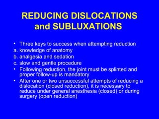
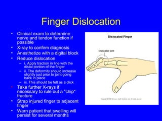
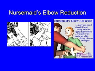
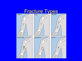
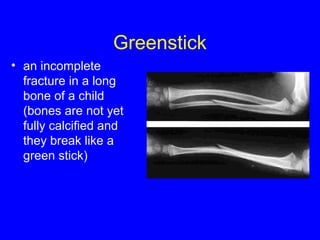
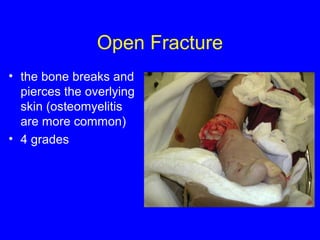
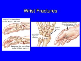
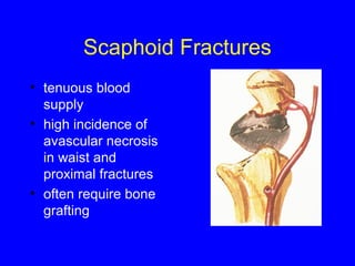
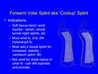
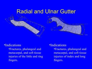
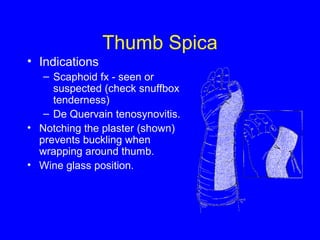
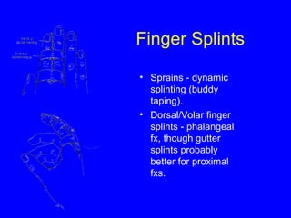
Orthopedic Injuries and 
Immobilization 
Stanford University 
Division of Emergency Medicine
History and Physical Exam 
• Immediately upon presentation with a dislocation or 
fracture, the neurovascular and circulatory status 
must be checked. 
• Attempt to ascertain the mechanism of injury. 
- may alert physician to other possibly associated 
injuries 
- as well as provide clues as to the type of injury 
involved 
• Radiographs should be obtained if fracture OR 
DISLOCATION is suspected 
• Radiographs should be obtained after reduction and 
IMMOBILIZATION of a fracture or dislocation.
How do you Describe This? 
• Named by where the 
distal articulating 
surface ends up relative 
to the proximal 
articulating surface 
• e.g. Anterior shoulder 
dislocation 
- Humeral head is anterior 
to the glenoid fossa 
Left Forearm fracture wwhhiicchh iiss DDoorrssaallllyy DDiissppllaacceedd
REDUCING DISLOCATIONS 
and SUBLUXATIONS 
• Three keys to success when attempting reduction 
a. knowledge of anatomy 
b. analgesia and sedation 
c. slow and gentle procedure 
• Following reduction, the joint must be splinted and 
proper follow-up is mandatory 
• After one or two unsuccessful attempts of reducing a 
dislocation (closed reduction), it is necessary to 
reduce under general anesthesia (closed) or during 
surgery (open reduction)
Finger Dislocation 
• Clinical exam to determine 
nerve and tendon function if 
possible 
• X-ray to confirm diagnosis 
• Anesthetize with a digital block 
• Reduce dislocation 
– i. Apply traction in line with the 
distal portion of the finger 
– ii. The deformity should increase 
slightly just prior to joint going 
back in place 
– iii. This should be felt as a click 
• Take further X-rays if 
necessary to rule out a "chip" 
fracture 
• Strap injured finger to adjacent 
finger 
• Warn patient that swelling will 
persist for several months
Shoulder Dislocation 
• Take a past medical history (i.e. 
has this happened before?) 
• Clinical exam (check for 
circumflex nerve function) 
• X-ray to rule out possible 
fracture (i.e. head of the 
humerus) 
• Several methods for reduction 
- Scapular rotation 
- Traction/counter traction
Subluxation of the Radial Head 
(Nursemaid’s Elbow) 
Definition of subluxation = a joint disruption 
in which the joint surfaces are 
maintained in some degree of 
apposition. 
Description: the radial head slips out from 
under the annular ligament. 
i. Generally caused by sudden traction of the 
forearm that extends and pronates the 
elbow (like the motion of pulling a child 
off the ground by his/her wrist). 
ii. Most common in children aging 1 - 4 years 
old, because the lip of the radial head is 
not well formed and may slip out from 
under the annular ligament with more 
ease. 
iii. Minimal pain if the arm is stationary but 
pain is felt upon flexing or supinating 
arm, (parents often think it is merely a 
sprain and wait 24 - 36 hours before 
seeking medical help) 
iv. No associated swelling, ecchymosis, or 
neurovascular deficit 
Radiography - Normal findings
Nursemaid’s Elbow Reduction
Fracture Types
Greenstick 
• an incomplete 
fracture in a long 
bone of a child 
(bones are not yet 
fully calcified and 
they break like a 
green stick)
Open Fracture 
• the bone breaks and 
pierces the overlying 
skin (osteomyelitis 
are more common) 
• 4 grades
Spiral Fracture 
• a fracture that 
spirals part of the 
length of a long 
bone
Wrist Fractures
Scaphoid Fractures 
• tenuous blood 
supply 
• high incidence of 
avascular necrosis 
in waist and 
proximal fractures 
• often require bone 
grafting
Scaphoid Fractures 
• high clinical suspicion 
even with normal x-ray 
• follow up important 
- repeat x-rays and 
early bone scan in 
patients with persistent 
pain 
• thumb spica with 
prolonged 
immobilization
Hey Kids, 
As Seen On 
TV!! 
Learn How to Splint in 
10 Easy Lessons!!!! 
Amaze Your 
Friends !!! 
WOW !!! 
Be the First on 
your Block !!!
Introduction 
• Evidence of rudimentary splints found as early as 500 
BC. 
• Used to temporarily immobilize fractures, 
dislocations, and soft tissue injuries. 
• Circumferential casts abandoned in the ED 
- increased compartment syndrome and other 
complications 
- ideal for the ED – allow swelling 
- splints easier to apply
Indications for Splinting 
• Fractures 
• Sprains 
• Joint infections 
• Tenosynovitis 
• Acute arthritis / gout 
• Lacerations over joints 
• Puncture wounds and animal 
bites of the hands or feet
Splinting Equipment 
• Plaster of Paris 
– Made from gypsum - calcium sulfate dihydrate 
– Exothermic reaction when wet - recrystallizes (can 
burn patient) 
– Warm water - faster set, but increases risk of burns 
– Fast drying - 5 - 8 minutes to set 
– Extra fast-drying - 2 - 4 minutes to set - less time to 
mold 
– Can take up to 1 day to cure (reach maximum 
strength) 
– Upper extremities - use 8-10 layers 
– Lower extremities - 12-15 layers, up to 20 if big 
person (increased risk of burn!)
Splinting Equipment 
• Ready Made Splinting Material 
– Plaster (OCL) 
• 10 -20 sheets of plaster with padding and cloth 
cover 
– Fiberglass (Orthoglass) 
• Cure rapidly (20 minutes) 
• Less messy 
• Stronger, lighter, wicks moisture better 
• Less moldable
Splinting Equipment 
• Stockinette 
• protects skin, looks nifty (often not necessary) 
• cut longer than splint 
• 2,3,4,8,10,12-in. widths 
• Padding - Webril 
• 2-3 layers, more if anticipate lots of swelling 
• Extra over elbows, heels 
• Be generous over bony prominences 
• Always pad between digits when splinting hands/feet or when 
buddy taping 
• Avoid wrinkles 
• Do not tighten - ischemia! 
• Avoid circumfrential use 
• Ace wraps
Specific Splints and Orthoses 
Upper Extremity 
• Elbow/Forearm 
– Long Arm Posterior 
– Double Sugar - Tong 
• Forearm/Wrist 
– Volar Forearm / Cockup 
– Sugar - Tong 
• Hand/Fingers 
– Ulnar Gutter 
– Radial Gutter 
– Thumb Spica 
– Finger Splints 
Lower Extremity 
• Knee 
– Knee Immobilizer / Bledsoe 
– Bulky Jones 
– Posterior Knee Splint 
• Ankle 
– Posterior Ankle 
– Stirrup 
• Foot 
– Hard Shoe
Long Arm Posterior Splint 
• Indications 
– Elbow and forearm injuries: 
– Distal humerus fx 
– Both-bone forearm fx 
– Unstable proximal radius or 
ulna fx (sugar-tong better) 
• Doesn’t completely eliminate 
supination / pronation -either 
add an anterior splint or use 
a double sugar-tong if 
complex or unstable distal 
forearm fx.
Double Sugar Tong 
• Indications 
– Elbow and forearm fx - 
prox/mid/distal radius and 
ulnar fx. 
– Better for most distal 
forearm and elbow fx 
because limits 
flex/extension and 
pronation / supination. 
10 
90
Forearm Volar Splint aka ‘Cockup’ Splint 
• Indications 
– Soft tissue hand / wrist 
injuries - sprain, carpal 
tunnel night splints, etc 
– Most wrist fx, 2nd -5th 
metacarpal fx. 
– Most add a dorsal splint for 
increased stability - 
‘sandwich splint’ (B). 
– Not used for distal radius or 
ulnar fx - can still supinate 
and pronate.
Forearm Sugar Tong 
• Indications 
– Distal radius and 
ulnar fx. 
• Prevents pronation / 
supination and 
immobilizes elbow.
Hand Splinting 
• The correct position for most hand splints 
is the position of function, a.k.a. the 
neutral position. 
• This is with the the hand in the “beer can” 
position (which may have contributed to 
the injury in the first place) : wrist slightly 
extended (10-25°) with fingers flexed as 
shown. 
• When immobilizing metacarpal neck 
fractures, the MCP joint should be flexed 
to 90°. 
• Have the patient hold an ace wrap (or a 
beer can if available) until the splint 
hardens. 
• For thumb fx, immobilize the thumb as if 
holding a wine glass.
Radial and Ulnar Gutter 
•Indications 
•Fractures, phalangeal and 
metacarpal, and soft tissue 
injuries of the little and ring 
fingers. 
•Indications 
•Fractures, phalangeal and 
metacarpal, and soft tissue 
injuries of index and long 
fingers.
Thumb Spica 
• Indications 
– Scaphoid fx - seen or 
suspected (check snuffbox 
tenderness) 
– De Quervain tenosynovitis. 
• Notching the plaster (shown) 
prevents buckling when 
wrapping around thumb. 
• Wine glass position.
Finger Splints 
• Sprains - dynamic 
splinting (buddy 
taping). 
• Dorsal/Volar finger 
splints - phalangeal 
fx, though gutter 
splints probably 
better for proximal 
fxs.
Jones Compression Dressing 
- aka Bulky Jones 
• Indications 
– Short term immobilization 
of soft tissue and 
ligamentous injuries to 
the knee or calf. 
• Allows slight flexion and 
extension - may add posterior 
knee splint to further 
immobilize the knee. 
• Procedure 
– Stockinette and 
Webril. 
– 1-2 layers of thick 
cotton padding. 
– 6 inch ace wrap.
Posterior Ankle Splint 
• Indications 
– Distal tibia/fibula fx. 
– Reduced dislocations 
– Severe sprains 
– Tarsal / metatarsal fx 
• Use at least 12-15 layers of 
plaster. 
• Adding a coaptation splint 
(stirrup) to the posterior splint 
eliminates inversion / 
eversion - especially useful 
for unstable fx and sprains.
Stirrup Splint 
• Indications 
– Similiar to posterior splint. 
– Less inversion /eversion 
and actually less plantar 
flexion compared to 
posterior splint. 
– Great for ankle sprains. 
– 12-15 layers of 4-6 inch 
plaster.
Other Orthoses 
• Knee Immobilizer 
– Semirigid brace, many models 
– Fastens with Velcro 
– Worn over clothing 
• Bledsoe Brace 
– Articulated knee brace 
– Amount of allowed flexion and extension can be adjusted 
– Used for ligamentous knee injuries and post-op 
• AirCast/ Airsplint 
– Resembles a stirrup splint with air bladders 
– Worn inside shoe 
• Hard Shoe 
– Used for foot fractures or soft tissue injuries
Complications 
• Burns 
– Thermal injury as plaster dries 
– Hot water, Increased number of 
layers, extra fast-drying, poor 
padding - all increase risk 
– If significant pain - remove splint 
to cool 
• Ischemia 
– Reduced risk compared to 
casting but still a possibility 
– Do not apply Webril and ace 
wraps tightly 
– Instruct to ice and elevate 
extremity 
– Close follow up if high risk for 
swelling, ischemia. 
– When in doubt, cut it off and look 
– Remember - pulses lost late. 
• Pressure sores 
– Smooth Webril and plaster well 
• Infection 
– Clean, debride and dress all 
wounds before splint 
application 
– Recheck if significant wound or 
increasing pain 
Any complaints of 
worsening pain - 
Take the splint off 
and look!
Questions?

Splints

  • 1.
    Orthopedic Injuries and Immobilization Stanford University Division of Emergency Medicine
  • 2.
    History and PhysicalExam • Immediately upon presentation with a dislocation or fracture, the neurovascular and circulatory status must be checked. • Attempt to ascertain the mechanism of injury. - may alert physician to other possibly associated injuries - as well as provide clues as to the type of injury involved • Radiographs should be obtained if fracture OR DISLOCATION is suspected • Radiographs should be obtained after reduction and IMMOBILIZATION of a fracture or dislocation.
  • 3.
    How do youDescribe This? • Named by where the distal articulating surface ends up relative to the proximal articulating surface • e.g. Anterior shoulder dislocation - Humeral head is anterior to the glenoid fossa Left Forearm fracture wwhhiicchh iiss DDoorrssaallllyy DDiissppllaacceedd
  • 4.
    REDUCING DISLOCATIONS andSUBLUXATIONS • Three keys to success when attempting reduction a. knowledge of anatomy b. analgesia and sedation c. slow and gentle procedure • Following reduction, the joint must be splinted and proper follow-up is mandatory • After one or two unsuccessful attempts of reducing a dislocation (closed reduction), it is necessary to reduce under general anesthesia (closed) or during surgery (open reduction)
  • 5.
    Finger Dislocation •Clinical exam to determine nerve and tendon function if possible • X-ray to confirm diagnosis • Anesthetize with a digital block • Reduce dislocation – i. Apply traction in line with the distal portion of the finger – ii. The deformity should increase slightly just prior to joint going back in place – iii. This should be felt as a click • Take further X-rays if necessary to rule out a "chip" fracture • Strap injured finger to adjacent finger • Warn patient that swelling will persist for several months
  • 6.
    Shoulder Dislocation •Take a past medical history (i.e. has this happened before?) • Clinical exam (check for circumflex nerve function) • X-ray to rule out possible fracture (i.e. head of the humerus) • Several methods for reduction - Scapular rotation - Traction/counter traction
  • 7.
    Subluxation of theRadial Head (Nursemaid’s Elbow) Definition of subluxation = a joint disruption in which the joint surfaces are maintained in some degree of apposition. Description: the radial head slips out from under the annular ligament. i. Generally caused by sudden traction of the forearm that extends and pronates the elbow (like the motion of pulling a child off the ground by his/her wrist). ii. Most common in children aging 1 - 4 years old, because the lip of the radial head is not well formed and may slip out from under the annular ligament with more ease. iii. Minimal pain if the arm is stationary but pain is felt upon flexing or supinating arm, (parents often think it is merely a sprain and wait 24 - 36 hours before seeking medical help) iv. No associated swelling, ecchymosis, or neurovascular deficit Radiography - Normal findings
  • 8.
  • 9.
  • 10.
    Greenstick • anincomplete fracture in a long bone of a child (bones are not yet fully calcified and they break like a green stick)
  • 11.
    Open Fracture •the bone breaks and pierces the overlying skin (osteomyelitis are more common) • 4 grades
  • 12.
    Spiral Fracture •a fracture that spirals part of the length of a long bone
  • 13.
  • 14.
    Scaphoid Fractures •tenuous blood supply • high incidence of avascular necrosis in waist and proximal fractures • often require bone grafting
  • 15.
    Scaphoid Fractures •high clinical suspicion even with normal x-ray • follow up important - repeat x-rays and early bone scan in patients with persistent pain • thumb spica with prolonged immobilization
  • 16.
    Hey Kids, AsSeen On TV!! Learn How to Splint in 10 Easy Lessons!!!! Amaze Your Friends !!! WOW !!! Be the First on your Block !!!
  • 17.
    Introduction • Evidenceof rudimentary splints found as early as 500 BC. • Used to temporarily immobilize fractures, dislocations, and soft tissue injuries. • Circumferential casts abandoned in the ED - increased compartment syndrome and other complications - ideal for the ED – allow swelling - splints easier to apply
  • 18.
    Indications for Splinting • Fractures • Sprains • Joint infections • Tenosynovitis • Acute arthritis / gout • Lacerations over joints • Puncture wounds and animal bites of the hands or feet
  • 19.
    Splinting Equipment •Plaster of Paris – Made from gypsum - calcium sulfate dihydrate – Exothermic reaction when wet - recrystallizes (can burn patient) – Warm water - faster set, but increases risk of burns – Fast drying - 5 - 8 minutes to set – Extra fast-drying - 2 - 4 minutes to set - less time to mold – Can take up to 1 day to cure (reach maximum strength) – Upper extremities - use 8-10 layers – Lower extremities - 12-15 layers, up to 20 if big person (increased risk of burn!)
  • 20.
    Splinting Equipment •Ready Made Splinting Material – Plaster (OCL) • 10 -20 sheets of plaster with padding and cloth cover – Fiberglass (Orthoglass) • Cure rapidly (20 minutes) • Less messy • Stronger, lighter, wicks moisture better • Less moldable
  • 21.
    Splinting Equipment •Stockinette • protects skin, looks nifty (often not necessary) • cut longer than splint • 2,3,4,8,10,12-in. widths • Padding - Webril • 2-3 layers, more if anticipate lots of swelling • Extra over elbows, heels • Be generous over bony prominences • Always pad between digits when splinting hands/feet or when buddy taping • Avoid wrinkles • Do not tighten - ischemia! • Avoid circumfrential use • Ace wraps
  • 22.
    Specific Splints andOrthoses Upper Extremity • Elbow/Forearm – Long Arm Posterior – Double Sugar - Tong • Forearm/Wrist – Volar Forearm / Cockup – Sugar - Tong • Hand/Fingers – Ulnar Gutter – Radial Gutter – Thumb Spica – Finger Splints Lower Extremity • Knee – Knee Immobilizer / Bledsoe – Bulky Jones – Posterior Knee Splint • Ankle – Posterior Ankle – Stirrup • Foot – Hard Shoe
  • 23.
    Long Arm PosteriorSplint • Indications – Elbow and forearm injuries: – Distal humerus fx – Both-bone forearm fx – Unstable proximal radius or ulna fx (sugar-tong better) • Doesn’t completely eliminate supination / pronation -either add an anterior splint or use a double sugar-tong if complex or unstable distal forearm fx.
  • 24.
    Double Sugar Tong • Indications – Elbow and forearm fx - prox/mid/distal radius and ulnar fx. – Better for most distal forearm and elbow fx because limits flex/extension and pronation / supination. 10 90
  • 25.
    Forearm Volar Splintaka ‘Cockup’ Splint • Indications – Soft tissue hand / wrist injuries - sprain, carpal tunnel night splints, etc – Most wrist fx, 2nd -5th metacarpal fx. – Most add a dorsal splint for increased stability - ‘sandwich splint’ (B). – Not used for distal radius or ulnar fx - can still supinate and pronate.
  • 26.
    Forearm Sugar Tong • Indications – Distal radius and ulnar fx. • Prevents pronation / supination and immobilizes elbow.
  • 27.
    Hand Splinting •The correct position for most hand splints is the position of function, a.k.a. the neutral position. • This is with the the hand in the “beer can” position (which may have contributed to the injury in the first place) : wrist slightly extended (10-25°) with fingers flexed as shown. • When immobilizing metacarpal neck fractures, the MCP joint should be flexed to 90°. • Have the patient hold an ace wrap (or a beer can if available) until the splint hardens. • For thumb fx, immobilize the thumb as if holding a wine glass.
  • 28.
    Radial and UlnarGutter •Indications •Fractures, phalangeal and metacarpal, and soft tissue injuries of the little and ring fingers. •Indications •Fractures, phalangeal and metacarpal, and soft tissue injuries of index and long fingers.
  • 29.
    Thumb Spica •Indications – Scaphoid fx - seen or suspected (check snuffbox tenderness) – De Quervain tenosynovitis. • Notching the plaster (shown) prevents buckling when wrapping around thumb. • Wine glass position.
  • 30.
    Finger Splints •Sprains - dynamic splinting (buddy taping). • Dorsal/Volar finger splints - phalangeal fx, though gutter splints probably better for proximal fxs.
  • 31.
    Jones Compression Dressing - aka Bulky Jones • Indications – Short term immobilization of soft tissue and ligamentous injuries to the knee or calf. • Allows slight flexion and extension - may add posterior knee splint to further immobilize the knee. • Procedure – Stockinette and Webril. – 1-2 layers of thick cotton padding. – 6 inch ace wrap.
  • 32.
    Posterior Ankle Splint • Indications – Distal tibia/fibula fx. – Reduced dislocations – Severe sprains – Tarsal / metatarsal fx • Use at least 12-15 layers of plaster. • Adding a coaptation splint (stirrup) to the posterior splint eliminates inversion / eversion - especially useful for unstable fx and sprains.
  • 33.
    Stirrup Splint •Indications – Similiar to posterior splint. – Less inversion /eversion and actually less plantar flexion compared to posterior splint. – Great for ankle sprains. – 12-15 layers of 4-6 inch plaster.
  • 34.
    Other Orthoses •Knee Immobilizer – Semirigid brace, many models – Fastens with Velcro – Worn over clothing • Bledsoe Brace – Articulated knee brace – Amount of allowed flexion and extension can be adjusted – Used for ligamentous knee injuries and post-op • AirCast/ Airsplint – Resembles a stirrup splint with air bladders – Worn inside shoe • Hard Shoe – Used for foot fractures or soft tissue injuries
  • 35.
    Complications • Burns – Thermal injury as plaster dries – Hot water, Increased number of layers, extra fast-drying, poor padding - all increase risk – If significant pain - remove splint to cool • Ischemia – Reduced risk compared to casting but still a possibility – Do not apply Webril and ace wraps tightly – Instruct to ice and elevate extremity – Close follow up if high risk for swelling, ischemia. – When in doubt, cut it off and look – Remember - pulses lost late. • Pressure sores – Smooth Webril and plaster well • Infection – Clean, debride and dress all wounds before splint application – Recheck if significant wound or increasing pain Any complaints of worsening pain - Take the splint off and look!
  • 36.