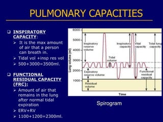
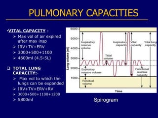
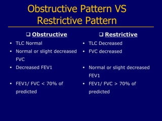
Spirometry
By: Ahmed Sohail Kothawala
Spirometry - Introduction
• SPIROMETRY:- It is a method of assessing lung function by measuring
the total volume of air the patient can expel from the lungs after a
maximal inhalation.
-Simple method for studying pulmonary ventilation
• SPIROMETER:- Instrument used to perform spirometry.
• SPIROGRAM: It is the graphical representation of changes in lung
volume under different conditions of breathing
-Pulmonary volume and capacities vary according to height, age sex
and physical training
 All volumes and capacities
 Increase in males
 Decrease with age
 Increase with height
 Increase in athletes and physically active individuals
Functioning of the Spirometer
A typical basic spirometer consists of a drum inverted over a
chamber of water, with the drum counterbalanced by a weight. In
the drum is a breathing gas, usually air or oxygen; a tube connects
the mouth with the gas chamber. When one breathes into and out
of the chamber, the drum rises and falls, and an appropriate
recording is made on a moving sheet of paper.
Why Perform Spirometry?
• Determine your lung capacity
• Measure airflow obstruction to help make a definitive diagnosis
of COPD
• Confirm presence of airway obstruction
• Assess severity of airflow obstruction in COPD
• Decide how likely it is that inhaled medications may help with
your syntoms
• Monitor disease progression in COPD
• Assess one aspect of response to therapy
• Perform pre-operative assessment
How to perform spirometry?
• For this test, you'll sit in front of a machine and be fitted
with a mouthpiece.
• It's important that the mouthpiece fits snugly so that all the
air you breathe into the machine.
• You'll also wear a nose clip to keep you from breathing air
out through your nose.
• The patient is then instructed to inhale as much as possible
and exhale rapidly & forcefully for as long as flow can be
maintained. The patient should exhale for atleast 6 seconds
PULMONARY VOLUMES
 TIDAL VOLUME :
 Volume of air inspired
or expired with each
normal breath
• Value 500ml (0.5 L)
 INSPIRATORY
RESERVE VOLUME:
 Max extra volume of
air that can be inspired
over normal tidal
volume.
• Value 3000ml (2.5-
3 L) Spirogram
PULMONARY VOLUMES
 EXPIRATORY
RESERVE VOLUME:
 Max extra volume of
air that can be expired
forcefully after the
normal tidal expiration
• Value 1100ml.
 RESIDUAL VOLUME:
 Vol of air remaining l
in the lungs after the
most forceful
expiration
• Value 1200ml. Spirogram
PULMONARY CAPACITIES
 INSPIRATORY
CAPACITY:
 It is the max amount
of air that a person
can breath in.
 Tidal vol +insp res vol
 500+3000=3500ml.
 FUNCTIONAL
RESIDUAL CAPACITY
(FRC):
 Amount of air that
remains in the lung
after normal tidal
expiration
 ERV+RV
 1100+1200=2300ml.
Spirogram
PULMONARY CAPACITIES
•VITAL CAPACITY :
 Max vol of air expired
after max insp
 IRV+TV+ERV
 3000+500+1100
 4600ml (4.5-5L)
 TOTAL LUNG
CAPACITY:-
 Max vol to which the
lungs can be expanded
 IRV+TV+ERV+RV
 3000+500+1100+1200
 5800ml Spirogram
DYNAMIC FUNCTION TEST
 Forced Vital Capacity (FVC)
 Max vol of air expired forcefully and rapidly (as hard and
as far as possible) after a full inspiration.
 Forced Expiratory Volume in first sec
 it is the volume of air expired in the first sec during the
FVC maneuver
 FEV1/FVC
 Imp in differentiating obstructive and restrictive lung
diseases.
Obstructive & Restrictive Lung
Diseases
 Obstructive Airways Disease –
where your ability to breathe out quickly is affected by
narrowing of the airways, but the amount of air you can hold
in your lungs is normal.
 Examples: Asthma, COPD, Emphysema, Cystic Fibrosis
 Restrictive Lung Disease –
where the amount of air you can breathe in is reduced
because your lungs are unable to fully expand (such as in
pulmonary fibrosis)
 Examples: Obesity, Pleural effusion, Myasthenia Gravis
Obstructive Pattern VS
Restrictive Pattern
 Obstructive
 TLC Normal
 Normal or slight decreased
FVC
 Decreased FEV1
 FEV1/ FVC < 70% of
predicted
 Restrictive
 TLC Decreased
 FVC decreased
 Normal or slight decreased
FEV1
 FEV1/ FVC > 70% of
predicted
Disadvantages of Spirometry
 Feel Dizzy
 Feel lightheaded
 Feel tired
 coughing
THANK YOU

Spirometry - Ahmed Sohail.ppt

  • 1.
  • 2.
    Spirometry - Introduction •SPIROMETRY:- It is a method of assessing lung function by measuring the total volume of air the patient can expel from the lungs after a maximal inhalation. -Simple method for studying pulmonary ventilation • SPIROMETER:- Instrument used to perform spirometry. • SPIROGRAM: It is the graphical representation of changes in lung volume under different conditions of breathing -Pulmonary volume and capacities vary according to height, age sex and physical training  All volumes and capacities  Increase in males  Decrease with age  Increase with height  Increase in athletes and physically active individuals
  • 3.
    Functioning of theSpirometer A typical basic spirometer consists of a drum inverted over a chamber of water, with the drum counterbalanced by a weight. In the drum is a breathing gas, usually air or oxygen; a tube connects the mouth with the gas chamber. When one breathes into and out of the chamber, the drum rises and falls, and an appropriate recording is made on a moving sheet of paper.
  • 4.
    Why Perform Spirometry? •Determine your lung capacity • Measure airflow obstruction to help make a definitive diagnosis of COPD • Confirm presence of airway obstruction • Assess severity of airflow obstruction in COPD • Decide how likely it is that inhaled medications may help with your syntoms • Monitor disease progression in COPD • Assess one aspect of response to therapy • Perform pre-operative assessment
  • 5.
    How to performspirometry? • For this test, you'll sit in front of a machine and be fitted with a mouthpiece. • It's important that the mouthpiece fits snugly so that all the air you breathe into the machine. • You'll also wear a nose clip to keep you from breathing air out through your nose. • The patient is then instructed to inhale as much as possible and exhale rapidly & forcefully for as long as flow can be maintained. The patient should exhale for atleast 6 seconds
  • 6.
    PULMONARY VOLUMES  TIDALVOLUME :  Volume of air inspired or expired with each normal breath • Value 500ml (0.5 L)  INSPIRATORY RESERVE VOLUME:  Max extra volume of air that can be inspired over normal tidal volume. • Value 3000ml (2.5- 3 L) Spirogram
  • 7.
    PULMONARY VOLUMES  EXPIRATORY RESERVEVOLUME:  Max extra volume of air that can be expired forcefully after the normal tidal expiration • Value 1100ml.  RESIDUAL VOLUME:  Vol of air remaining l in the lungs after the most forceful expiration • Value 1200ml. Spirogram
  • 8.
    PULMONARY CAPACITIES  INSPIRATORY CAPACITY: It is the max amount of air that a person can breath in.  Tidal vol +insp res vol  500+3000=3500ml.  FUNCTIONAL RESIDUAL CAPACITY (FRC):  Amount of air that remains in the lung after normal tidal expiration  ERV+RV  1100+1200=2300ml. Spirogram
  • 9.
    PULMONARY CAPACITIES •VITAL CAPACITY:  Max vol of air expired after max insp  IRV+TV+ERV  3000+500+1100  4600ml (4.5-5L)  TOTAL LUNG CAPACITY:-  Max vol to which the lungs can be expanded  IRV+TV+ERV+RV  3000+500+1100+1200  5800ml Spirogram
  • 10.
    DYNAMIC FUNCTION TEST Forced Vital Capacity (FVC)  Max vol of air expired forcefully and rapidly (as hard and as far as possible) after a full inspiration.  Forced Expiratory Volume in first sec  it is the volume of air expired in the first sec during the FVC maneuver  FEV1/FVC  Imp in differentiating obstructive and restrictive lung diseases.
  • 11.
    Obstructive & RestrictiveLung Diseases  Obstructive Airways Disease – where your ability to breathe out quickly is affected by narrowing of the airways, but the amount of air you can hold in your lungs is normal.  Examples: Asthma, COPD, Emphysema, Cystic Fibrosis  Restrictive Lung Disease – where the amount of air you can breathe in is reduced because your lungs are unable to fully expand (such as in pulmonary fibrosis)  Examples: Obesity, Pleural effusion, Myasthenia Gravis
  • 12.
    Obstructive Pattern VS RestrictivePattern  Obstructive  TLC Normal  Normal or slight decreased FVC  Decreased FEV1  FEV1/ FVC < 70% of predicted  Restrictive  TLC Decreased  FVC decreased  Normal or slight decreased FEV1  FEV1/ FVC > 70% of predicted
  • 13.
    Disadvantages of Spirometry Feel Dizzy  Feel lightheaded  Feel tired  coughing
  • 14.