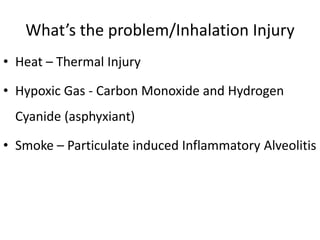
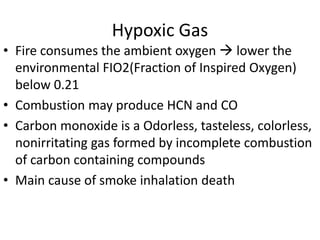
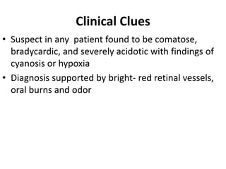
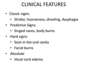
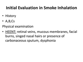
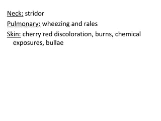
Smoke Inhalation
Rahul AP
BPT,MPT (CRD&ICU)
Asst Proff: LIAHS
Kannur,Kerala
Definition
• Smoke inhalation injury refers to an injury due
to inhalation or exposure to hot gaseous products
of combustion.
• This can cause serious respiratory complications.
• Smoke inhalation is the primary cause of death
for victims of indoor fires.
What’s the problem/Inhalation Injury
• Heat – Thermal Injury
• Hypoxic Gas - Carbon Monoxide and Hydrogen
Cyanide (asphyxiant)
• Smoke – Particulate induced Inflammatory Alveolitis
Thermal Injury
• Obviously patients may have massive burn injury
with or without trauma
• Heat causes erythema, ulceration and edema
– It May progress to significant upper airway edema
and obstruction
– Usually it resolves in 3-5 days
– If external face, neck burns are present this may
create airway difficulties
• Heat inhalation cause damage above the carina
• Hoarseness will be present (abnormal voice changes
and difficulty in making sounds when trying to
speak)Vocal sounds may be weak, breathy, scratchy,
or husky, and the pitch or quality of the voice may
change
• Stridor- harsh vibrating noise when breathing,
caused by obstruction of the windpipe or larynx.
• Dry Heat (from house fire) it has very poor
conveyance of heat beyond vocal cords
• Flash burns usually only singe nasal hair and
mucosa
Hypoxic Gas
• Fire consumes the ambient oxygen  lower the
environmental FIO2(Fraction of Inspired Oxygen)
below 0.21
• Combustion may produce HCN and CO
• Carbon monoxide is a Odorless, tasteless, colorless,
nonirritating gas formed by incomplete combustion
of carbon containing compounds
• Main cause of smoke inhalation death
• Cyanide is Formed when polyurethane(seen in
flexible foam), acrylonitrile(seen in plastic),
nylon, wool and cotton materials when burnd
Smoke/Particulate matter
• Smoke is visible hot air and it contain small
particulate matter and toxic gas, soot and ash,
dust, pollen etc….
• It Causes obstruction and sloughing in the small
airways leading to atelectasis
• Bronchospasm – due to direct toxic effects
• Asphyxiants: (fromed from burning synthetic
substances)when Inhaled cause injury to the
lower airway
• ARDS Acute respiratory distress syndrome
• Low molecular weight toxins in smoke alter
the pH and create free radicals in distal airways
causing tissue destruction, mainly nitrous oxide
• Acute neutrophilic airway inflammation occurs,
but symptoms may be delayed 12-36 hours
• Progression to pulmonary edema,
bronchopulmonary Pneumonia /or ARDs
Physiologic Changes
• Destruction of ciliated epithelium in trachea and
bronchial tree
• Increased vascular permeability causing
pulmonary edema
• Alveolar hemorrhage & hyaline membrane
formation
• Airway obstruction facilitates distal airway collapse
and atelectasis
Clinical Clues
• Suspect in any patient found to be comatose,
bradycardic, and severely acidotic with findings of
cyanosis or hypoxia
• Diagnosis supported by bright- red retinal vessels,
oral burns and odor
CLINICAL FEATURES
• Classic signs:
• Stridor, hoarseness, drooling, dysphagia
• Predictive Signs:
• Singed nares, body burns
• Hard signs:
• Soot in the oral cavity
• Facial burns
• Absolute
• Vocal cord edema
Initial Evaluation in Smoke Inhalation
• History
• A,B,Cs
Physical examination
• HEENT: retinal veins, mucous membranes, facial
burns, singed nasal hairs or presence of
carbonaceous sputum, dysphonia
Neck: stridor
Pulmonary: wheezing and rales
Skin: cherry red discoloration, burns, chemical
exposures, bullae
Diagnostic Studies for
Inhalation Injury
• CXR
• ABG with co-oximetry (A decreased partial
pressure of arterial oxygen&fraction of inspired
oxygen (PaO2&FIO2 )
• Pulse oximetry check (COHb level)
• Upper endoscopy
• Bronchoscopy
• Electrolytes
• CT
CXR is a Poor Indicator of Inhalation Injury
• 56% of patients had normal chest xrays
• Most frequent abnormal findings is diffuse
alveolar infiltrates (35%)
• Focal abnormalities, consolidation or atelectasis,
12.5% on admission and 25.6% on day 2
• Other studies have shown (41%) of patients with
acute inhalation injury and burns to have normal
chest radiographs (Wittram, 1994).
ABG & Fluid Requirements
• A decreased partial pressure of arterial oxygen and
fraction of inspired oxygen (PaO2:FIO2 ratio) less
than 100 is very sever
• Indicates increased fluid needs
• Bronchoscopy
• Invasive, and limited access at some cases
• Useful in evaluating bacterial contamination and
disease progression
CT scans
• CT scans at 6, 12, 24 hours
• Hyperinflated fraction of abnormal lung tissue
were present
Management
• Humidified Oxygen (high flow 100 % Oxygen)
• Intubate if necessary
• Stridor
• Respiratory distress
• Hypoxia or hypercapnea
• Deep burns to face or neck
• Blistering or edema in oropharynx
–Place a large ETT tube
• Help manage secretions and debris
• Hyperbaric Oxygen
• Bronchodilators (IV or nebulized)
• Mucolytics and/or expectorants
• Intubation, mechanical ventilation
• IV fluid therapy
• Antibiotics
• Supportive care with treatment of burns
• “Lily kit” or cyanide antidote kit
• Amyl nitrite, IV sodium nitrite and IV sodium
thiosulfate
Thank You

Smoke Inhalation Physiotherapy Management

  • 1.
    Smoke Inhalation Rahul AP BPT,MPT(CRD&ICU) Asst Proff: LIAHS Kannur,Kerala
  • 2.
    Definition • Smoke inhalationinjury refers to an injury due to inhalation or exposure to hot gaseous products of combustion. • This can cause serious respiratory complications. • Smoke inhalation is the primary cause of death for victims of indoor fires.
  • 3.
    What’s the problem/InhalationInjury • Heat – Thermal Injury • Hypoxic Gas - Carbon Monoxide and Hydrogen Cyanide (asphyxiant) • Smoke – Particulate induced Inflammatory Alveolitis
  • 4.
    Thermal Injury • Obviouslypatients may have massive burn injury with or without trauma • Heat causes erythema, ulceration and edema – It May progress to significant upper airway edema and obstruction – Usually it resolves in 3-5 days – If external face, neck burns are present this may create airway difficulties
  • 5.
    • Heat inhalationcause damage above the carina • Hoarseness will be present (abnormal voice changes and difficulty in making sounds when trying to speak)Vocal sounds may be weak, breathy, scratchy, or husky, and the pitch or quality of the voice may change • Stridor- harsh vibrating noise when breathing, caused by obstruction of the windpipe or larynx.
  • 6.
    • Dry Heat(from house fire) it has very poor conveyance of heat beyond vocal cords • Flash burns usually only singe nasal hair and mucosa
  • 7.
    Hypoxic Gas • Fireconsumes the ambient oxygen  lower the environmental FIO2(Fraction of Inspired Oxygen) below 0.21 • Combustion may produce HCN and CO • Carbon monoxide is a Odorless, tasteless, colorless, nonirritating gas formed by incomplete combustion of carbon containing compounds • Main cause of smoke inhalation death
  • 8.
    • Cyanide isFormed when polyurethane(seen in flexible foam), acrylonitrile(seen in plastic), nylon, wool and cotton materials when burnd
  • 9.
    Smoke/Particulate matter • Smokeis visible hot air and it contain small particulate matter and toxic gas, soot and ash, dust, pollen etc…. • It Causes obstruction and sloughing in the small airways leading to atelectasis • Bronchospasm – due to direct toxic effects • Asphyxiants: (fromed from burning synthetic substances)when Inhaled cause injury to the lower airway • ARDS Acute respiratory distress syndrome
  • 10.
    • Low molecularweight toxins in smoke alter the pH and create free radicals in distal airways causing tissue destruction, mainly nitrous oxide • Acute neutrophilic airway inflammation occurs, but symptoms may be delayed 12-36 hours • Progression to pulmonary edema, bronchopulmonary Pneumonia /or ARDs
  • 11.
    Physiologic Changes • Destructionof ciliated epithelium in trachea and bronchial tree • Increased vascular permeability causing pulmonary edema • Alveolar hemorrhage & hyaline membrane formation • Airway obstruction facilitates distal airway collapse and atelectasis
  • 12.
    Clinical Clues • Suspectin any patient found to be comatose, bradycardic, and severely acidotic with findings of cyanosis or hypoxia • Diagnosis supported by bright- red retinal vessels, oral burns and odor
  • 13.
    CLINICAL FEATURES • Classicsigns: • Stridor, hoarseness, drooling, dysphagia • Predictive Signs: • Singed nares, body burns • Hard signs: • Soot in the oral cavity • Facial burns • Absolute • Vocal cord edema
  • 14.
    Initial Evaluation inSmoke Inhalation • History • A,B,Cs Physical examination • HEENT: retinal veins, mucous membranes, facial burns, singed nasal hairs or presence of carbonaceous sputum, dysphonia
  • 15.
    Neck: stridor Pulmonary: wheezingand rales Skin: cherry red discoloration, burns, chemical exposures, bullae
  • 16.
    Diagnostic Studies for InhalationInjury • CXR • ABG with co-oximetry (A decreased partial pressure of arterial oxygen&fraction of inspired oxygen (PaO2&FIO2 ) • Pulse oximetry check (COHb level) • Upper endoscopy • Bronchoscopy • Electrolytes • CT
  • 17.
    CXR is aPoor Indicator of Inhalation Injury • 56% of patients had normal chest xrays • Most frequent abnormal findings is diffuse alveolar infiltrates (35%) • Focal abnormalities, consolidation or atelectasis, 12.5% on admission and 25.6% on day 2 • Other studies have shown (41%) of patients with acute inhalation injury and burns to have normal chest radiographs (Wittram, 1994).
  • 18.
    ABG & FluidRequirements • A decreased partial pressure of arterial oxygen and fraction of inspired oxygen (PaO2:FIO2 ratio) less than 100 is very sever • Indicates increased fluid needs
  • 19.
    • Bronchoscopy • Invasive,and limited access at some cases • Useful in evaluating bacterial contamination and disease progression
  • 20.
    CT scans • CTscans at 6, 12, 24 hours • Hyperinflated fraction of abnormal lung tissue were present
  • 21.
    Management • Humidified Oxygen(high flow 100 % Oxygen) • Intubate if necessary • Stridor • Respiratory distress • Hypoxia or hypercapnea • Deep burns to face or neck • Blistering or edema in oropharynx –Place a large ETT tube • Help manage secretions and debris
  • 22.
    • Hyperbaric Oxygen •Bronchodilators (IV or nebulized) • Mucolytics and/or expectorants • Intubation, mechanical ventilation • IV fluid therapy • Antibiotics • Supportive care with treatment of burns • “Lily kit” or cyanide antidote kit • Amyl nitrite, IV sodium nitrite and IV sodium thiosulfate
  • 23.