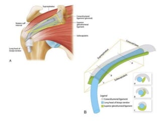
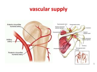
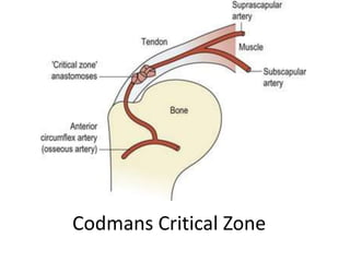
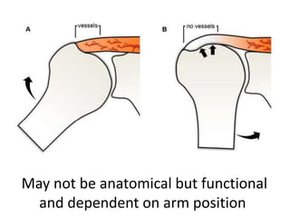
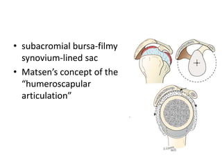
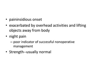
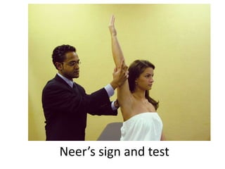
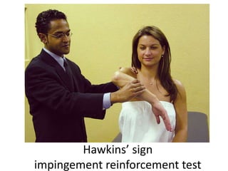
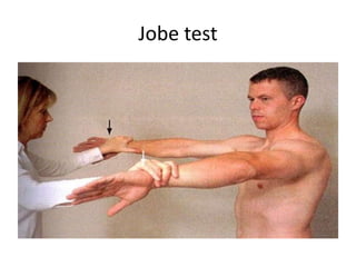
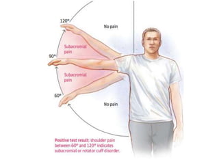
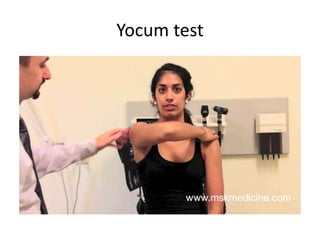
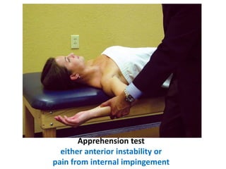
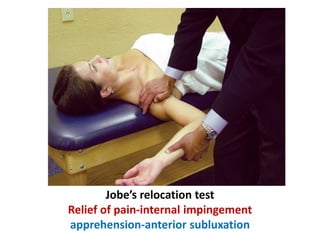
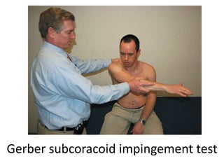
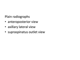
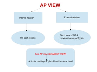
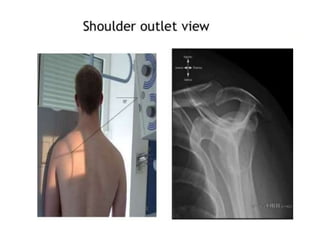
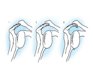
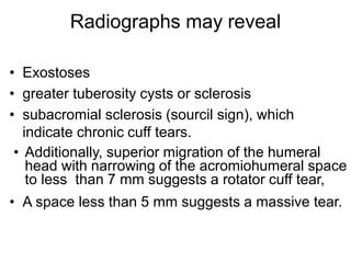
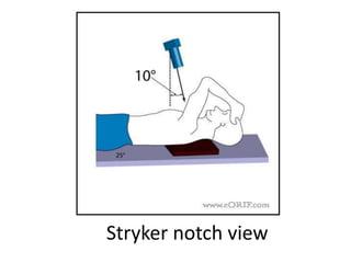
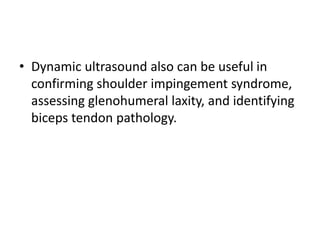
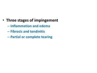
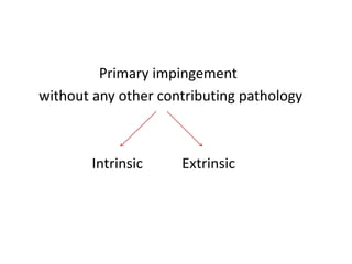
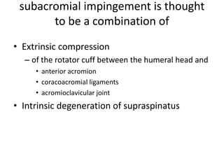
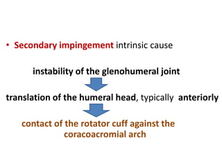
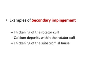
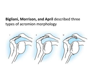
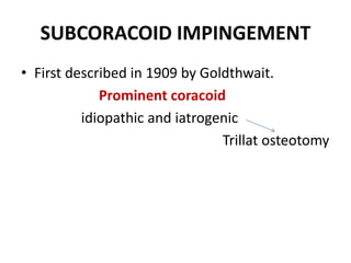
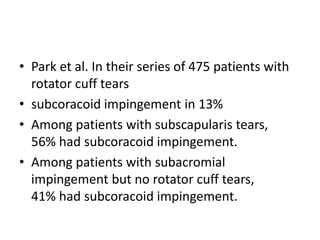
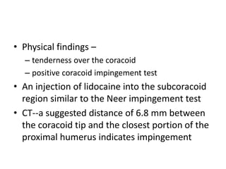
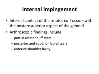
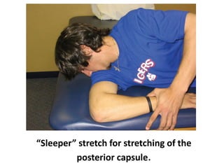
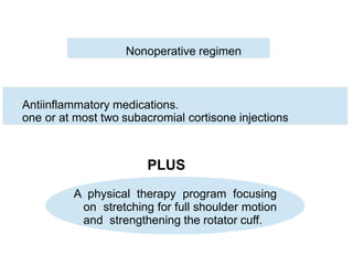
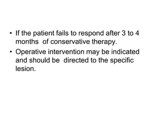
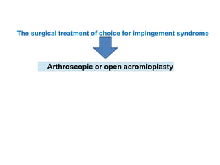
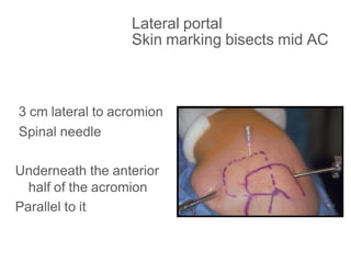
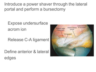
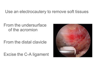
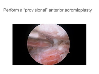
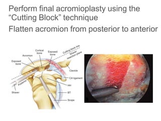
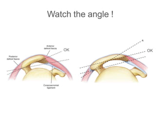
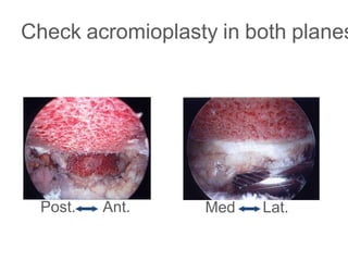
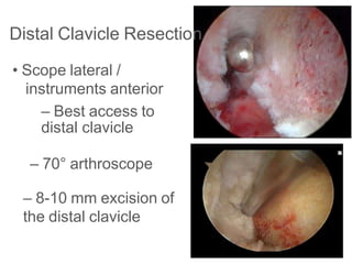
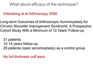
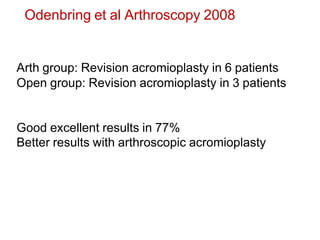
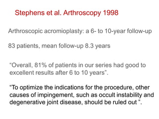
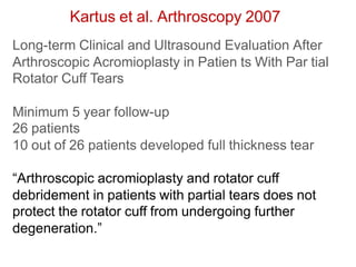
Shoulder impingement syndrome
By Praveen Kumar Reddy Gorantla
DNB trainee
Lisie hospital
• First description of subacromial bursitis in
1867—Jarjavay.
• Codman, in 1931--inability to abduct the arm
had incomplete or complete ruptures of the
supraspinatus tendon, rather than primary
bursal problems.
• The concept of outlet impingement first
elucidated by Meyer in 1937
In 1972, Neer described impingement syndrome
• characterized by a ridge of proliferative spurs and
excrescences on the undersurface of the anterior
process of the acromion, apparently caused by
repeated impingement of the rotator cuff and the
humeral head with traction of the coracoacromial
ligament.
• Anterior third of the acromion and its anterior lip
seemed to be the offending structure in most cases
• Impingement test
• Neer eventually came to argue that a vast
majority, if not all, lesions of the rotator cuff
were due to subacromial impingement.
• The debate of extrinsic impingement versus
intrinsic degeneration as the etiology of
rotator cuff tears continues to this day.
• Most mobile joint in the body
• The cuff envelops and blends with the
glenohumeral capsule on all sides except at
the redundant inferior pouch.
• The tendon of the long head of the biceps-
“fifth tendon” of the cuff-intraarticular but
extrasynovial structure-passes deep in the
“rotator interval”
The rotator interval
• The superficial roof of the rotator interval is
the coracohumeral (CH) ligament and the floor
of the interval is the superior glenohumeral
ligament (SGHL).
• suspensory structure for the humeral head
vascular supply
Codmans Critical Zone
May not be anatomical but functional
and dependent on arm position
• subacromial bursa-filmy
synovium-lined sac
• Matsen’s concept of the
“humeroscapular
articulation”
function primarily as
a stabilizer
• paininsidious onset
• exacerbated by overhead activities and lifting
objects away from body
• night pain
– poor indicator of successful nonoperative
management
• Strength--usually normal
Neer’s sign and test
Hawkins’ sign
impingement reinforcement test
Jobe test
Yocum test
Apprehension test
either anterior instability or
pain from internal impingement
Jobe’s relocation test
Relief of pain-internal impingement
apprehension-anterior subluxation
Gerber subcoracoid impingement test
Plain radiographs
• anteroposterior view
• axillary lateral view
• supraspinatus outlet view
AP VIEW
Internal rotation External rotation
Hill sach lesions
Good view of GT &
proximal humerusphysis
Ture AP view (GRASHEY VIEW)
Articular cartilage of glenoid and humeral head
Radiographs may reveal
• Exostoses
• greater tuberosity cysts or sclerosis
• subacromial sclerosis (sourcil sign), which
indicate chronic cuff tears.
• Additionally, superior migration of the humeral
head with narrowing of the acromiohumeral space
to less than 7 mm suggests a rotator cuff tear,
• A space less than 5 mm suggests a massive tear.
Stryker notch view
ARTHROGRAM
• Traditionally, an arthrogram has been used to
document full- thickness rotator cuff tears.
• Leakage of contrast material into the
subacromial and subdeltoid spaces after
injection into the glenohumeral joint indicates
a full-thickness tear.
• Arthrography is still useful for patients
in whom MRI is contraindicated
• Arthrography can be combined with MRI
to improve the diagnostic accuracy
MRI
• A patient with symptoms of
subacromial impingement may show
increased signal in the supraspinatus
tendon on T2-weighted MRI consistent
with tendinopathy;
• Increased fluid in the subacromial
bursa also is a sign of subacromial
impingement.
• Dynamic ultrasound also can be useful in
confirming shoulder impingement syndrome,
assessing glenohumeral laxity, and identifying
biceps tendon pathology.
• Three stages of impingement
– Inflammation and edema
– Fibrosis and tendinitis
– Partial or complete tearing
Developmental Stages of
Impingement Syndrome
Stage 1: Edema and Hemorrhage
• Typical age of patient: <25 years old
• Differential diagnosis:
– subluxation
– AC joint arthritis
• Clinical course: reversible
• Treatment: conservative
Stage 2: Fibrosis and Tendinitis
• Typical age of patient: 25 to 40 years old
• Differential diagnosis:
– frozen shoulder
– calcium deposits
• Clinical course: recurrent pain with activity
• Treatment: consider bursectomy or division of
coracoacromial ligament
Stage 3: Bone Spurs and Tendon Rupture
• Typical age of patient: >40 years old
• Differential diagnosis:
– cervical radiculitis
– neoplasm
• Clinical course: progressive disability
• Treatment: anterior acromioplasty, rotator
cuff repair
Four types of impingement
• Primary impingement
• Secondary impingement
• subcoracoid impingement
• Internal impingement
Primary impingement
without any other contributing pathology
Intrinsic Extrinsic
subacromial impingement is thought
to be a combination of
• Extrinsic compression
– of the rotator cuff between the humeral head and
• anterior acromion
• coracoacromial ligaments
• acromioclavicular joint
• Intrinsic degeneration of supraspinatus
• Secondary impingement intrinsic cause
instability of the glenohumeral joint
translation of the humeral head, typically anteriorly
contact of the rotator cuff against the
coracoacromial arch
• Examples of Secondary impingement
– Thickening of the rotator cuff
– Calcium deposits within the rotator cuff
– Thickening of the subacromial bursa
Bigliani, Morrison, and April described three
types of acromion morphology
Associated conditionshook-shaped acromion
• os acromiale
• posterior capsular contracture
• scapular dyskinesia
• tuberosity fracture malunion
• Instability
os acromiale
SUBCORACOID IMPINGEMENT
• First described in 1909 by Goldthwait.
Prominent coracoid
idiopathic and iatrogenic
Trillat osteotomy
• Park et al. In their series of 475 patients with
rotator cuff tears
• subcoracoid impingement in 13%
• Among patients with subscapularis tears,
56% had subcoracoid impingement.
• Among patients with subacromial
impingement but no rotator cuff tears,
41% had subcoracoid impingement.
• Physical findings –
– tenderness over the coracoid
– positive coracoid impingement test
• An injection of lidocaine into the subcoracoid
region similar to the Neer impingement test
• CT--a suggested distance of 6.8 mm between
the coracoid tip and the closest portion of the
proximal humerus indicates impingement
Gerber subcoracoid impingement test
Internal impingement
• Internal contact of the rotator cuff occurs with
the posterosuperior aspect of the glenoid
• Arthroscopic findings include
– partial rotator cuff tears
– posterior and superior labral tears
– anterior shoulder laxity
“Sleeper” stretch for stretching of the
posterior capsule.
TREATMENT
Nonoperative regimen
Antiinflammatory medications.
one or at most two subacromial cortisone injections
PLUS
A physical therapy program focusing
on stretching for full shoulder motion
and strengthening the rotator cuff.
• If the patient fails to respond after 3 to 4
months of conservative therapy.
• Operative intervention may be indicated
and should be directed to the specific
lesion.
The surgical treatment of choice for impingement syndrome
Arthroscopic or open acromioplasty
End-point Assessment
CA ligament released?
Acromion flat in A-P plane?
Acromion flat in M-L plane?
AC joint inspected?
Rotator cuff inspected?
Bony Landmarks
ACJ
Coracoid
Acromion
Posterior portal
Same skin
incision for GHJ
arthroscopy
Redirect cannula
Aim and advance the scope
beneath the anteriolateral
corner
Room with a view
Clear visualization
4 Walls ,
floor,
ceiling
CA ligament is landmark
Lateral portal
Skin marking bisects mid AC
3 cm lateral to acromion
Spinal needle
Underneath the anterior
half of the acromion
Parallel to it
Introduce a power shaver through the lateral
portal and perform a bursectomy
Expose undersurface
acrom ion
Release C-A ligament
Define anterior & lateral
edges
Use an electrocautery to remove soft tissues
From the undersurface
of the acromion
From the distal clavicle
Excise the C-A ligament
Perform a “provisional” anterior acromioplasty
Perform final acromioplasty using the
“Cutting Block” technique
Flatten acromion from posterior to anterior
Watch the angle !
ΟΚ ΟΚ
Check acromioplasty in both planes
Post. Ant. Med. Lat.
Distal Clavicle Resection
• Scope lateral /
instruments anterior
– Best access to
distal clavicle
– 70° arthroscope
– 8-10 mm excision of
the distal clavicle
What about efficacy of the technique?
Odenbring et al Arthroscopy 2008
Long-term Outcomes of Arthroscopic Acromioplasty for
Chronic Shoulder Impingement Syndrome: A Prospective
Cohort Study With a Minimum of 12 Years' Follow-up
31 patients
12-14 years follow-up
29 patients (open acromioplasty) as a control group
No full thickness cuff tears
Odenbring et al Arthroscopy 2008
Arth group: Revision acromioplasty in 6 patients
Open group: Revision acromioplasty in 3 patients
Good excellent results in 77%
Better results with arthroscopic acromioplasty
Stephens et al. Arthroscopy 1998
Arthroscopic acromioplasty: a 6- to 10-year follow-up
83 patients, mean follow-up 8.3 years
“Overall, 81% of patients in our series had good to
excellent results after 6 to 10 years”.
“To optimize the indications for the procedure, other
causes of impingement, such as occult instability and
degenerative joint disease, should be ruled out ”.
Kartus et al. Arthroscopy 2007
Long-term Clinical and Ultrasound Evaluation After
Arthroscopic Acromioplasty in Patien ts With Par tial
Rotator Cuff Tears
Minimum 5 year follow-up
26 patients
10 out of 26 patients developed full thickness tear
“Arthroscopic acromioplasty and rotator cuff
debridement in patients with partial tears does not
protect the rotator cuff from undergoing further
degeneration.”
Chahal et al. Arthroscopy 2012
The Ro le of Subacromial Decompression in Patien ts
Undergoing Arthroscopic Repair of Full-Thickness Tears
the Rotator Cuff: A Systematic Review and Meta-analysi
“On the basis of the currently available literature,
there is no statistically significant difference in
subjective outcome after arthroscopic rotator cuff
repair with or without acromioplasty at intermediate
follow-up.”
Complications after
acromioplasty
Not limited to infection
i. Seroma formation,
ii. Hematoma
iii.Synovial fistula
iv.Biceps rupture
v.Pulmonary embolus
vi.Acromial fracture
Familiari et al. J Orthop Traum atol 2012
Is acromioplasty necessary in the setting of full-thickness
rotator cuff tears? A systematic review.
• 354 patients: SAD and Scope Cuff Repair
• 4 Studies: 2 Level I and 2 Level II
• Conlusions:
– “does not support the routine use of partial
acromioplasty or CA ligament release in the
surgical treatment of rotator cuff disease”
– “in some instances, partial acromioplasty and
release of the CA ligament can result in anterior
escape and worsening symptoms”
Clement et al. Arthroscopy 2015 (September)
Short-Term Outcome After Arthroscopic Bursectomy
Debridement of Rotator Cuff Calcific Tendonopathy Wit
and Without Subacromial Decompression: A Prospectiv
Randomized Controlled Tr ial
13 MONTHS FOLLOW-UP
“Functional outcome of patients with calcific
tendonitis after arthroscopic bursectomy and
debridement of the calcific deposit is not influenced if
performed in combination with or without a
subacromial decompression.”
“The emphasis of treatment is shifting from that of
decompression to restoring the health of the rotator cuff ”
F. Fu, 1991

Shoulder impingement syndrome

  • 1.
    Shoulder impingement syndrome ByPraveen Kumar Reddy Gorantla DNB trainee Lisie hospital
  • 2.
    • First descriptionof subacromial bursitis in 1867—Jarjavay. • Codman, in 1931--inability to abduct the arm had incomplete or complete ruptures of the supraspinatus tendon, rather than primary bursal problems. • The concept of outlet impingement first elucidated by Meyer in 1937
  • 3.
    In 1972, Neerdescribed impingement syndrome • characterized by a ridge of proliferative spurs and excrescences on the undersurface of the anterior process of the acromion, apparently caused by repeated impingement of the rotator cuff and the humeral head with traction of the coracoacromial ligament. • Anterior third of the acromion and its anterior lip seemed to be the offending structure in most cases • Impingement test
  • 4.
    • Neer eventuallycame to argue that a vast majority, if not all, lesions of the rotator cuff were due to subacromial impingement. • The debate of extrinsic impingement versus intrinsic degeneration as the etiology of rotator cuff tears continues to this day.
  • 5.
    • Most mobilejoint in the body • The cuff envelops and blends with the glenohumeral capsule on all sides except at the redundant inferior pouch. • The tendon of the long head of the biceps- “fifth tendon” of the cuff-intraarticular but extrasynovial structure-passes deep in the “rotator interval”
  • 6.
    The rotator interval •The superficial roof of the rotator interval is the coracohumeral (CH) ligament and the floor of the interval is the superior glenohumeral ligament (SGHL). • suspensory structure for the humeral head
  • 11.
  • 12.
  • 13.
    May not beanatomical but functional and dependent on arm position
  • 14.
    • subacromial bursa-filmy synovium-linedsac • Matsen’s concept of the “humeroscapular articulation”
  • 15.
  • 16.
    • paininsidious onset •exacerbated by overhead activities and lifting objects away from body • night pain – poor indicator of successful nonoperative management • Strength--usually normal
  • 17.
  • 18.
  • 19.
  • 21.
  • 22.
    Apprehension test either anteriorinstability or pain from internal impingement
  • 23.
    Jobe’s relocation test Reliefof pain-internal impingement apprehension-anterior subluxation
  • 24.
  • 25.
    Plain radiographs • anteroposteriorview • axillary lateral view • supraspinatus outlet view
  • 26.
    AP VIEW Internal rotationExternal rotation Hill sach lesions Good view of GT & proximal humerusphysis Ture AP view (GRASHEY VIEW) Articular cartilage of glenoid and humeral head
  • 29.
    Radiographs may reveal •Exostoses • greater tuberosity cysts or sclerosis • subacromial sclerosis (sourcil sign), which indicate chronic cuff tears. • Additionally, superior migration of the humeral head with narrowing of the acromiohumeral space to less than 7 mm suggests a rotator cuff tear, • A space less than 5 mm suggests a massive tear.
  • 30.
  • 33.
    ARTHROGRAM • Traditionally, anarthrogram has been used to document full- thickness rotator cuff tears. • Leakage of contrast material into the subacromial and subdeltoid spaces after injection into the glenohumeral joint indicates a full-thickness tear. • Arthrography is still useful for patients in whom MRI is contraindicated • Arthrography can be combined with MRI to improve the diagnostic accuracy
  • 34.
    MRI • A patientwith symptoms of subacromial impingement may show increased signal in the supraspinatus tendon on T2-weighted MRI consistent with tendinopathy; • Increased fluid in the subacromial bursa also is a sign of subacromial impingement.
  • 38.
    • Dynamic ultrasoundalso can be useful in confirming shoulder impingement syndrome, assessing glenohumeral laxity, and identifying biceps tendon pathology.
  • 39.
    • Three stagesof impingement – Inflammation and edema – Fibrosis and tendinitis – Partial or complete tearing
  • 40.
    Developmental Stages of ImpingementSyndrome Stage 1: Edema and Hemorrhage • Typical age of patient: <25 years old • Differential diagnosis: – subluxation – AC joint arthritis • Clinical course: reversible • Treatment: conservative
  • 41.
    Stage 2: Fibrosisand Tendinitis • Typical age of patient: 25 to 40 years old • Differential diagnosis: – frozen shoulder – calcium deposits • Clinical course: recurrent pain with activity • Treatment: consider bursectomy or division of coracoacromial ligament
  • 42.
    Stage 3: BoneSpurs and Tendon Rupture • Typical age of patient: >40 years old • Differential diagnosis: – cervical radiculitis – neoplasm • Clinical course: progressive disability • Treatment: anterior acromioplasty, rotator cuff repair
  • 43.
    Four types ofimpingement • Primary impingement • Secondary impingement • subcoracoid impingement • Internal impingement
  • 44.
    Primary impingement without anyother contributing pathology Intrinsic Extrinsic
  • 45.
    subacromial impingement isthought to be a combination of • Extrinsic compression – of the rotator cuff between the humeral head and • anterior acromion • coracoacromial ligaments • acromioclavicular joint • Intrinsic degeneration of supraspinatus
  • 46.
    • Secondary impingementintrinsic cause instability of the glenohumeral joint translation of the humeral head, typically anteriorly contact of the rotator cuff against the coracoacromial arch
  • 47.
    • Examples ofSecondary impingement – Thickening of the rotator cuff – Calcium deposits within the rotator cuff – Thickening of the subacromial bursa
  • 48.
    Bigliani, Morrison, andApril described three types of acromion morphology
  • 49.
    Associated conditionshook-shaped acromion •os acromiale • posterior capsular contracture • scapular dyskinesia • tuberosity fracture malunion • Instability
  • 51.
  • 52.
    SUBCORACOID IMPINGEMENT • Firstdescribed in 1909 by Goldthwait. Prominent coracoid idiopathic and iatrogenic Trillat osteotomy
  • 53.
    • Park etal. In their series of 475 patients with rotator cuff tears • subcoracoid impingement in 13% • Among patients with subscapularis tears, 56% had subcoracoid impingement. • Among patients with subacromial impingement but no rotator cuff tears, 41% had subcoracoid impingement.
  • 54.
    • Physical findings– – tenderness over the coracoid – positive coracoid impingement test • An injection of lidocaine into the subcoracoid region similar to the Neer impingement test • CT--a suggested distance of 6.8 mm between the coracoid tip and the closest portion of the proximal humerus indicates impingement
  • 55.
  • 56.
    Internal impingement • Internalcontact of the rotator cuff occurs with the posterosuperior aspect of the glenoid • Arthroscopic findings include – partial rotator cuff tears – posterior and superior labral tears – anterior shoulder laxity
  • 57.
    “Sleeper” stretch forstretching of the posterior capsule.
  • 58.
  • 59.
    Nonoperative regimen Antiinflammatory medications. oneor at most two subacromial cortisone injections PLUS A physical therapy program focusing on stretching for full shoulder motion and strengthening the rotator cuff.
  • 60.
    • If thepatient fails to respond after 3 to 4 months of conservative therapy. • Operative intervention may be indicated and should be directed to the specific lesion.
  • 61.
    The surgical treatmentof choice for impingement syndrome Arthroscopic or open acromioplasty
  • 62.
    End-point Assessment CA ligamentreleased? Acromion flat in A-P plane? Acromion flat in M-L plane? AC joint inspected? Rotator cuff inspected?
  • 64.
  • 65.
    Posterior portal Same skin incisionfor GHJ arthroscopy Redirect cannula Aim and advance the scope beneath the anteriolateral corner
  • 66.
    Room with aview Clear visualization 4 Walls , floor, ceiling CA ligament is landmark
  • 67.
    Lateral portal Skin markingbisects mid AC 3 cm lateral to acromion Spinal needle Underneath the anterior half of the acromion Parallel to it
  • 68.
    Introduce a powershaver through the lateral portal and perform a bursectomy Expose undersurface acrom ion Release C-A ligament Define anterior & lateral edges
  • 69.
    Use an electrocauteryto remove soft tissues From the undersurface of the acromion From the distal clavicle Excise the C-A ligament
  • 70.
    Perform a “provisional”anterior acromioplasty
  • 71.
    Perform final acromioplastyusing the “Cutting Block” technique Flatten acromion from posterior to anterior
  • 72.
    Watch the angle! ΟΚ ΟΚ
  • 73.
    Check acromioplasty inboth planes Post. Ant. Med. Lat.
  • 74.
    Distal Clavicle Resection •Scope lateral / instruments anterior – Best access to distal clavicle – 70° arthroscope – 8-10 mm excision of the distal clavicle
  • 75.
    What about efficacyof the technique?
  • 76.
    What about efficacyof the technique? Odenbring et al Arthroscopy 2008 Long-term Outcomes of Arthroscopic Acromioplasty for Chronic Shoulder Impingement Syndrome: A Prospective Cohort Study With a Minimum of 12 Years' Follow-up 31 patients 12-14 years follow-up 29 patients (open acromioplasty) as a control group No full thickness cuff tears
  • 77.
    Odenbring et alArthroscopy 2008 Arth group: Revision acromioplasty in 6 patients Open group: Revision acromioplasty in 3 patients Good excellent results in 77% Better results with arthroscopic acromioplasty
  • 78.
    Stephens et al.Arthroscopy 1998 Arthroscopic acromioplasty: a 6- to 10-year follow-up 83 patients, mean follow-up 8.3 years “Overall, 81% of patients in our series had good to excellent results after 6 to 10 years”. “To optimize the indications for the procedure, other causes of impingement, such as occult instability and degenerative joint disease, should be ruled out ”.
  • 79.
    Kartus et al.Arthroscopy 2007 Long-term Clinical and Ultrasound Evaluation After Arthroscopic Acromioplasty in Patien ts With Par tial Rotator Cuff Tears Minimum 5 year follow-up 26 patients 10 out of 26 patients developed full thickness tear “Arthroscopic acromioplasty and rotator cuff debridement in patients with partial tears does not protect the rotator cuff from undergoing further degeneration.”
  • 80.
    Chahal et al.Arthroscopy 2012 The Ro le of Subacromial Decompression in Patien ts Undergoing Arthroscopic Repair of Full-Thickness Tears the Rotator Cuff: A Systematic Review and Meta-analysi “On the basis of the currently available literature, there is no statistically significant difference in subjective outcome after arthroscopic rotator cuff repair with or without acromioplasty at intermediate follow-up.”
  • 81.
    Complications after acromioplasty Not limitedto infection i. Seroma formation, ii. Hematoma iii.Synovial fistula iv.Biceps rupture v.Pulmonary embolus vi.Acromial fracture
  • 82.
    Familiari et al.J Orthop Traum atol 2012 Is acromioplasty necessary in the setting of full-thickness rotator cuff tears? A systematic review. • 354 patients: SAD and Scope Cuff Repair • 4 Studies: 2 Level I and 2 Level II • Conlusions: – “does not support the routine use of partial acromioplasty or CA ligament release in the surgical treatment of rotator cuff disease” – “in some instances, partial acromioplasty and release of the CA ligament can result in anterior escape and worsening symptoms”
  • 83.
    Clement et al.Arthroscopy 2015 (September) Short-Term Outcome After Arthroscopic Bursectomy Debridement of Rotator Cuff Calcific Tendonopathy Wit and Without Subacromial Decompression: A Prospectiv Randomized Controlled Tr ial 13 MONTHS FOLLOW-UP “Functional outcome of patients with calcific tendonitis after arthroscopic bursectomy and debridement of the calcific deposit is not influenced if performed in combination with or without a subacromial decompression.”
  • 84.
    “The emphasis oftreatment is shifting from that of decompression to restoring the health of the rotator cuff ” F. Fu, 1991