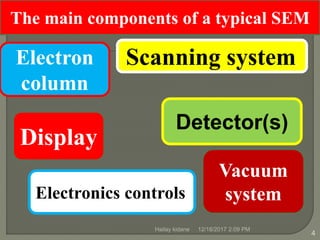
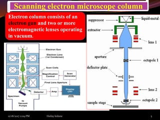
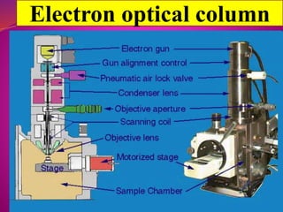
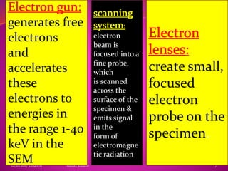
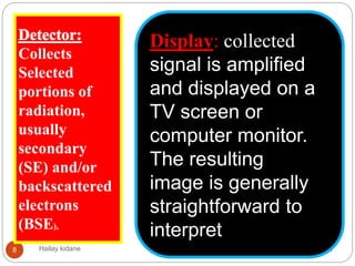
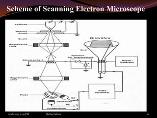
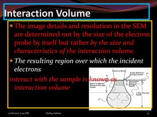
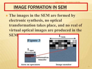
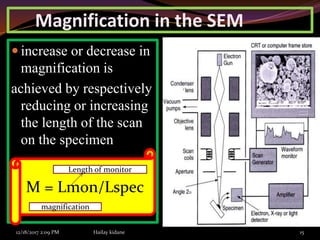
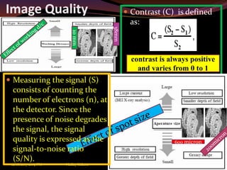
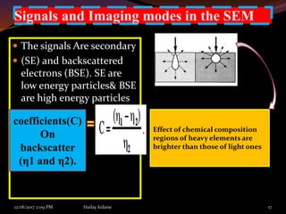
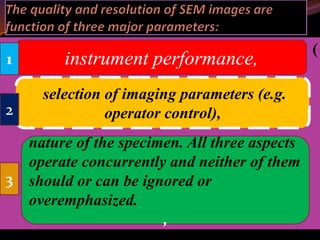
Prepared by: Hailay Kidane
hailaykidane@gmail.com
Submitted to:Adane Haile
(Asst.Professor)
1st year Ms.c
semester 1
ID:SGSR/0178/09
13/11/2009 E.c
The SEM instrument is made up of two main
components, the electronic console and the
electron column. The electronic console provides
control knobs and switches that allow for
instrument adjustments such as filament
current, accelerating voltage, focus,
magnification, brightness and contrast
12/18/2017 2:09 PM Hailay kidane 3
An instrument for observing and
Analyzing the surface micro
structure of a bulk sample using
a finely focused beam of
energetic electrons
What does the SEM do?
12/18/2017 2:09 PMHailay kidane
4
The main components of a typical SEM
Electron
column
Scanning system,
Display
Vacuum
system
Detector(s)
Electronics controls,
12/18/2017 2:09 PM Hailay kidane 5
Electron column consists of an
electron gun and two or more
electromagnetic lenses operating
in vacuum.
Scanning electron microscope column
12/18/2017 2:09 PM Hailay kidane 6
Electron optical column
12/18/2017 2:09 PM Hailay kidane 7
generates free
electrons
and
accelerates
these
electrons to
energies in
the range 1-40
keV in the
SEM
:
create small,
focused
electron
probe on the
specimen.
electron
beam is
focused into a
fine probe,
which
is scanned
across the
surface of the
specimen &
emits signal
in the
form of
electromagne
tic radiation
12/18/2017 2:09 PMHailay kidane8
Collects
Selected
portions of
radiation,
usually
secondary
(SE) and/or
backscattered
electrons
(BSE),
Display: collected
signal is amplified
and displayed on a
TV screen or
computer monitor.
The resulting
image is generally
straightforward to
interpret
Vacuum System
12/18/2017 2:09 PM Hailay kidane 9
The ability for a SEM to provide a controlled electron
beam requires that the electronic column be under
vacuum at a pressure of at least 5x10⁻⁵ Torr. The
reasons are the following.
 A hot tungsten filament will oxidize and burn out in
the presence of air at atmospheric pressure
 Column optics requires clean, dust-free environment.
 air particles and dust inside the column can interfere
and block the electrons before the ever
reach the specimen in the sample chamber
Scheme of Scanning Electron Microscope
12/18/2017 2:09 PM Hailay kidane 10
modes of imaging in the SEM
Beam accelerating voltage
(kV):
Probe convergence angle (αp):
Probe current (ip):
Probe diameter or spot
size (dp):
12/18/2017 2:09 PM Hailay kidane
11
1
2
3
4
Brightness(B) = 4ip / (π dp αp)2
Sample requirements in SEM
Samples must be compatible with high vacuum(~ 10-5 mbar).
Non-conductive materials need coating by thin
conductive film such as Au, Pt, Pd, their alloys, as well as
carbon
12/18/2017 2:09 PM Hailay kidane 12
Special
types of
SEM
variable-pressure SEM (VPSEM)
and environmental SEM(ESEM)
Samples containing water and other volatile
components cannot be studied directly. Also fine
powder samples need to be fixed firmly to a specimen
holder substrate so that they will not contaminate the
SEM specimen chamber.
Interaction Volume
12/18/2017 2:09 PM Hailay kidane 13
 The image details and resolution in the SEM
are determined not by the size of the electron
probe by itself but rather by the size and
characteristics of the interaction volume.
 The resulting region over which the incident
electrons
interact with the sample is known as
interaction volume
IMAGE FORMATION IN SEM
 The images in the SEM are formed by
electronic synthesis, no optical
transformation takes place, and no real of
virtual optical images are produced in the
SEM.
12/18/2017 2:09 PMHailay kidane
14
Magnification in the SEM
 increase or decrease in
magnification is
achieved by respectively
reducing or increasing
the length of the scan
on the specimen
.
12/18/2017 2:09 PM Hailay kidane 15
M = Lmon/Lspec
Length of monitor
magnification
Image Quality  Contrast (C) is defined
as:
12/18/2017 2:09 PM Hailay kidane 16
contrast is always positive
and varies from 0 to 1
 Measuring the signal (S)
consists of counting the
number of electrons (n), at
the detector. Since the
presence of noise degrades
the signal, the signal
quality is expressed as the
signal-to-noise ratio
(S/N).
10mm
38mm
600 micron
Signals and Imaging modes in the SEM
 The signals Are secondary
 (SE) and backscattered
electrons (BSE). SE are
low energy particles& BSE
are high energy particles
12/18/2017 2:09 PM Hailay kidane 17
coefficients(C)
On
backscatter
(η1 and η2).
Effect of chemical composition
regions of heavy elements are
brighter than those of light ones
12/18/2017 2:09 PM Hailay kidane 18
Image disturbances and their possible causes in SEM
(
instrument performance,
nature of the specimen. All three aspects
operate concurrently and neither of them
should or can be ignored or
overemphasized.
,
selection of imaging parameters (e.g.
operator control),
1
2
3
reference
1) Bob Hafner ;Scanning Electron Microscopy Primer
2) Brandon Cheney Ant’s ;Introduction to Scanning Electron
Microscopy
3) http://www.jeol.com/sem/docs/sem_guide/tbcontd.html
4) http://en.wikipedia.org/wiki/Turbomolecular_pump
5) D.S. Su: guide of Scanning electron microscopy (SEM)
12/18/2017 2:09 PM Hailay kidane 20
12/18/2017 2:09 PM Hailay kidane 21
THANK
YOU
12/18/2017 2:09 PM Hailay kidane 22

presentation on scanning electron microscopy

  • 1.
    Prepared by: HailayKidane hailaykidane@gmail.com Submitted to:Adane Haile (Asst.Professor) 1st year Ms.c semester 1 ID:SGSR/0178/09 13/11/2009 E.c
  • 2.
    The SEM instrumentis made up of two main components, the electronic console and the electron column. The electronic console provides control knobs and switches that allow for instrument adjustments such as filament current, accelerating voltage, focus, magnification, brightness and contrast
  • 3.
    12/18/2017 2:09 PMHailay kidane 3 An instrument for observing and Analyzing the surface micro structure of a bulk sample using a finely focused beam of energetic electrons What does the SEM do?
  • 4.
    12/18/2017 2:09 PMHailaykidane 4 The main components of a typical SEM Electron column Scanning system, Display Vacuum system Detector(s) Electronics controls,
  • 5.
    12/18/2017 2:09 PMHailay kidane 5 Electron column consists of an electron gun and two or more electromagnetic lenses operating in vacuum. Scanning electron microscope column
  • 6.
    12/18/2017 2:09 PMHailay kidane 6 Electron optical column
  • 7.
    12/18/2017 2:09 PMHailay kidane 7 generates free electrons and accelerates these electrons to energies in the range 1-40 keV in the SEM : create small, focused electron probe on the specimen. electron beam is focused into a fine probe, which is scanned across the surface of the specimen & emits signal in the form of electromagne tic radiation
  • 8.
    12/18/2017 2:09 PMHailaykidane8 Collects Selected portions of radiation, usually secondary (SE) and/or backscattered electrons (BSE), Display: collected signal is amplified and displayed on a TV screen or computer monitor. The resulting image is generally straightforward to interpret
  • 9.
    Vacuum System 12/18/2017 2:09PM Hailay kidane 9 The ability for a SEM to provide a controlled electron beam requires that the electronic column be under vacuum at a pressure of at least 5x10⁻⁵ Torr. The reasons are the following.  A hot tungsten filament will oxidize and burn out in the presence of air at atmospheric pressure  Column optics requires clean, dust-free environment.  air particles and dust inside the column can interfere and block the electrons before the ever reach the specimen in the sample chamber
  • 10.
    Scheme of ScanningElectron Microscope 12/18/2017 2:09 PM Hailay kidane 10
  • 11.
    modes of imagingin the SEM Beam accelerating voltage (kV): Probe convergence angle (αp): Probe current (ip): Probe diameter or spot size (dp): 12/18/2017 2:09 PM Hailay kidane 11 1 2 3 4 Brightness(B) = 4ip / (π dp αp)2
  • 12.
    Sample requirements inSEM Samples must be compatible with high vacuum(~ 10-5 mbar). Non-conductive materials need coating by thin conductive film such as Au, Pt, Pd, their alloys, as well as carbon 12/18/2017 2:09 PM Hailay kidane 12 Special types of SEM variable-pressure SEM (VPSEM) and environmental SEM(ESEM) Samples containing water and other volatile components cannot be studied directly. Also fine powder samples need to be fixed firmly to a specimen holder substrate so that they will not contaminate the SEM specimen chamber.
  • 13.
    Interaction Volume 12/18/2017 2:09PM Hailay kidane 13  The image details and resolution in the SEM are determined not by the size of the electron probe by itself but rather by the size and characteristics of the interaction volume.  The resulting region over which the incident electrons interact with the sample is known as interaction volume
  • 14.
    IMAGE FORMATION INSEM  The images in the SEM are formed by electronic synthesis, no optical transformation takes place, and no real of virtual optical images are produced in the SEM. 12/18/2017 2:09 PMHailay kidane 14
  • 15.
    Magnification in theSEM  increase or decrease in magnification is achieved by respectively reducing or increasing the length of the scan on the specimen . 12/18/2017 2:09 PM Hailay kidane 15 M = Lmon/Lspec Length of monitor magnification
  • 16.
    Image Quality Contrast (C) is defined as: 12/18/2017 2:09 PM Hailay kidane 16 contrast is always positive and varies from 0 to 1  Measuring the signal (S) consists of counting the number of electrons (n), at the detector. Since the presence of noise degrades the signal, the signal quality is expressed as the signal-to-noise ratio (S/N). 10mm 38mm 600 micron
  • 17.
    Signals and Imagingmodes in the SEM  The signals Are secondary  (SE) and backscattered electrons (BSE). SE are low energy particles& BSE are high energy particles 12/18/2017 2:09 PM Hailay kidane 17 coefficients(C) On backscatter (η1 and η2). Effect of chemical composition regions of heavy elements are brighter than those of light ones
  • 18.
    12/18/2017 2:09 PMHailay kidane 18 Image disturbances and their possible causes in SEM
  • 19.
    ( instrument performance, nature ofthe specimen. All three aspects operate concurrently and neither of them should or can be ignored or overemphasized. , selection of imaging parameters (e.g. operator control), 1 2 3
  • 20.
    reference 1) Bob Hafner;Scanning Electron Microscopy Primer 2) Brandon Cheney Ant’s ;Introduction to Scanning Electron Microscopy 3) http://www.jeol.com/sem/docs/sem_guide/tbcontd.html 4) http://en.wikipedia.org/wiki/Turbomolecular_pump 5) D.S. Su: guide of Scanning electron microscopy (SEM) 12/18/2017 2:09 PM Hailay kidane 20
  • 21.
    12/18/2017 2:09 PMHailay kidane 21 THANK YOU
  • 22.
    12/18/2017 2:09 PMHailay kidane 22