
Recommended
More Related Content
What's hot
What's hot (20)
Viewers also liked
Viewers also liked (14)
Similar to CNS Meninges & Ventricles (39
Similar to CNS Meninges & Ventricles (39 (20)
More from Michael Walls
More from Michael Walls (19)
Recently uploaded
Recently uploaded (20)
CNS Meninges & Ventricles (39
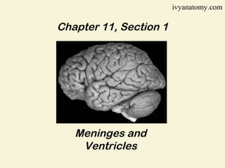
- 1. ivyanatomy.com Chapter 11, Section 1 Meninges and Ventricles
- 2. The central nervous system (CNS) consists of the brain and spinal cord introduction A nucleus (sing. nucleus) refers to a group of cell bodies within the CNS e.g. dentate nucleus A tract is a group of axons within the CNS e.g. corticospinal tract The peripheral nervous system (PNS) consists of cranial and spinal nerves A ganglion is a group of cell bodies within the PNS e.g. dorsal root ganglion A nerve is a group of axons, along with their protective sheaths in the PNS e.g. sciatic nerve
- 3. meninges The meninges is a three-layered membrane that surrounds the brain and spinal cord. 1. Dura Mater “tough mother” • Tough outer layer of meninges • Dense connective tissue with many blood vessels and nerves • Forms dural sinuses that drain venous blood from the brain. 2. Arachnoid Mater “spider-web like” • Subarachnoid space • Space between arachnoid and pia mater filled with Cerebral Spinal Fluid (CSF) 3. Pia Mater “gentle mother” • Thin membrane with many nerves and blood vessels • Membrane is attached to surface of brain and spinal cord.
- 4. Figure 11.1 Meninges (a) membranes called meninges enclose the brain and Figure 11.1 Meninges (a) membranes called meninges enclose the brain and spinal cord. (b) the meninges include three layers: dura mater, arachnoid spinal cord. (b) the meninges include three layers: dura mater, arachnoid mater, and pia mater. Note the dural sinus formed by the dura mater. mater, and pia mater. Note the dural sinus formed by the dura mater.
- 5. Figure 11.2 Meninges of the spinal cord. (a)three layers of the meninges . (b)a small space (epidural space) fills the space between the dura mater and the vertebra.
- 6. VENTRICLES Interconnected cavities, called ventricles lie within the cerebral hemispheres and the brainstem. Ventricles are continuous with the central canal of the spinal cord and are filled with Cerebrospinal Fluid (CSF). 4 Ventricles: 2 Lateral ventricles 1st ventricle is in left cerebral hemisphere 2nd ventricle is in the right cerebral hemisphere Lateral ventricles are connected by interventricular foramina. Third ventricle Surrounds the diencephalon Cerebral aqueduct connects 3rd and 4th ventricles Fourth ventricle Within brainstem, just anterior to cerebellum
- 7. Figure 11.3 Ventricles in the brain. (a) anterior view. (b) lateral view.
- 8. Cerebrospinal Fluid CSF is secreted by specialized capillaries called Choroid Plexuses • CSF complete surrounds brain and spinal cord • Ependymal cells regulate the composition of CSF • CSF functions as a nutritive and protective fluid. Figure 10.4 (a) choroid plexuses in ventricle walls secrete CSF. The fluid circulates through ventricles and central canal.
- 9. Spinal Tap The spinal cord ends near the 2nd Lumbar vertebra, but the arachnoid and dura maters continue to the 2nd Sacral vertebra. Thecal Sac – space beyond spinal cord filled with CSF. Spinal taps (lumbar punctures) and spinal blocks are usually given below the 4th lumbar vertebra to access the CSF without puncturing the spinal cord. End of section 1, chapter 11
- 10. Spinal Tap The spinal cord ends near the 2nd Lumbar vertebra, but the arachnoid and dura maters continue to the 2nd Sacral vertebra. Thecal Sac – space beyond spinal cord filled with CSF. Spinal taps (lumbar punctures) and spinal blocks are usually given below the 4th lumbar vertebra to access the CSF without puncturing the spinal cord. End of section 1, chapter 11