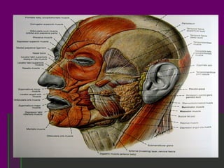
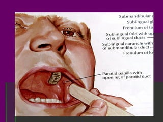
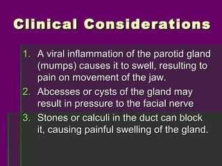
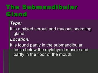
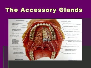
T he Salivar y
Glands and
Accessor y
Glands of the
Or al Cavity
The Salivary Glands of
the
Oral Cavity
 1. Parotid Gland

 2. Submandibular Gland

 3. Sublingual Gland
T he Parotid
Gland
The Parotid Gland

 The parotid gland is one of the three major
  salivary glands that produce and secrete
  saliva for the oral cavity.
      weight           :    about 14 to 28 g.
      color            :    yellowish in the
                            fresh state
      composition :         serous secreting
                            units
Location and Relations
of the Parotid Gland
 A. Anterior surface: lies against the posterior
    border of the ramus of the mandible
 B. Posterior surface: on the external auditory
    meatus and sternocleidomastoid muscle
 C. Superficial surface: lobulated, covered by
    skin, fascia, lymph nodes, and facial
    branches of the great auricular nerve.
 D. Deep surface: styloid process and its
    muscles as well as under the mastoid and
    sternocleidomastoid muscles.
The Parotid Duct

 From the rostral border of the gland,
   crosses masseter muscle, turns inward
   to pierce the fat pad of the cheek and
   then the buccinator muscle, to open into
   the mouth opposite the second maxillary
   molar. Approximately 5 cm. long.
Clinical Considerations

 1. A viral inflammation of the parotid gland
    (mumps) causes it to swell, resulting to
    pain on movement of the jaw.
 2. Abcesses or cysts of the gland may
    result in pressure to the facial nerve
 3. Stones or calculi in the duct can block
    it, causing painful swelling of the gland.
T he
Submandibular
Gland
The Submandibular
Gland
Type:
It is a mixed serous and mucous secreting
   gland.
Location:
It is found partly in the submandibular
   fossa below the mylohyoid muscle and
   partly in the floor of the mouth.
The Submandibular
Gland
Duct:
The submandibular duct (Wharton’s) arises from
  the medial surface of the gland and
  accompanies it under the mlohyoid muscle; it
  passes diagonally across the medial aspect of
  the sublingual gland and adheres to it. It
  opens at the sublingual papillae beside the
  base of the lingual frenulum.
The Submandibular
Gland
The Submandibular
Gland
T he Sublingual
Gland
The Sublingual Gland
 The smallest of the three major salivary
  glands and rests upon the mylohyoid
  muscle in the sublingual fossa close to
  the symphysis. It is primarily a mucous
  secreting gland.
The Sublingual Gland
 Location:
   The gland, indicated by the subligual fold is
   found between the alveolus and the anterior
   part of the tongue.
 Duct:
   The gland has many lesser sublingual ducts
   (Rivini’s) that opens separately at the floor of
   the mouth and a greater sublingual duct
   (Bartholin’s) that opens on the sublingual
   caruncle.
The Sublingual Gland
T he Accessor y
Glands
The Accessory Glands

 Beside the main salivary glands, many
 others exists: some in the tongue, others
 around and in the palatine tonsil between
 its crypts, with the large number in the
 soft palate, the posterior part of the hard
 palate, the lips and the cheeks. These
 are similar in structure to larger salivary
 glands and are mainly mucous type.
The Accessory Glands

Salivary Glands

  • 1.
    T he Salivary Glands and Accessor y Glands of the Or al Cavity
  • 2.
    The Salivary Glandsof the Oral Cavity 1. Parotid Gland 2. Submandibular Gland 3. Sublingual Gland
  • 3.
  • 4.
    The Parotid Gland The parotid gland is one of the three major salivary glands that produce and secrete saliva for the oral cavity. weight : about 14 to 28 g. color : yellowish in the fresh state composition : serous secreting units
  • 5.
    Location and Relations ofthe Parotid Gland A. Anterior surface: lies against the posterior border of the ramus of the mandible B. Posterior surface: on the external auditory meatus and sternocleidomastoid muscle C. Superficial surface: lobulated, covered by skin, fascia, lymph nodes, and facial branches of the great auricular nerve. D. Deep surface: styloid process and its muscles as well as under the mastoid and sternocleidomastoid muscles.
  • 7.
    The Parotid Duct From the rostral border of the gland, crosses masseter muscle, turns inward to pierce the fat pad of the cheek and then the buccinator muscle, to open into the mouth opposite the second maxillary molar. Approximately 5 cm. long.
  • 10.
    Clinical Considerations 1.A viral inflammation of the parotid gland (mumps) causes it to swell, resulting to pain on movement of the jaw. 2. Abcesses or cysts of the gland may result in pressure to the facial nerve 3. Stones or calculi in the duct can block it, causing painful swelling of the gland.
  • 11.
  • 12.
    The Submandibular Gland Type: It isa mixed serous and mucous secreting gland. Location: It is found partly in the submandibular fossa below the mylohyoid muscle and partly in the floor of the mouth.
  • 13.
    The Submandibular Gland Duct: The submandibularduct (Wharton’s) arises from the medial surface of the gland and accompanies it under the mlohyoid muscle; it passes diagonally across the medial aspect of the sublingual gland and adheres to it. It opens at the sublingual papillae beside the base of the lingual frenulum.
  • 14.
  • 15.
  • 16.
  • 17.
    The Sublingual Gland The smallest of the three major salivary glands and rests upon the mylohyoid muscle in the sublingual fossa close to the symphysis. It is primarily a mucous secreting gland.
  • 18.
    The Sublingual Gland Location: The gland, indicated by the subligual fold is found between the alveolus and the anterior part of the tongue. Duct: The gland has many lesser sublingual ducts (Rivini’s) that opens separately at the floor of the mouth and a greater sublingual duct (Bartholin’s) that opens on the sublingual caruncle.
  • 19.
  • 20.
    T he Accessory Glands
  • 21.
    The Accessory Glands Beside the main salivary glands, many others exists: some in the tongue, others around and in the palatine tonsil between its crypts, with the large number in the soft palate, the posterior part of the hard palate, the lips and the cheeks. These are similar in structure to larger salivary glands and are mainly mucous type.
  • 22.