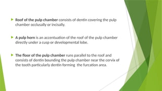
Endodontics is the dental specialty focused on treating diseases and injuries of the dental pulp and periradicular tissues. It encompasses various techniques and technologies for diagnosing and preserving tooth vitality, and has evolved significantly since its early practices, eventually becoming officially recognized as a dental specialty in 1963. The document details the anatomy, functions, and historical development of endodontics, including the introduction of innovative techniques and tools that have improved treatment outcomes.


![BLOOD VESSELS
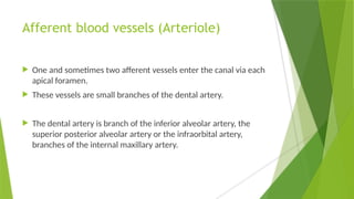
afferent(af‚er-ent) [L. afferens, fr. af-fero, to bring to] centripetal
(1); esodic;toward a center, denoting certain arteries, veins,
lymphatics, and nerves.
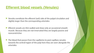
efferent(ef‚er-ent) [L. efferens, fr. effero, to bring out]
Conducting (fluid or a nerve impulse) outward from a given
organ or part thereof; e.g., the efferent connections of a group
of nerve cells, efferent blood vessels, or the excretory duct of an
organ.](https://image.slidesharecdn.com/1-250212140236-56cfa69c/85/1-Introduction-to-endodontics-pptx-31-320.jpg)