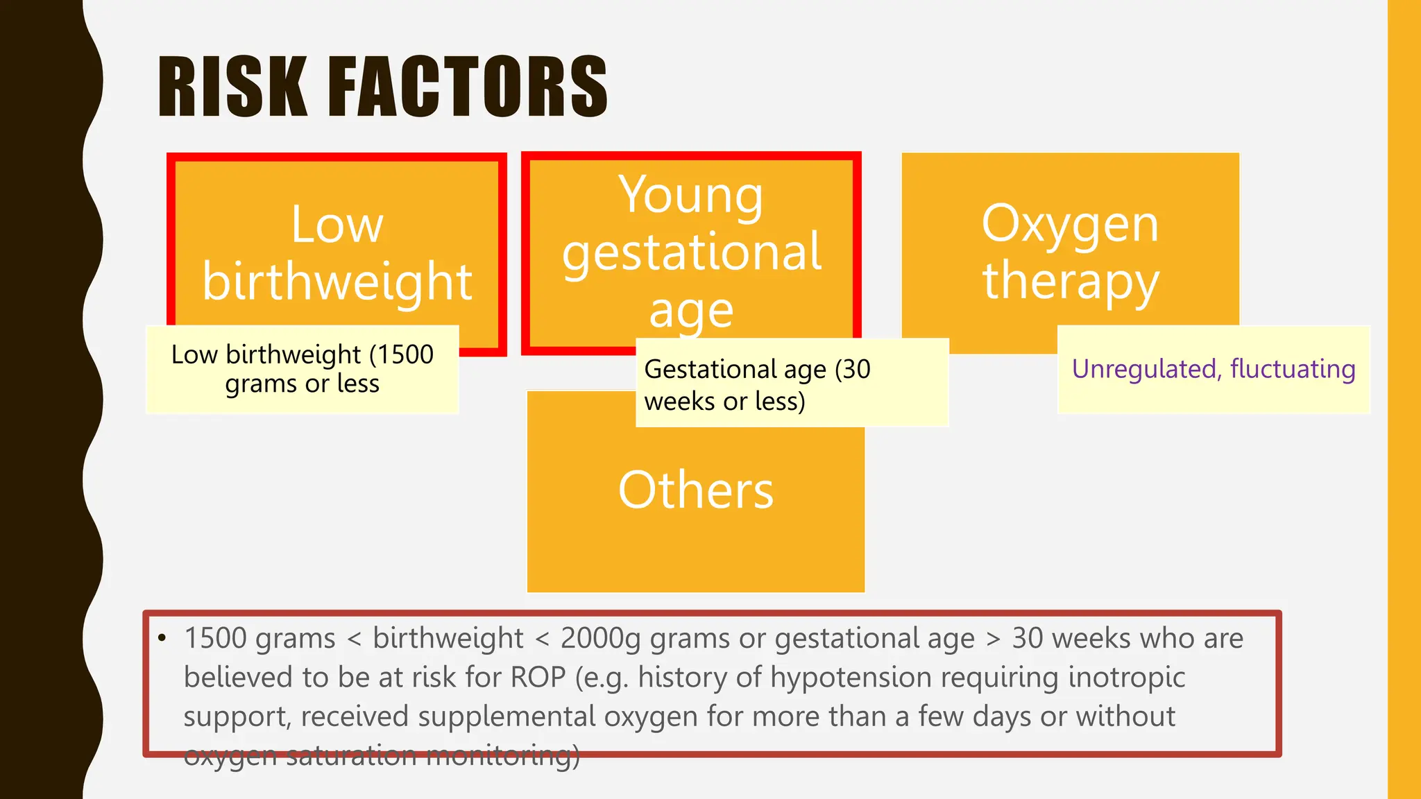
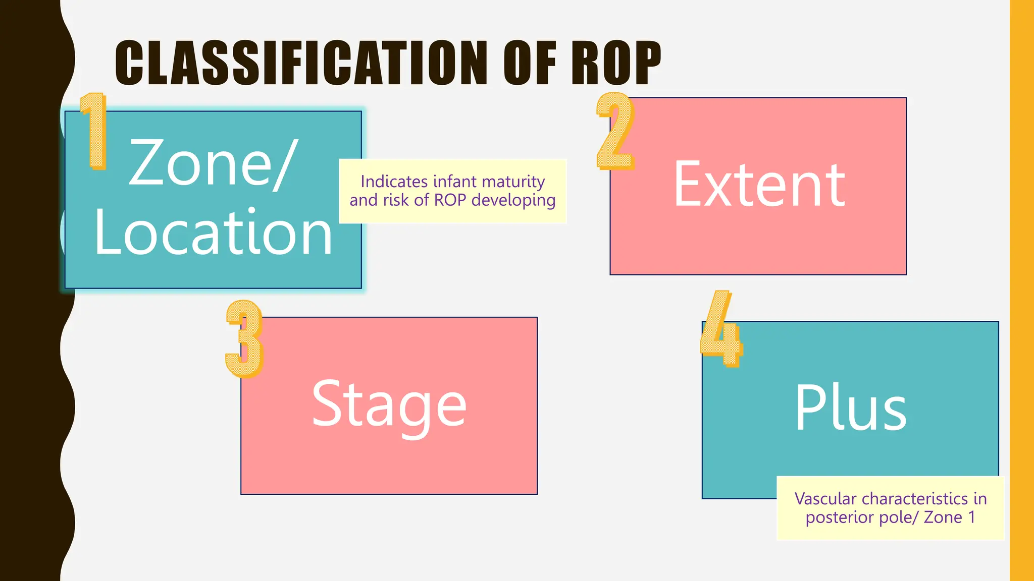
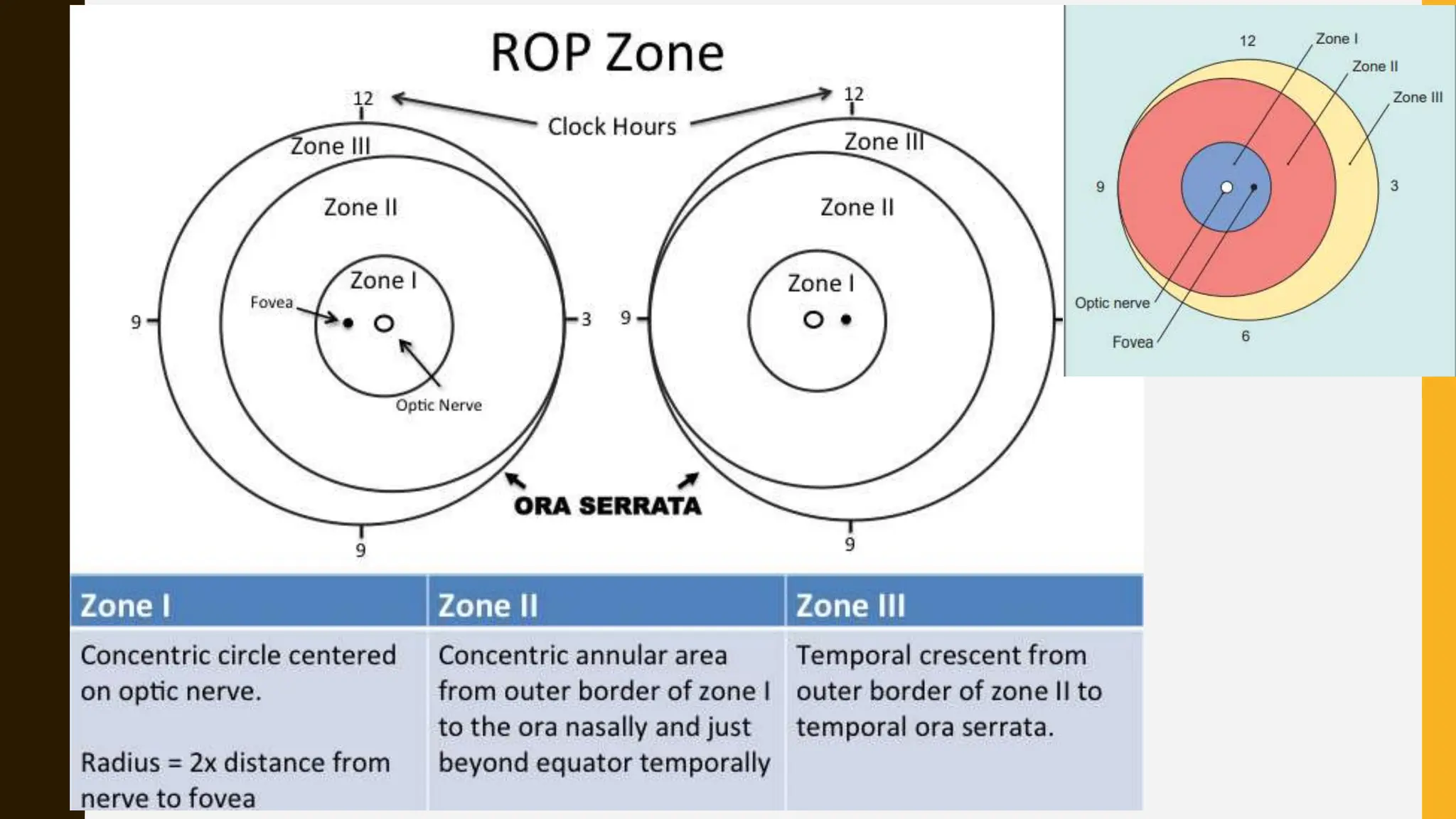
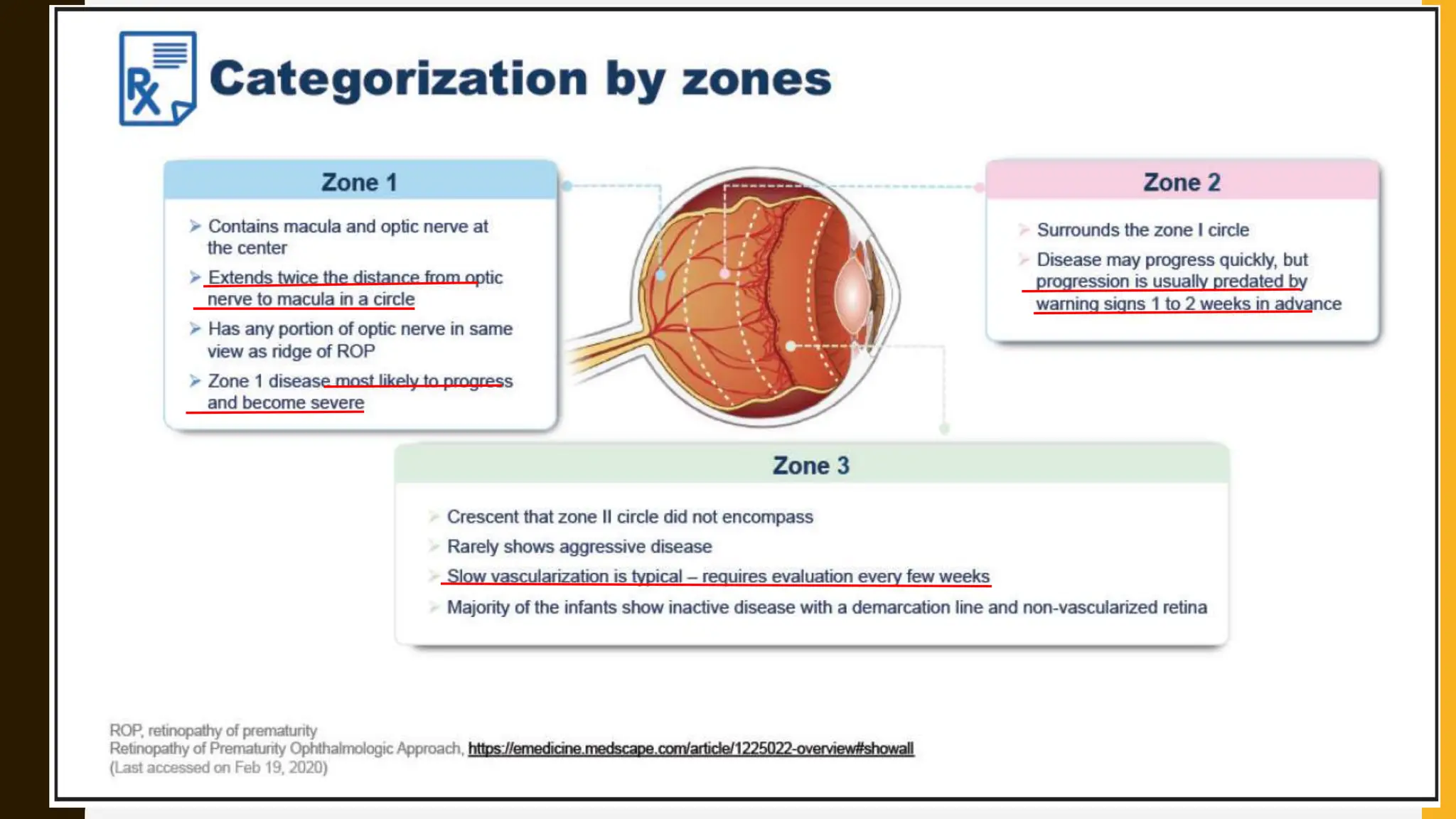
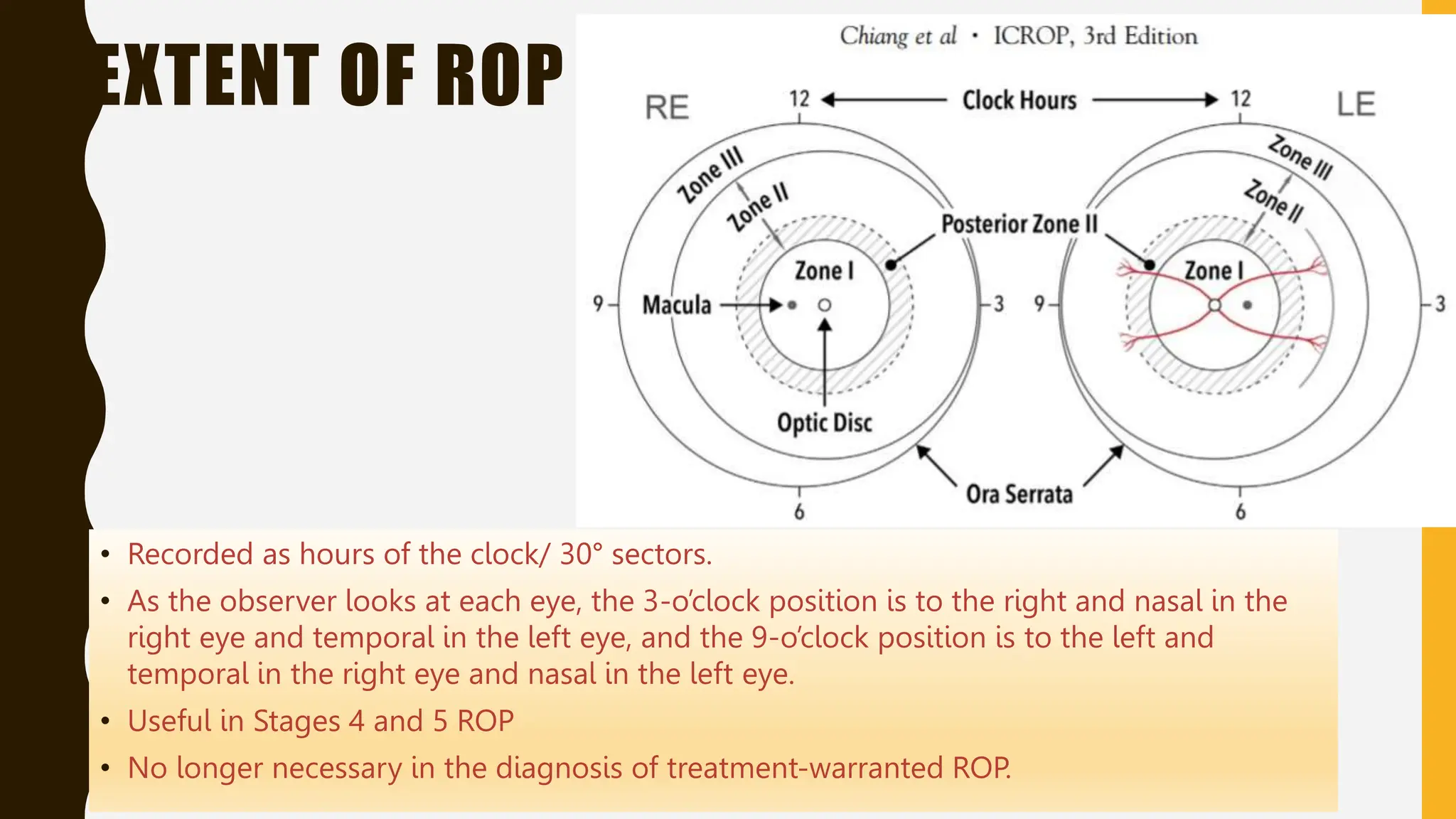
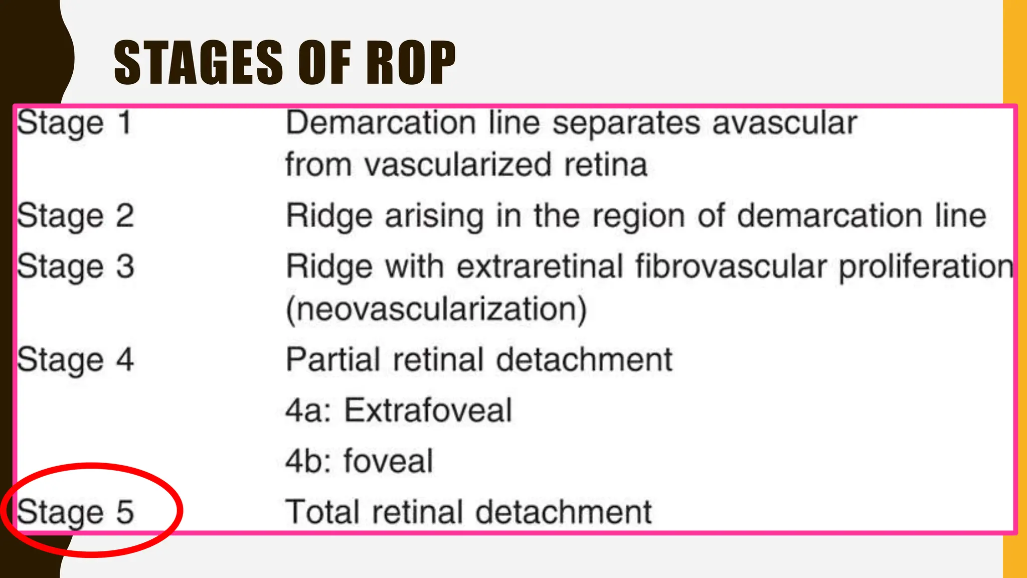
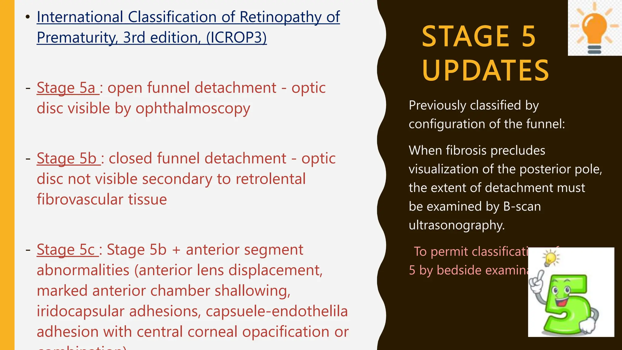
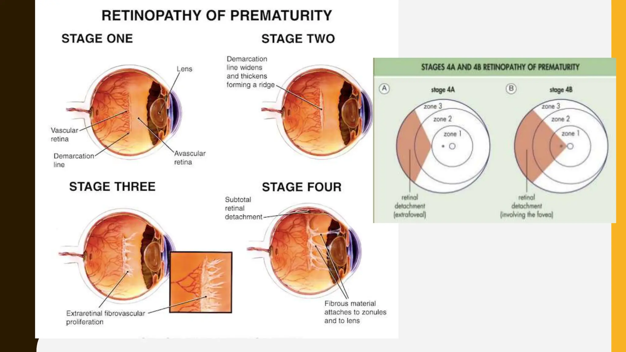
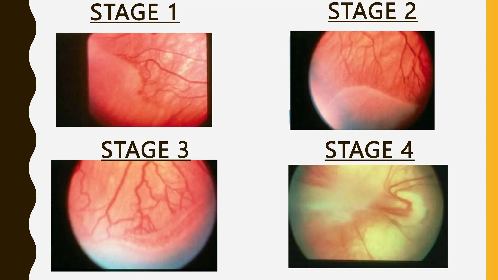
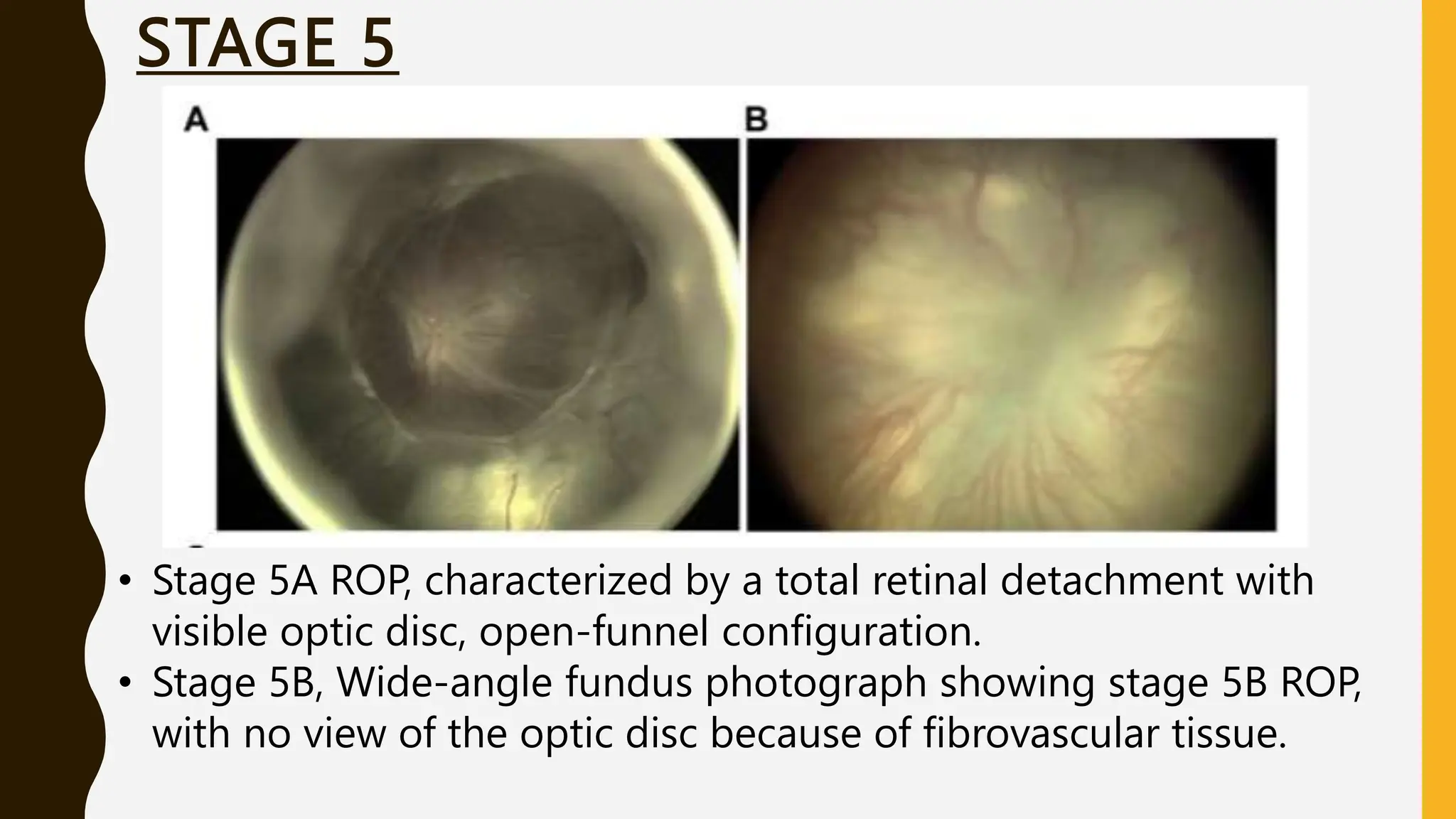
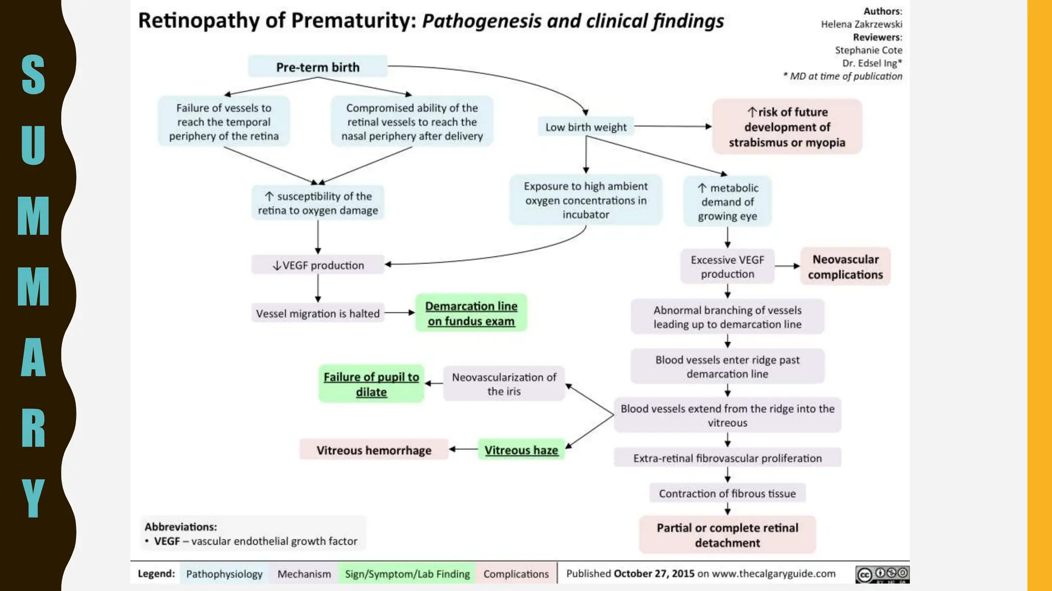
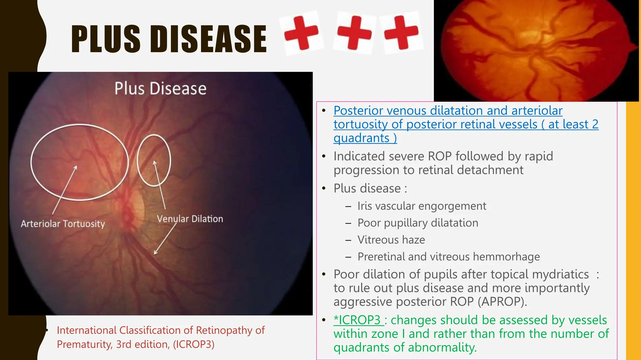
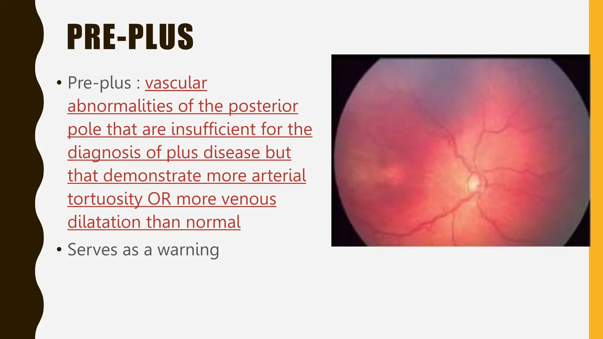
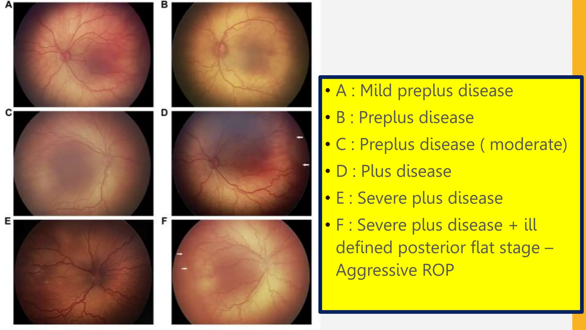
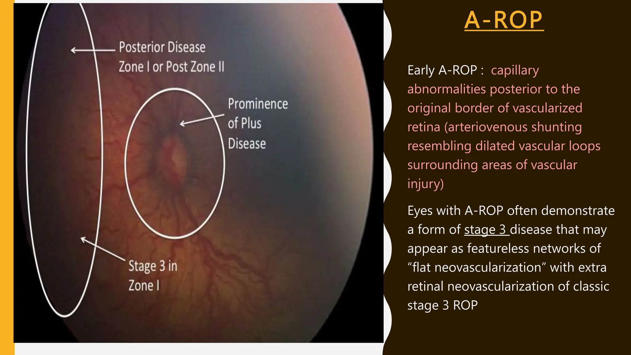
RETINOPATHY OF
PREMATURITY
(ROP)
T E A M PA E D I AT R I C O P H T H A L
ROP
• Vasoproliferative disorder of the retina which occurs principally in the
preterm infant
• Only in infants with immature, incompletely vascularized retina
• Outcome ? – minimal sequalae with no effect on vision in milder case
OR bilateral irreversible total blindness in advanced cases with
complications
DISEASE ENTITY
• Initially described as retrolental fibroplasia by Terry in 1942 was the leading cause
of
blindness in children in the United States (US).
• 3 epidemic waves
• 1st wave : 1940s-1950s in industrialized countries primarily due to unmonitored
supplemental oxygen.
• Regulation and monitoring of high oxygen at birth caused ROP to virtually
disappear,
• 2nd wave : 1970’s ,premature infants survived at earlier gestational ages and lower
birth weights, and ROP re-emerged to be a serious problem
• 3rd wave : Mid-1990s in low and middle income countries due to both high rates
of preterm birth and varying levels of neonatal care in these countries similar to
that in the US in the 1940's and 50's).
RISK FACTORS
Low
birthweight
Young
gestational
age
Oxygen
therapy
Others
Unregulated, fluctuating
Gestational age (30
weeks or less)
Low birthweight (1500
grams or less
• 1500 grams < birthweight < 2000g grams or gestational age > 30 weeks who are
believed to be at risk for ROP (e.g. history of hypotension requiring inotropic
support, received supplemental oxygen for more than a few days or without
oxygen saturation monitoring)
OTHERS RISK FACTORS
• Drugs : Glucocorticoids, Surfactant (RDS), Indomethacin, Xanthine,
Dopamine
• Addition : Intra-ventricular haemorrhage, ante-natal blood loss
requiring blood transfusions and surgery under general anaesthesia
• Sepsis
• Candidemia
• Raised serum bilirubin levels
However, there is insufficient evidence to determine the degree of
importance of these risk factors in contributing to the pathogenesis of
ROP.
PATHOPHYSIOLOGY?
ROP occurs in premature infants
who are born before the retinal
vessels complete their normal
growth.
• Retina avascular up to 4 months gestation
– vascular complexes come out from
hyaloids
• 14 – 16 weeks : vessels at optic disc grow
towards periphery ( from precursor
endothelial cells)
• 22 weeks : posterior pole vascularization
• Following this, angiogenesis occurs via
budding from existing vessels to extend
retinal vessels to the periphery - migrating
endothelial cells are attracted by a gradient
of vascular endothelial growth factor
(VEGF) toward the ora serrata.
• 8 months : vessels reach nasal periphery
• 40 weeks : vessels reach temporal
periphery (complete vascularization)
THEORIES
Biphasic theory of Aston and Patz
• High ambient oxygen -> vasoconstriction and toxic obliteration of retinal capillaries ->
on returning to room air, relative ischaemia develops -> angiogenic factors secreted ->
vascular proliferation
Spindle cell theory of Kretzer and Hittner
• blood vessels form in normally hypoxic uterine condition by canalization and
endothelial cells differentiating behind a migrating sheet of of spindle cells
• High ambient oxygen triggers extensive gap junction formation between spindle cells
which interferes with migration and canalization of blood vessels
• Spindle cells secrete angiogenic factors -> vascular proliferation
OXYGEN THERAPY
• Oxygen therapy has been previously implicated in the
aetiology of ROP
• Repeated hypoxic and hyperoxic episodes may be an
important factor in the pathogenesis of ROP
• Biphasic theory of Aston and Patz
- Early investigations in animals that vascularize their retinas
after birth - kittens no older than 12 days exposed to
continuously delivered 70% to 80% oxygen experienced
constriction of newly formed retinal capillaries, a
phenomenon termed “vaso-obliteration”
- On returning to room air, the kittens experienced a
“vasoproliferative” effect
In conclusion, the best oxygen levels for reducing ROP /
reducing pulmonary morbidity, and increasing survival and
cognitive development have not been determined.
Premature
• Down regulated VEGF by relative hypoxia + vessel migration
halted.
• Then ↑metabolic demand → excessive VEGF → ROP
CLASSIFICATION OF ROP
Zone/
Location
Extent
Stage
Indicates infant maturity
and risk of ROP developing
Plus
Vascular characteristics in
posterior pole/ Zone 1
EXTENT OF ROP
• Recorded as hours of the clock/ 30° sectors.
• As the observer looks at each eye, the 3-o’clock position is to the right and nasal in the
right eye and temporal in the left eye, and the 9-o’clock position is to the left and
temporal in the right eye and nasal in the left eye.
• Useful in Stages 4 and 5 ROP
• No longer necessary in the diagnosis of treatment-warranted ROP.
STAGES OF ROP
STAGE 5
UPDATES
• International Classification of Retinopathy of
Prematurity, 3rd edition, (ICROP3)
- Stage 5a : open funnel detachment - optic
disc visible by ophthalmoscopy
- Stage 5b : closed funnel detachment - optic
disc not visible secondary to retrolental
fibrovascular tissue
- Stage 5c : Stage 5b + anterior segment
abnormalities (anterior lens displacement,
marked anterior chamber shallowing,
iridocapsular adhesions, capsuele-endothelila
adhesion with central corneal opacification or
Previously classified by
configuration of the funnel:
When fibrosis precludes
visualization of the posterior pole,
the extent of detachment must
be examined by B-scan
ultrasonography.
-To permit classification of stage
5 by bedside examination
STAGE 2
STAGE 1
STAGE 4
STAGE 3
STAGE 5
• Stage 5A ROP, characterized by a total retinal detachment with
visible optic disc, open-funnel configuration.
• Stage 5B, Wide-angle fundus photograph showing stage 5B ROP,
with no view of the optic disc because of fibrovascular tissue.
S
U
M
M
A
R
Y
PLUS DISEASE
• Posterior venous dilatation and arteriolar
tortuosity of posterior retinal vessels ( at least 2
quadrants )
• Indicated severe ROP followed by rapid
progression to retinal detachment
• Plus disease :
– Iris vascular engorgement
– Poor pupillary dilatation
– Vitreous haze
– Preretinal and vitreous hemmorhage
• Poor dilation of pupils after topical mydriatics :
to rule out plus disease and more importantly
aggressive posterior ROP (APROP).
• *ICROP3 : changes should be assessed by vessels
within zone I and rather than from the number of
quadrants of abnormality.
• International Classification of Retinopathy of
Prematurity, 3rd edition, (ICROP3)
PRE-PLUS
• Pre-plus : vascular
abnormalities of the posterior
pole that are insufficient for the
diagnosis of plus disease but
that demonstrate more arterial
tortuosity OR more venous
dilatation than normal
• Serves as a warning
• A : Mild preplus disease
• B : Preplus disease
• C : Preplus disease ( moderate)
• D : Plus disease
• E : Severe plus disease
• F : Severe plus disease + ill
defined posterior flat stage –
Aggressive ROP
AGGRESSIVE ROP (A- ROP)
• Previously known as Rush disease or APROP
• Florid acute ROP seen in the 1940s in extremely premature infants
• Previously to describe a severe, rapidly progressive form of ROP
located in posterior zones I or II.
• Increasing recognition that may occur beyond the posterior retina and
in larger preterm infants, Committee recommends the new term
Aggressive ROP.
• Hallmark of A-ROP : rapid development of pathologic
neovascularization and severe plus disease without progression being
observed through the typical stages of ROP.
A-ROP
Early A-ROP : capillary
abnormalities posterior to the
original border of vascularized
retina (arteriovenous shunting
resembling dilated vascular loops
surrounding areas of vascular
injury)
Eyes with A-ROP often demonstrate
a form of stage 3 disease that may
appear as featureless networks of
“flat neovascularization” with extra
retinal neovascularization of classic
stage 3 ROP
REGRESSION OF ROP
• Disease involution and resolution.
• May be complete or incomplete (including persistence of retinal
abnormalities)
• Characterized by thinning and whitening of neovascular tissue – first
visible sign typically vascular
• Signs of vascular regression includes
- decreased plus disease
- vascularization into the peripheral avascular retina (
spontaneously/ after anti-vegf treatment) – complete / incomplete (
persistent avascular retina – PAR)
- involution of the tunica vasculosa lentos,
- better pupillary dilation
- greater media clarity
- resolution of intraretinal hemorrhages
REACTIVATION
• Recurrence of acute phase features
• More frequently after anti-VEGF treatment than after spontaneous
regression and rarely if ever occurs after complete laser
photocoagulation
• Signs of reactivation range from development of a new self-limiting
demarcation line to reactivated stage 3 with plus disease.
• Signs of vascular reactivation includes
- recurrent vascular dilation, tortuosity, or both -similar to acute-phase preplus
or plus
- Extraretinal new vessels - may appear as a fibrovascular ridge, which may
progress to fibrosis, contraction and tractional detachment.
- Hemorrhages
LONG TERM SEQUALE
• Late tractional, rhegmatogenous, or, rarely exudative retinal detachments
- Retinal detachment occurring in the absence of signs of ROP activity should
not be designated as being the result of reactivation but rather as a sequela.
• Retinoschisis from chronic traction of involuted stage 3
• Persistent avascular retina - prone to retinal thinning, holes, and lattice like changes
• Macular anomalies - smaller foveal avascular zone and blunting or absence of the
foveal depression.
• Retinal vascular changes - persistent tortuosity, straightening of the vascular arcades
with macular dragging, falciform retinal fold, abnormal retinal vessel branching,
circumferential interconnecting vascular arcades, and telangiectatic vessels.
• Vitreous haemorrhage and Glaucoma (secondary angle-closure glaucoma)
THANK YOU

Retinopathy of Prematurity (ROP).pptx slides

  • 1.
    RETINOPATHY OF PREMATURITY (ROP) T EA M PA E D I AT R I C O P H T H A L
  • 2.
    ROP • Vasoproliferative disorderof the retina which occurs principally in the preterm infant • Only in infants with immature, incompletely vascularized retina • Outcome ? – minimal sequalae with no effect on vision in milder case OR bilateral irreversible total blindness in advanced cases with complications
  • 3.
    DISEASE ENTITY • Initiallydescribed as retrolental fibroplasia by Terry in 1942 was the leading cause of blindness in children in the United States (US). • 3 epidemic waves • 1st wave : 1940s-1950s in industrialized countries primarily due to unmonitored supplemental oxygen. • Regulation and monitoring of high oxygen at birth caused ROP to virtually disappear, • 2nd wave : 1970’s ,premature infants survived at earlier gestational ages and lower birth weights, and ROP re-emerged to be a serious problem • 3rd wave : Mid-1990s in low and middle income countries due to both high rates of preterm birth and varying levels of neonatal care in these countries similar to that in the US in the 1940's and 50's).
  • 4.
    RISK FACTORS Low birthweight Young gestational age Oxygen therapy Others Unregulated, fluctuating Gestationalage (30 weeks or less) Low birthweight (1500 grams or less • 1500 grams < birthweight < 2000g grams or gestational age > 30 weeks who are believed to be at risk for ROP (e.g. history of hypotension requiring inotropic support, received supplemental oxygen for more than a few days or without oxygen saturation monitoring)
  • 5.
    OTHERS RISK FACTORS •Drugs : Glucocorticoids, Surfactant (RDS), Indomethacin, Xanthine, Dopamine • Addition : Intra-ventricular haemorrhage, ante-natal blood loss requiring blood transfusions and surgery under general anaesthesia • Sepsis • Candidemia • Raised serum bilirubin levels However, there is insufficient evidence to determine the degree of importance of these risk factors in contributing to the pathogenesis of ROP.
  • 6.
    PATHOPHYSIOLOGY? ROP occurs inpremature infants who are born before the retinal vessels complete their normal growth.
  • 7.
    • Retina avascularup to 4 months gestation – vascular complexes come out from hyaloids • 14 – 16 weeks : vessels at optic disc grow towards periphery ( from precursor endothelial cells) • 22 weeks : posterior pole vascularization • Following this, angiogenesis occurs via budding from existing vessels to extend retinal vessels to the periphery - migrating endothelial cells are attracted by a gradient of vascular endothelial growth factor (VEGF) toward the ora serrata. • 8 months : vessels reach nasal periphery • 40 weeks : vessels reach temporal periphery (complete vascularization)
  • 8.
    THEORIES Biphasic theory ofAston and Patz • High ambient oxygen -> vasoconstriction and toxic obliteration of retinal capillaries -> on returning to room air, relative ischaemia develops -> angiogenic factors secreted -> vascular proliferation Spindle cell theory of Kretzer and Hittner • blood vessels form in normally hypoxic uterine condition by canalization and endothelial cells differentiating behind a migrating sheet of of spindle cells • High ambient oxygen triggers extensive gap junction formation between spindle cells which interferes with migration and canalization of blood vessels • Spindle cells secrete angiogenic factors -> vascular proliferation
  • 9.
    OXYGEN THERAPY • Oxygentherapy has been previously implicated in the aetiology of ROP • Repeated hypoxic and hyperoxic episodes may be an important factor in the pathogenesis of ROP • Biphasic theory of Aston and Patz - Early investigations in animals that vascularize their retinas after birth - kittens no older than 12 days exposed to continuously delivered 70% to 80% oxygen experienced constriction of newly formed retinal capillaries, a phenomenon termed “vaso-obliteration” - On returning to room air, the kittens experienced a “vasoproliferative” effect
  • 10.
    In conclusion, thebest oxygen levels for reducing ROP / reducing pulmonary morbidity, and increasing survival and cognitive development have not been determined.
  • 12.
    Premature • Down regulatedVEGF by relative hypoxia + vessel migration halted. • Then ↑metabolic demand → excessive VEGF → ROP
  • 13.
    CLASSIFICATION OF ROP Zone/ Location Extent Stage Indicatesinfant maturity and risk of ROP developing Plus Vascular characteristics in posterior pole/ Zone 1
  • 16.
    EXTENT OF ROP •Recorded as hours of the clock/ 30° sectors. • As the observer looks at each eye, the 3-o’clock position is to the right and nasal in the right eye and temporal in the left eye, and the 9-o’clock position is to the left and temporal in the right eye and nasal in the left eye. • Useful in Stages 4 and 5 ROP • No longer necessary in the diagnosis of treatment-warranted ROP.
  • 17.
  • 18.
    STAGE 5 UPDATES • InternationalClassification of Retinopathy of Prematurity, 3rd edition, (ICROP3) - Stage 5a : open funnel detachment - optic disc visible by ophthalmoscopy - Stage 5b : closed funnel detachment - optic disc not visible secondary to retrolental fibrovascular tissue - Stage 5c : Stage 5b + anterior segment abnormalities (anterior lens displacement, marked anterior chamber shallowing, iridocapsular adhesions, capsuele-endothelila adhesion with central corneal opacification or Previously classified by configuration of the funnel: When fibrosis precludes visualization of the posterior pole, the extent of detachment must be examined by B-scan ultrasonography. -To permit classification of stage 5 by bedside examination
  • 20.
  • 21.
    STAGE 5 • Stage5A ROP, characterized by a total retinal detachment with visible optic disc, open-funnel configuration. • Stage 5B, Wide-angle fundus photograph showing stage 5B ROP, with no view of the optic disc because of fibrovascular tissue.
  • 22.
  • 23.
    PLUS DISEASE • Posteriorvenous dilatation and arteriolar tortuosity of posterior retinal vessels ( at least 2 quadrants ) • Indicated severe ROP followed by rapid progression to retinal detachment • Plus disease : – Iris vascular engorgement – Poor pupillary dilatation – Vitreous haze – Preretinal and vitreous hemmorhage • Poor dilation of pupils after topical mydriatics : to rule out plus disease and more importantly aggressive posterior ROP (APROP). • *ICROP3 : changes should be assessed by vessels within zone I and rather than from the number of quadrants of abnormality. • International Classification of Retinopathy of Prematurity, 3rd edition, (ICROP3)
  • 24.
    PRE-PLUS • Pre-plus :vascular abnormalities of the posterior pole that are insufficient for the diagnosis of plus disease but that demonstrate more arterial tortuosity OR more venous dilatation than normal • Serves as a warning
  • 25.
    • A :Mild preplus disease • B : Preplus disease • C : Preplus disease ( moderate) • D : Plus disease • E : Severe plus disease • F : Severe plus disease + ill defined posterior flat stage – Aggressive ROP
  • 26.
    AGGRESSIVE ROP (A-ROP) • Previously known as Rush disease or APROP • Florid acute ROP seen in the 1940s in extremely premature infants • Previously to describe a severe, rapidly progressive form of ROP located in posterior zones I or II. • Increasing recognition that may occur beyond the posterior retina and in larger preterm infants, Committee recommends the new term Aggressive ROP. • Hallmark of A-ROP : rapid development of pathologic neovascularization and severe plus disease without progression being observed through the typical stages of ROP.
  • 27.
    A-ROP Early A-ROP :capillary abnormalities posterior to the original border of vascularized retina (arteriovenous shunting resembling dilated vascular loops surrounding areas of vascular injury) Eyes with A-ROP often demonstrate a form of stage 3 disease that may appear as featureless networks of “flat neovascularization” with extra retinal neovascularization of classic stage 3 ROP
  • 28.
    REGRESSION OF ROP •Disease involution and resolution. • May be complete or incomplete (including persistence of retinal abnormalities) • Characterized by thinning and whitening of neovascular tissue – first visible sign typically vascular • Signs of vascular regression includes - decreased plus disease - vascularization into the peripheral avascular retina ( spontaneously/ after anti-vegf treatment) – complete / incomplete ( persistent avascular retina – PAR) - involution of the tunica vasculosa lentos, - better pupillary dilation - greater media clarity - resolution of intraretinal hemorrhages
  • 29.
    REACTIVATION • Recurrence ofacute phase features • More frequently after anti-VEGF treatment than after spontaneous regression and rarely if ever occurs after complete laser photocoagulation • Signs of reactivation range from development of a new self-limiting demarcation line to reactivated stage 3 with plus disease. • Signs of vascular reactivation includes - recurrent vascular dilation, tortuosity, or both -similar to acute-phase preplus or plus - Extraretinal new vessels - may appear as a fibrovascular ridge, which may progress to fibrosis, contraction and tractional detachment. - Hemorrhages
  • 30.
    LONG TERM SEQUALE •Late tractional, rhegmatogenous, or, rarely exudative retinal detachments - Retinal detachment occurring in the absence of signs of ROP activity should not be designated as being the result of reactivation but rather as a sequela. • Retinoschisis from chronic traction of involuted stage 3 • Persistent avascular retina - prone to retinal thinning, holes, and lattice like changes • Macular anomalies - smaller foveal avascular zone and blunting or absence of the foveal depression. • Retinal vascular changes - persistent tortuosity, straightening of the vascular arcades with macular dragging, falciform retinal fold, abnormal retinal vessel branching, circumferential interconnecting vascular arcades, and telangiectatic vessels. • Vitreous haemorrhage and Glaucoma (secondary angle-closure glaucoma)
  • 31.