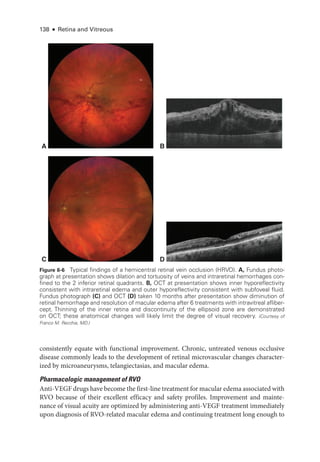
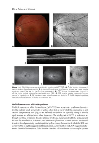
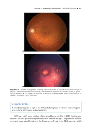
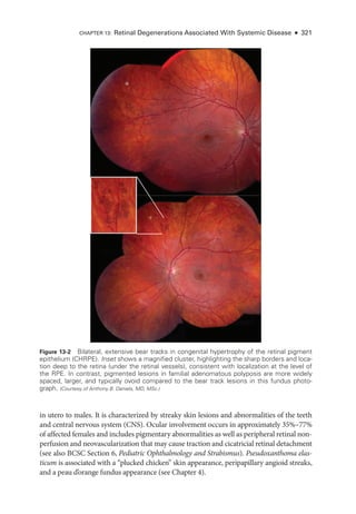
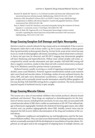
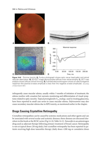
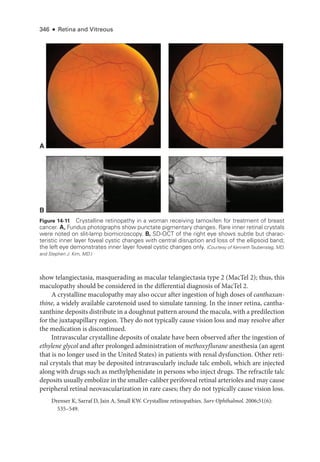
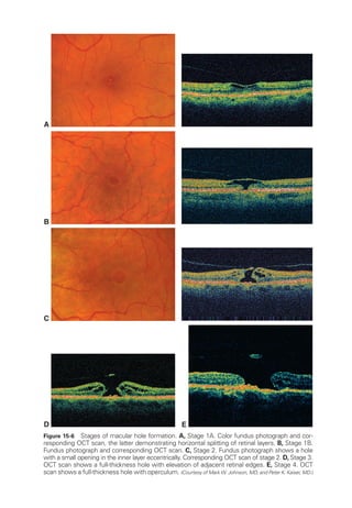
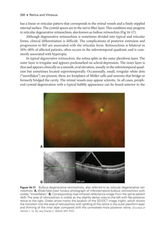
Downloaded 20 times


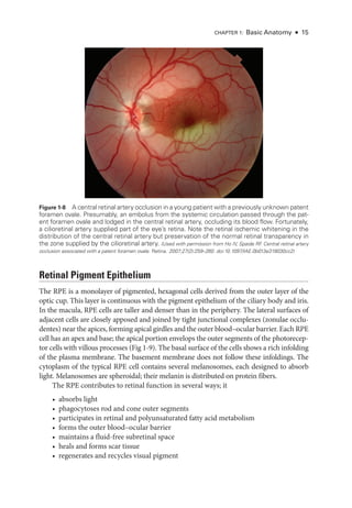


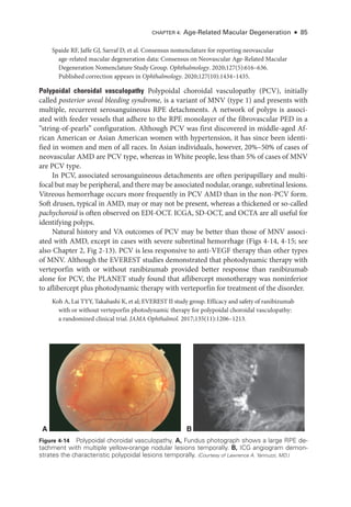
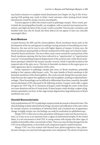
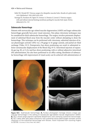
![Chapter 3: Ret
i
nal Physiology and Psychophysics ● 45
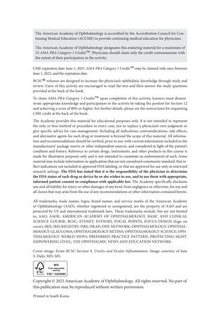
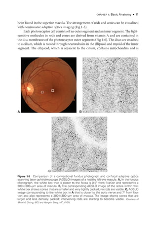
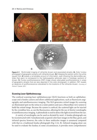
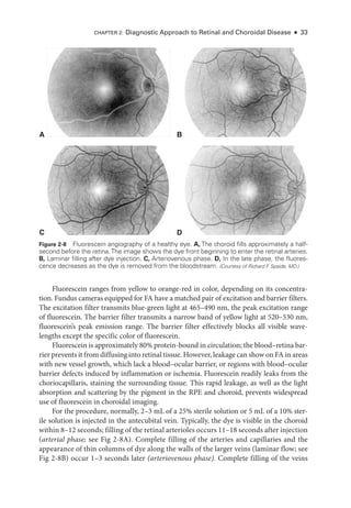
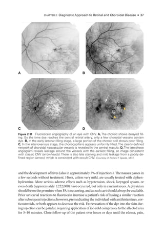
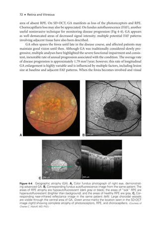
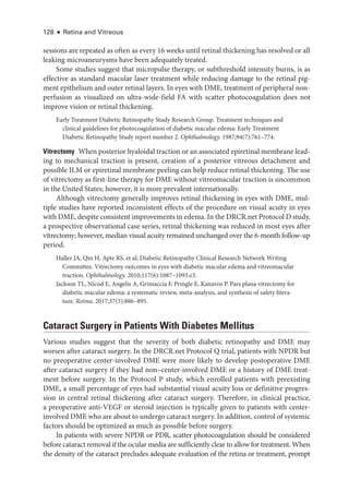
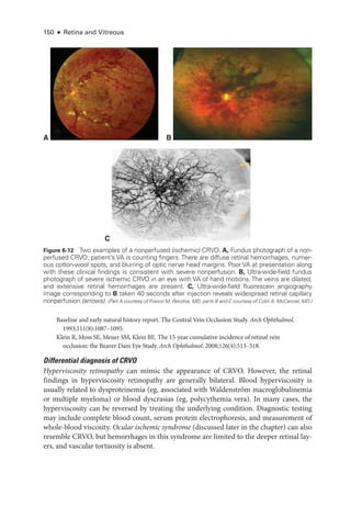
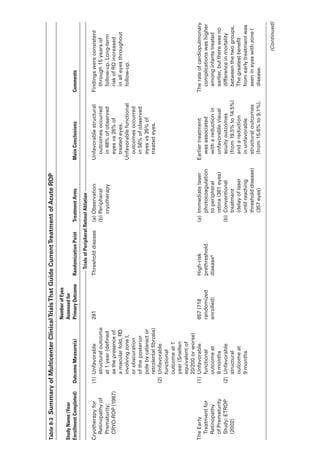
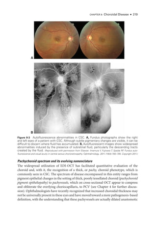
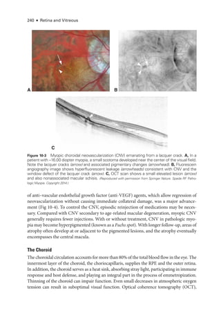
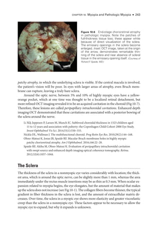
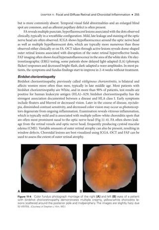
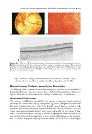
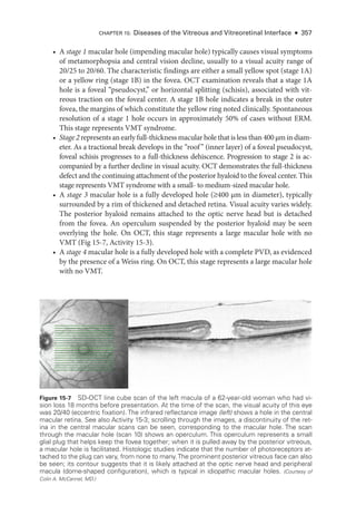
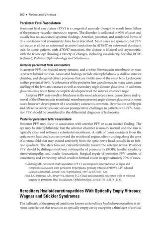
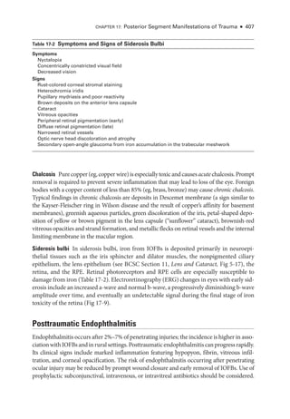
Full-
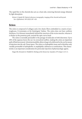
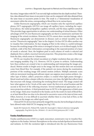
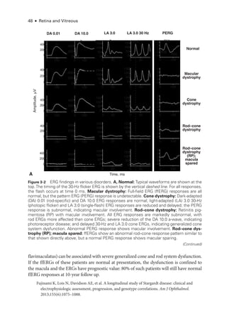
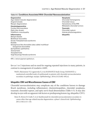
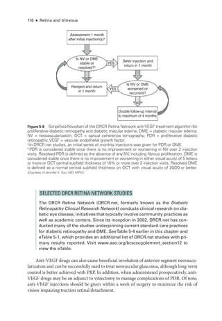
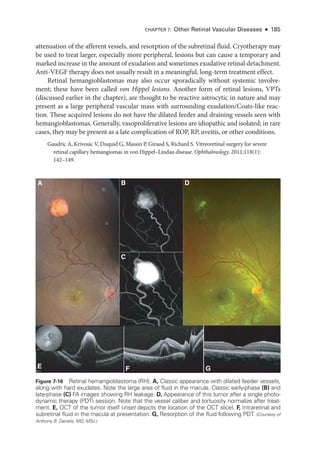
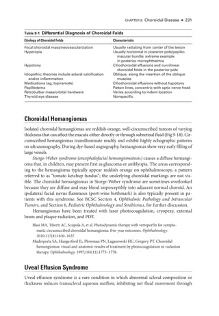
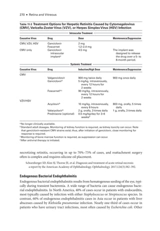
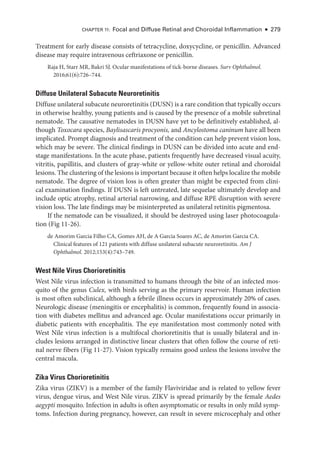
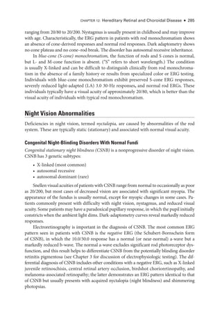
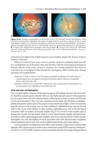
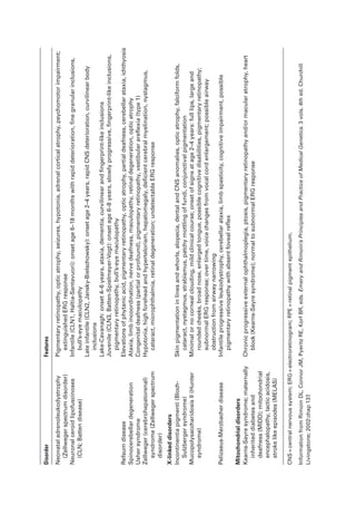
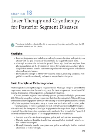
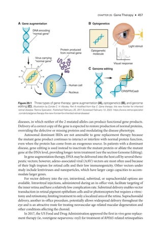
Field (Ganzfeld) Electroretinography
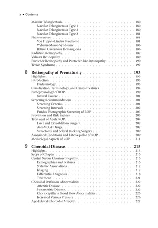
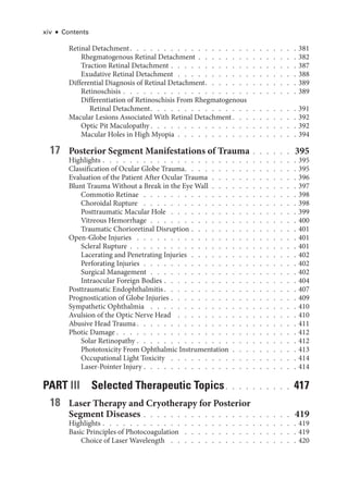
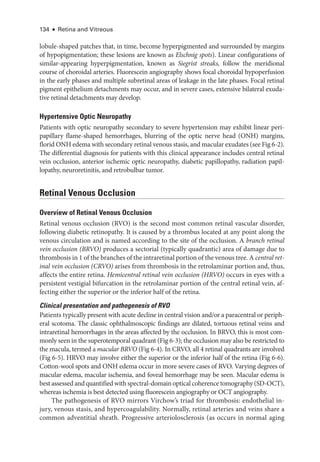
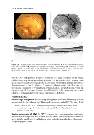
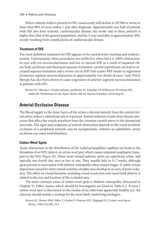
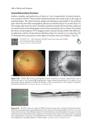
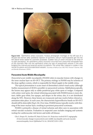
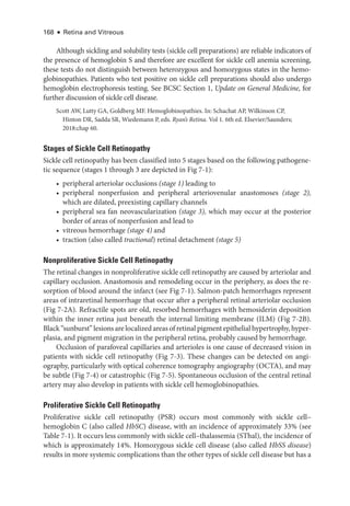
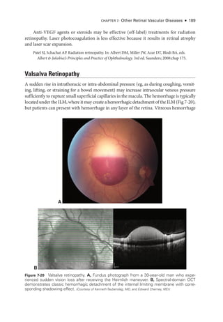
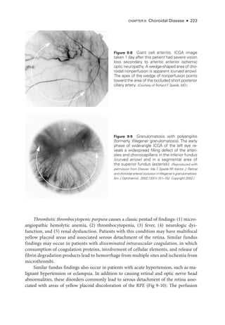
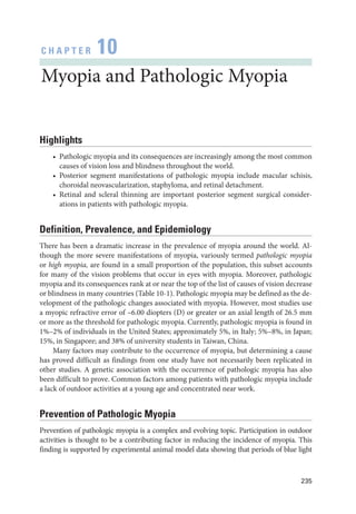
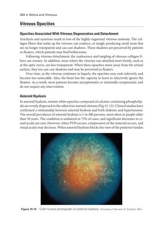
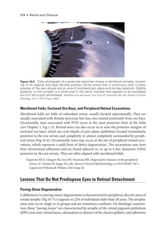
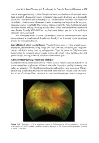
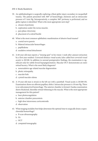
In full-
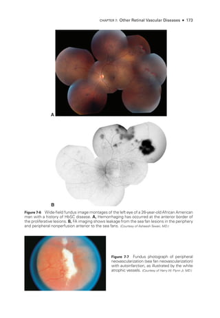
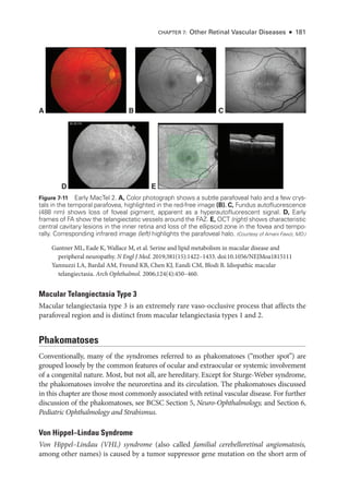
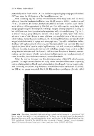
field electroretinography, a Ganzfeld bowl uniformly illuminates the entire ret
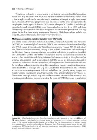
ina
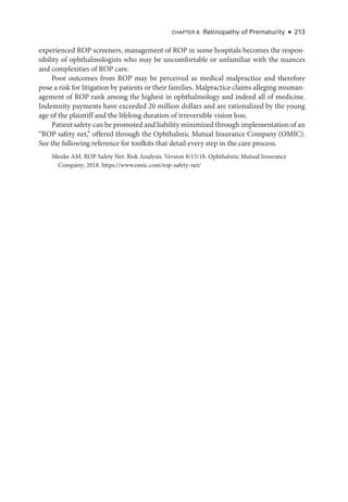
with a full-field luminance stimulus; the Ganzfeld also provides a uniform background for
photopic adaptation and photopic ERG recording. Regular calibration of flash strength is
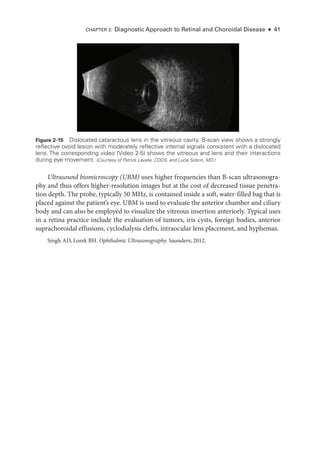
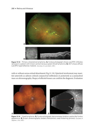
required for clinical accuracy. Figure 3 -1 shows typical full-
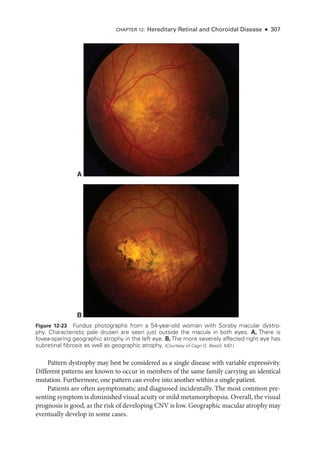
field ERG (ffERG) responses;
however, normal values vary with recording techniques, and each laboratory must estab
lish its own normative data (Video 3 -2). Even with standardization, variations in the type
of electrode and specific equipment used
will affect the test results.
VIDEO 3-2 Interpreting an electroretinogram.
Courtesy of Milam A. Brantley, MD, PhD.
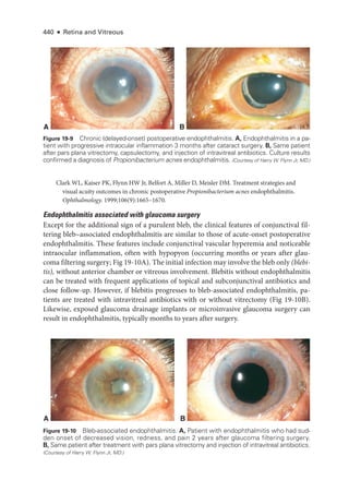
The pupils are dilated to maximize ret
i
nal illumination. In most laboratories, the pa
tient is dark-
adapted while the pupils are dilating, the electrodes are positioned
under
dim red light, and then stimulation commences by using an interstimulus interval sufficient
to allow the ret
ina to recover between flashes (from 2 seconds for low intensities up to
20 seconds for high intensities). Many laboratories rec
ord responses to a series of increas
ing stimulus strengths. The patient is then light-
adapted (using standardized background
intensity and adaptation time), and photopic testing is performed, in which the stimuli are
delivered
under rod-
suppressing background illumination.
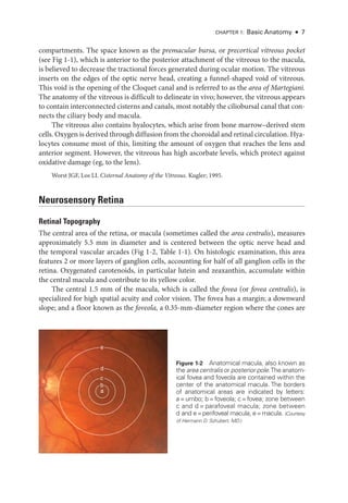
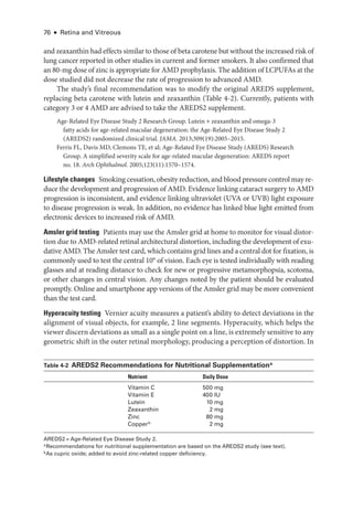
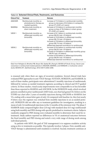
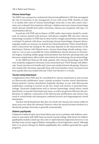
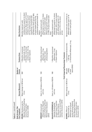
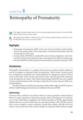
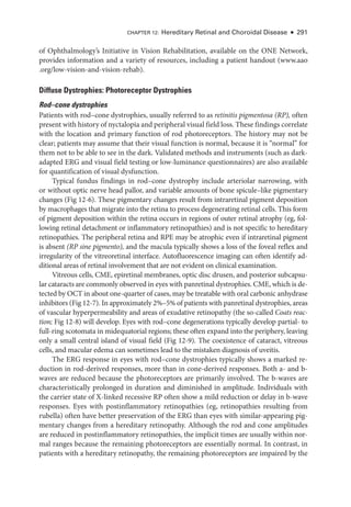
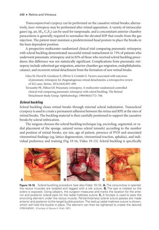
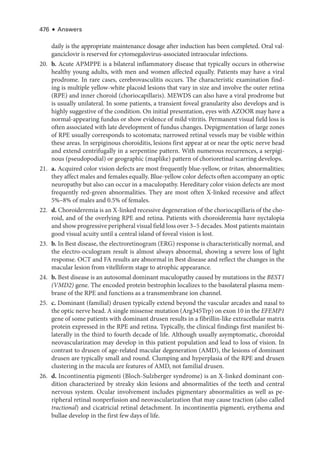
The ISCEV standard ffERG consists of 6 dif
fer
ent responses (see Fig 3 -1A). The no
menclature used for
these is based on the flash strength as mea
sured in candela-
seconds
per square meter and the adaptive state of the eye (ie, dark-
adapted [DA] or light-
adapted
[LA]); older terms are given in parentheses in the following list. Mea
sure
ment of the ERG
focuses on the size and timing of the major components, as indicated in Figure 3 -1.
1. DA 0.01 (rod-
specific): In this response, a b-wave arises in the on-bipolar cells (BPCs)
(inner nuclear layer) of the rod system. A reduction in this response identifies dys
function within the rod system, but
because it arises at an inner ret
i
nal level, this
response cannot differentiate between dysfunction at the level of the photoreceptor
and inner ret
i
nal dysfunction. It therefore acts as a mea
sure of rod system sensitivity.
2. DA 3.0 (mixed rod–
cone): This response consists of an a-
wave and a b-
wave. The
a-
wave at this flash strength usually has 2 peaks between approximately 15 and 21 milli
seconds,
either of which may be prominent.
Because only approximately the first
8 milliseconds of the DA a-
wave reflects photoreceptor hyperpolarization, the ISCEV
standard includes additional brighter flash testing for better diagnostic specificity.
3. DA 10.0/30.0: At
either of
these flash strengths, the a-
wave has an easily mea
sur
able
peak, and most of the a-
wave reflects photoreceptor function. Consequently, this
response can localize dysfunction to
either a photoreceptor or an inner ret
i
nal level.
Thus, a reduced DA 0.01 response accompanied by marked reduction in the a-
wave
of the DA 10.0/30.0 response indicates photoreceptor dysfunction. However, if
the a-
wave amplitude is normal (see Fig 3 -1A) or near normal and the b-
wave am
plitude is lower than that of the a-
wave (known as a negative or electronegative ERG
waveform), dysfunction occurs post-
phototransduction, at an inner ret
i
nal level.](https://image.slidesharecdn.com/12retinaandvitreous-250103182737-09041aee/85/Retina-and-Vitreous-64-320.jpg)
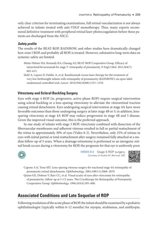
![Chapter 3: Ret
i
nal Physiology and Psychophysics ● 49
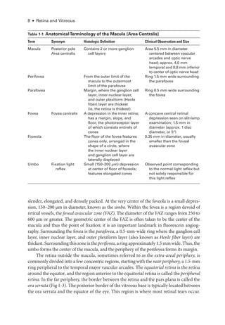
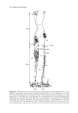
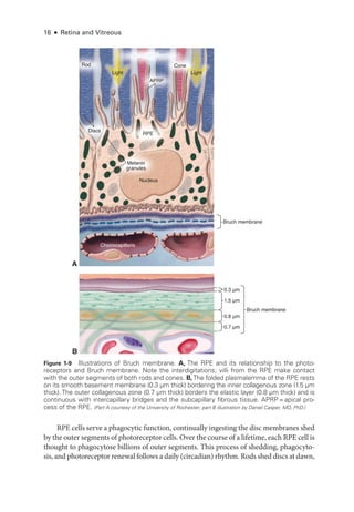
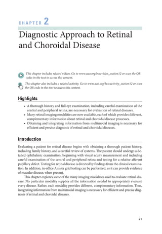
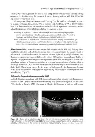
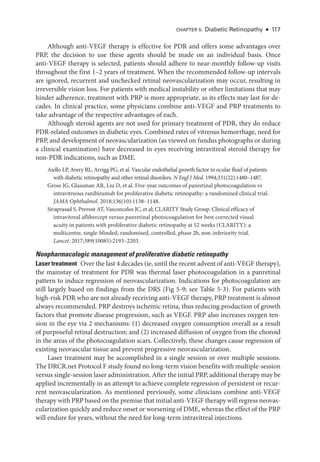
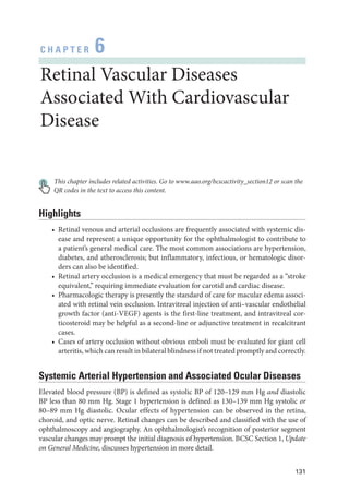
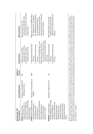
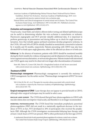
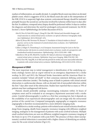
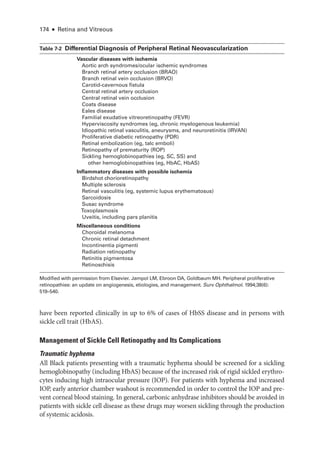
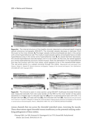
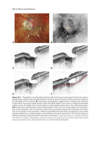
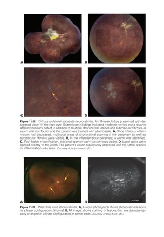
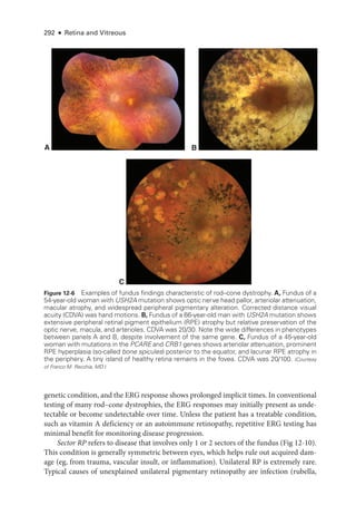
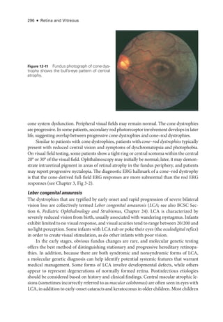
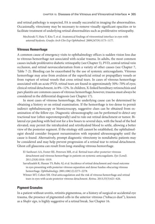
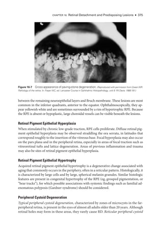
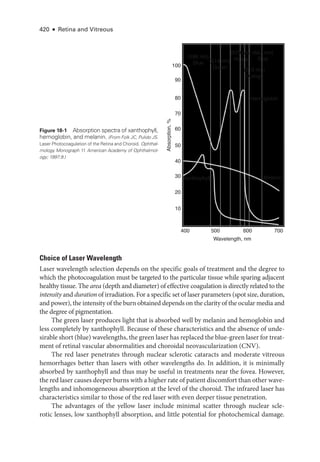
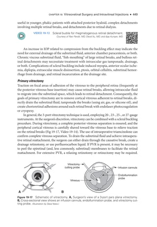
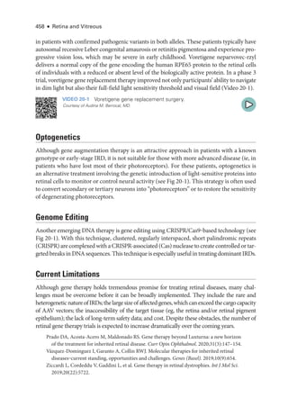
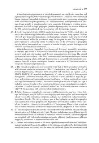
Multifocal Electroretinography
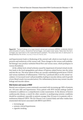
Multifocal electroretinography can produce a topographic ERG map of central ret
i
nal
cone system function, which can help the clinician diagnose macular dysfunction and
assess the extent of central ret
i
nal involvement in generalized ret
i
nal disease (Fig 3 -3).
The stimulus consists of multiple hexagons, smaller in the center than the periphery to
reflect cone photoreceptor density, each of which flashes in a pseudorandom sequence.
Time, ms
Amplitude,
μV
cCSNB
iCSNB
XLRS
BCM
0
0
0 0
0
0
0 0
0 0
0 100
–4
0 100
4
0
2
–2
–4
0 100
4
0
2
–2
–4
0 100
4
0
2
–2
–4
0 100
4
0
2
–2
–4
0 50
0
0 50
150
100
50
0
0 50
150
100
50
0
0 50
150
100
50
0
0 50
150
100
50
0
0 50
0
0 50
150
100
50
0
0 50
150
100
50
0
0 50
150
100
50
0
0 50
150
100
50
0
0
100
400
200
0
100
400
200
0
100
400
200
0
100
400
200
0
100
0
100
400
200
0
100
400
200
0
100
400
200
0
100
400
200
0
100
Time, ms
cCSNB
iCSNB
XLRS
BCM
0
0
0
0
0
0 100
–4
0 100
4
0
2
–2
–4
0 100
4
0
2
–2
–4
0 100
4
0
2
–2
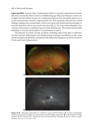
–4
0 100
4
0
2
–2
–4
0 50
0
0 50
150
100
50
0
0 50
150
100
50
0
0 50
150
100
50
0
0 50
150
100
50
0
0 50
0
0 50
150
100
50
0
0 50
150
100
50
0
0 50
150
100
50
0
0 50
150
100
50
0
0
100
400
200
0
100
400
200
0
100
400
200
0
100
400
200
0
100
100
100
100
100
100
Amplitude,
μV
0
0
0 0
0
0
0 0
0 0
0 100
–4
0 100
4
0
2
–2
–4
0 100
4
0
2
–2
–4
0 100
4
0
2
–2
–4
0 100
4
0
2
–2
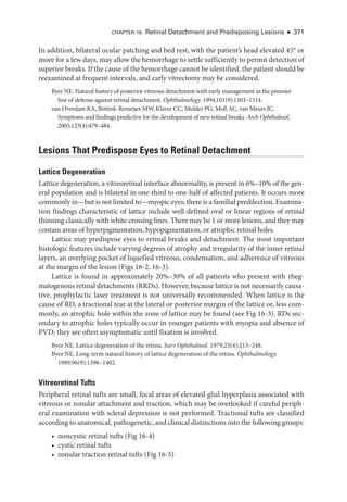
–4
0 50
0
0 50
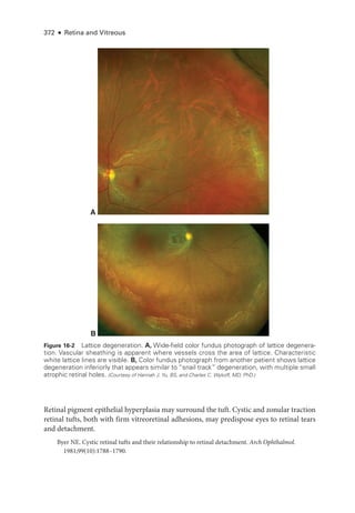
150
100
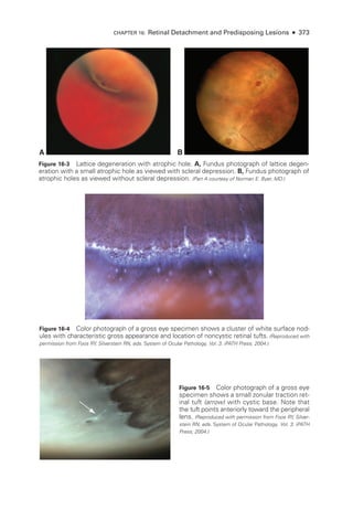
50
0
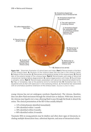
0 50
150
100
50
0
0 50
150
100
50
0
0 50
150
100
50
0
0 50
0
0 50
150
100
50
0
0 50
150
100
50
0
0 50
150
100
50
0
0 50
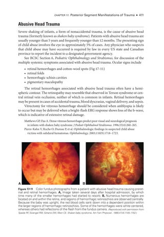
150
100
50
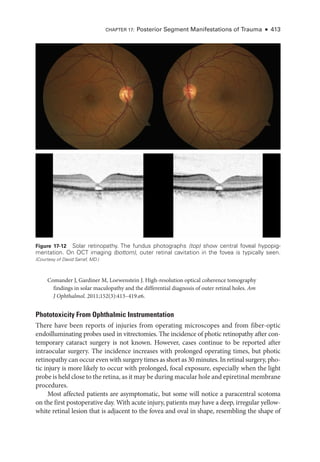
0
0
100
400
200
0
100
400
200
0
100
400
200
0
100
400
200
0
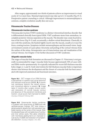
100
0
100
400
200
0
100
400
200
0
100
400
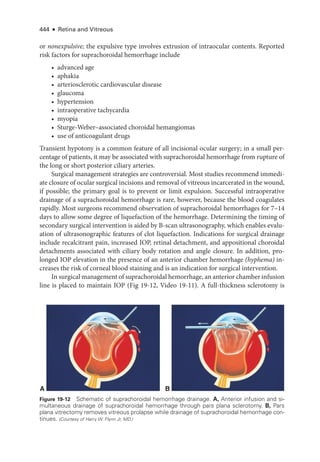
200
0
100
400
200
0
100
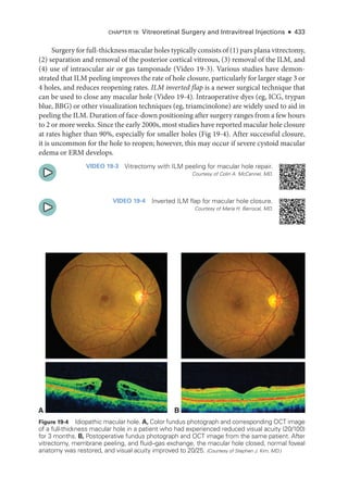
Time, ms
Amplitude,
μV
cCSNB
iCSNB
XLRS
BCM
0
0
0 0
0
0
0 0
0 0
0 100
–4
0 100
4
0
2
–2
–4
0 100
4
0
2
–2
–4
0 100
4
0
2
–2
–4
0 100
4
0
2
–2
–4
0 50
0
0 50
150
100
50
0
0 50
150
100
50
0
0 50
150
100
50
0
0 50
150
100
50
0
0 50
0
0 50
150
100
50
0
0 50
150
100
50
0
0 50
150
100
50
0
0 50
150
100
50
0
0
100
400
200
0
100
400
200
0
100
400
200
0
100
400
200
0
100
0
100
400
200
0
100
400
200
0
100
400
200
0
100
400
200
0
100
Time, ms
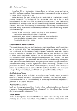
Rod–cone
dystrophy
(RP);
macula
spared
A
B
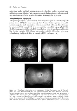
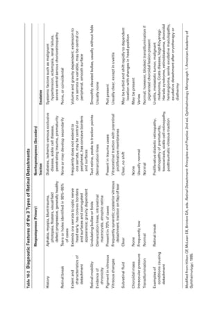
DA 0.01 DA 10.0 LA 3.0 30 Hz
LA 3.0 PERG
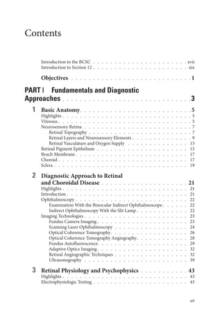
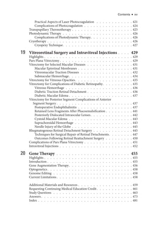
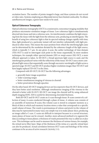
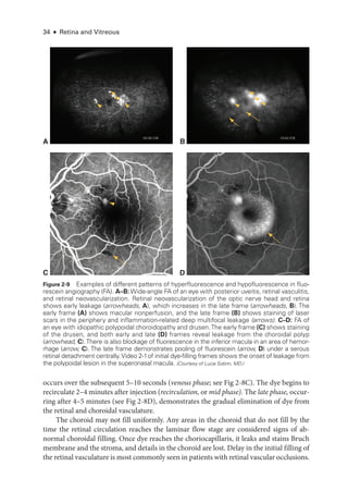
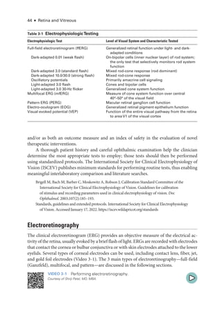
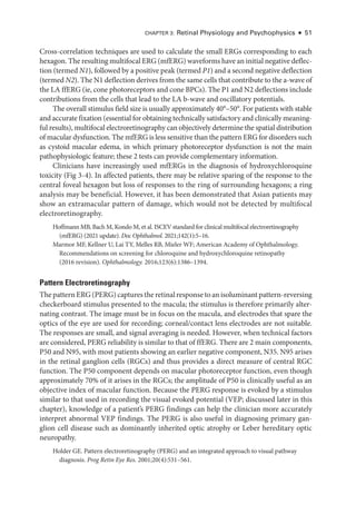
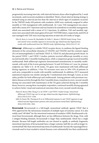
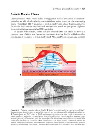
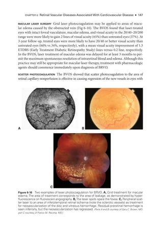
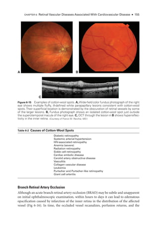
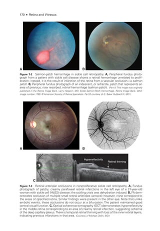
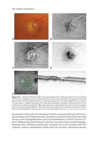
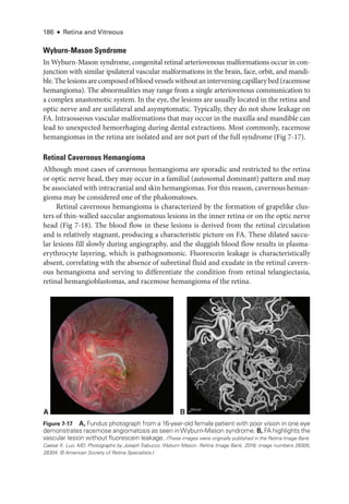
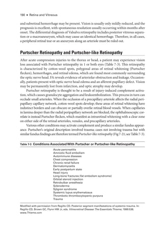
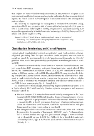
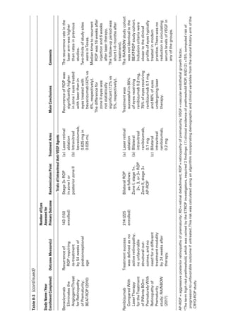
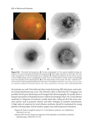
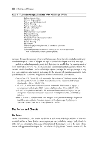
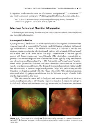
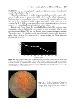
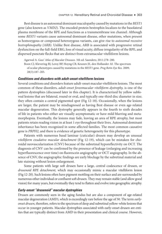
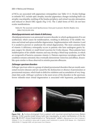
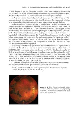
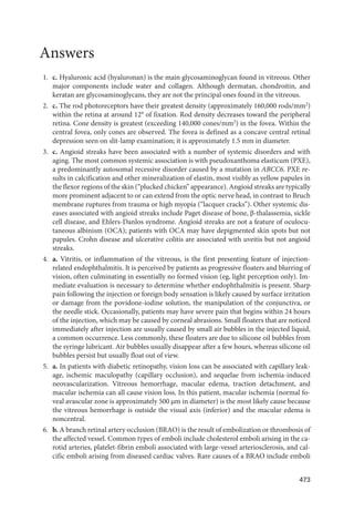
Figure 3-2 (continued) B, cCSNB (“complete” congenital stationary night blindness): Findings
show loss of on-
pathway function at a postreceptoral level. DA 0.01 response is undetectable;
DA 10.0 response is profoundly electronegative, with the normal a-
wave reflecting normal pho-
toreceptor function and the relatively marked b-
wave reduction indicating inner ret
i
nal disease;
30-
Hz flicker ERG shows only minor changes in waveform and mild delay; LA 3.0 ERG shows
changes diagnostic of loss of cone on-
pathway function but sparing of the off-
pathway. The
a-
wave commences normally but then shows a broadened trough.The b-
wave rises sharply, with
loss of the photopic oscillatory potentials, and marked reduction in the b-wave:a-wave ratio.
The PERG response is markedly subnormal. iCSNB (“incomplete” CSNB): DA 0.01 response
is subnormal but detectable; DA 10.0 response is markedly electronegative; 30-
Hz flicker ERG
response is markedly subnormal, showing delay and a characteristic triphasic waveform; LA 3.0
ERG shows a subnormal a-
wave and a markedly subnormal b-
wave, reflecting involvement of
both on-and off-
cone pathways; the PERG response is subnormal. XLRS (X-
linked retinoschisis):
DA 0.01 response is severely reduced; DA 10.0 response is profoundly electronegative; 30-
Hz
flicker ERG shows delay; LA 3.0 ERG shows delay and marked reduction in the b-wave:
a-wave
ratio; the PERG response is markedly subnormal. BCM (blue-
cone [S-
cone] monochromatism):
DA 0.01 and DA 10.0 ERG responses are normal; 30-
Hz flicker ERG response is virtually unde-
tectable; LA 3.0 response shows only a small b-
wave at approximately 50 ms, consistent with
an S-
cone origin; PERG response is undetectable. (Courtesy of Graham E. Holder, PhD.)](https://image.slidesharecdn.com/12retinaandvitreous-250103182737-09041aee/85/Retina-and-Vitreous-68-320.jpg)
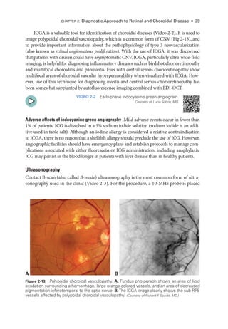
![E
1 µV
50 ms
Amp.P1 [nV/deg2
]
D
DA 0.01 DA 10.0
400
200
0
0 100
400
200
0
0
100
400
200
0
200
150
100
50
0
0 100 0 50
200
150
100
50
0
0 50
4
2
0
–2
–4
0 100
4
2
0
–2
–4
0 100
4
2
0
–2
–4
0 100
4
2
0
–2
–4
0 100
200
150
100
50
0
0 50
200
150
100
50
0
0 50
400
200
0
0 100
LA 3.0 30 Hz LA 3.0 PERG 15° PERG 30°
A B 200 µm
C
DA 0.01 DA 10.0
400
200
0
200
150
100
50
0
0 100 0 50
200
150
100
50
0
0 50
4
2
0
–2
–4
0 100
4
2
0
–2
–4
0 100
4
2
0
–2
–4
0 100
4
2
0
–2
–4
0 100
200
150
100
50
0
0 50
200
150
100
50
0
0 50
400
200
0
0 100
LA 3.0 30 Hz LA 3.0 PERG 15° PERG 30°
D
DA 0.01 DA 10.0
400
200
0
0 100
400
200
0
0
100
400
200
0
200
150
100
50
0
0 100 0 50
200
150
100
50
0
0 50
4
2
0
–2
–4
0 100
4
2
0
–2
–4
0 100
200
150
100
50
0
0 50
200
150
100
50
0
0 50
400
200
0
0 100
LA 3.0 30 Hz LA 3.0 PERG 15°
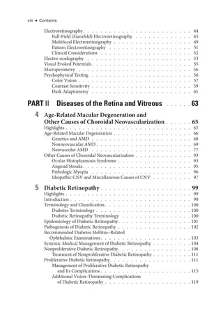
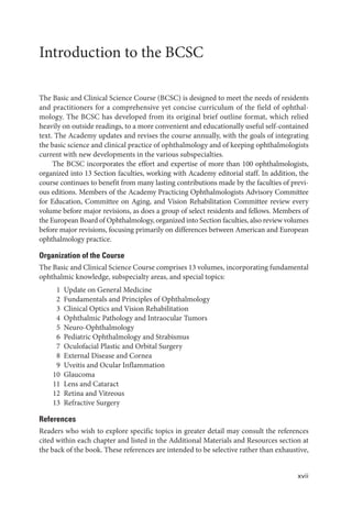
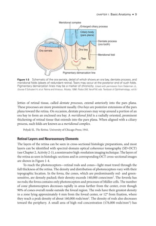
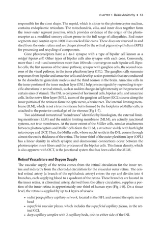
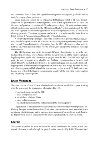
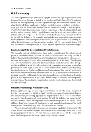
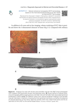
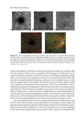
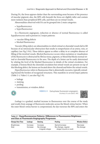
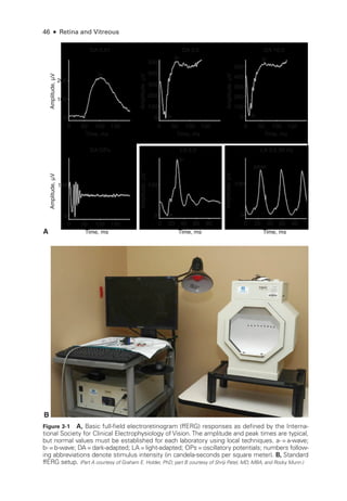
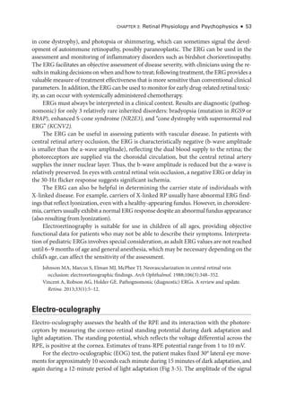
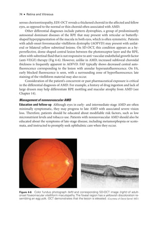
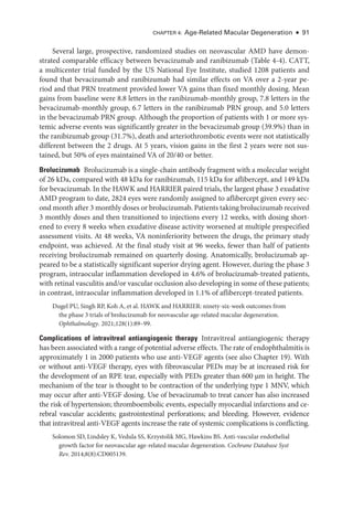
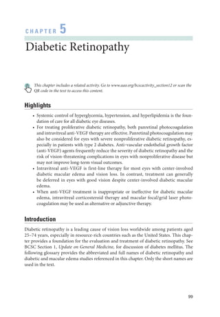
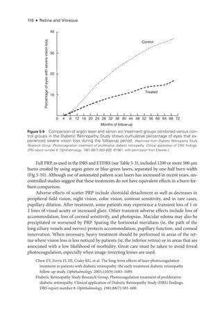
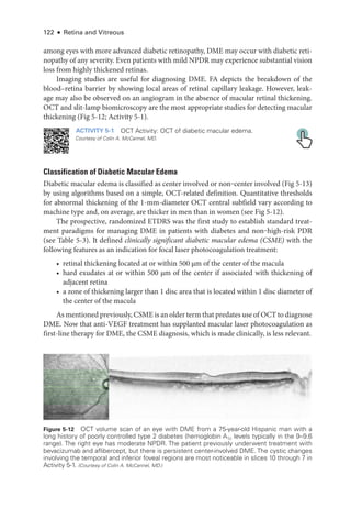
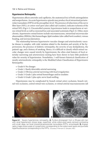
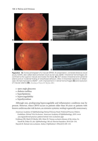
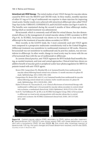
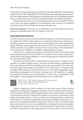
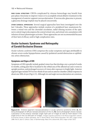
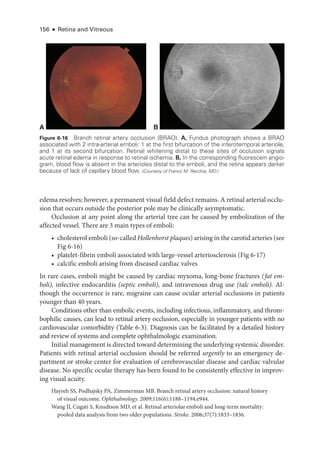
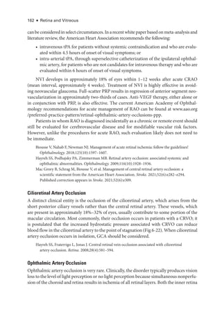
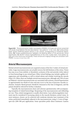
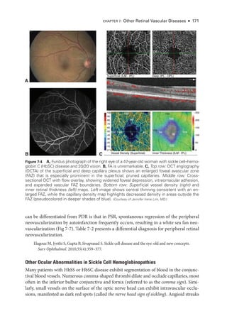
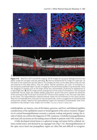
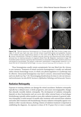
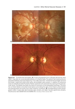
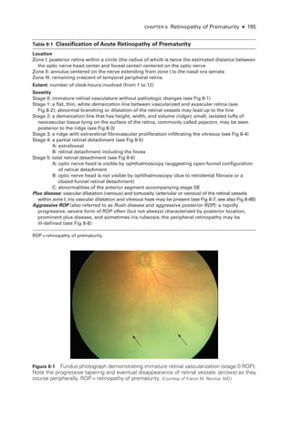
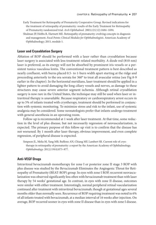
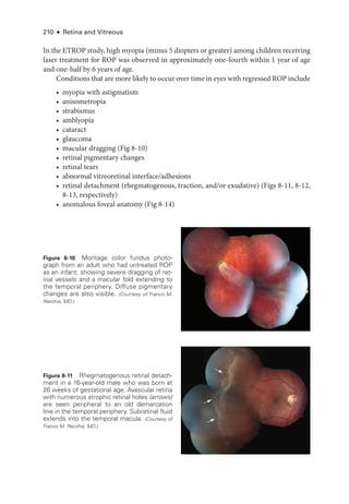
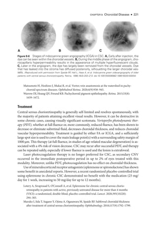
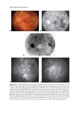
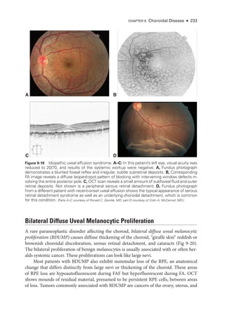
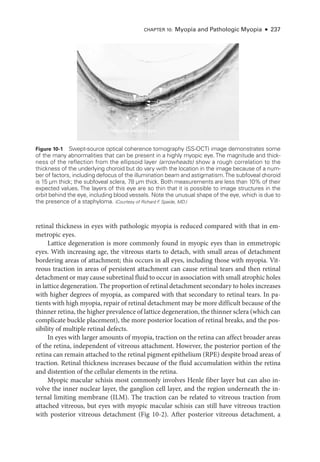
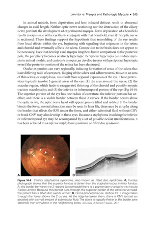
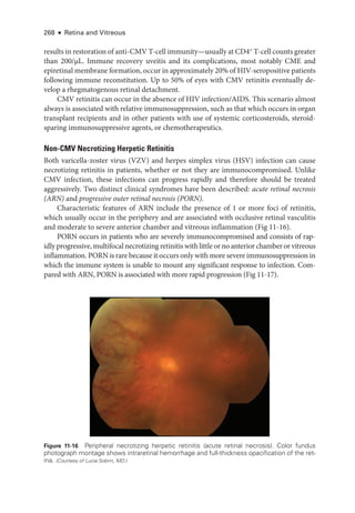
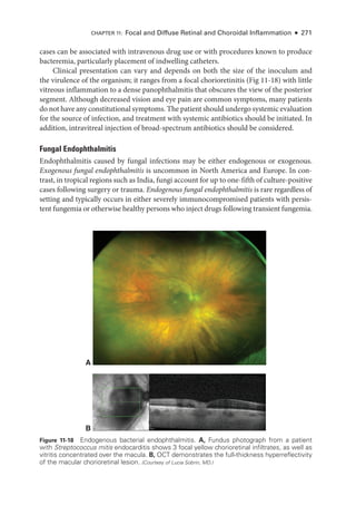
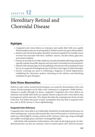
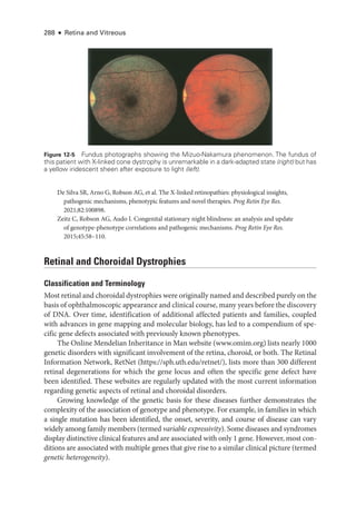
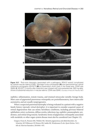
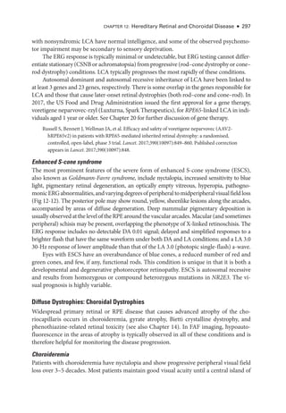
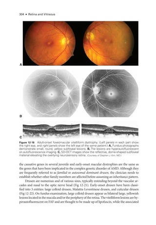
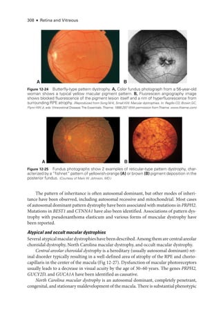
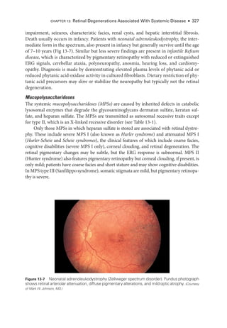
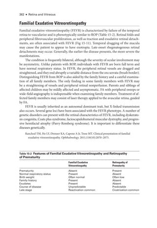
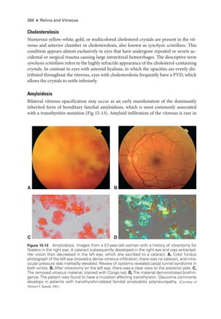
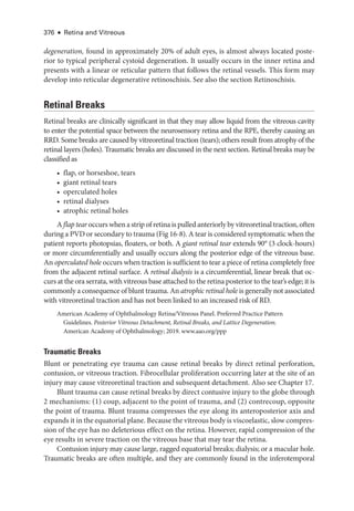
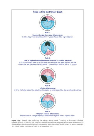
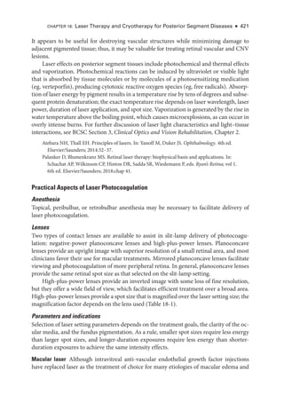
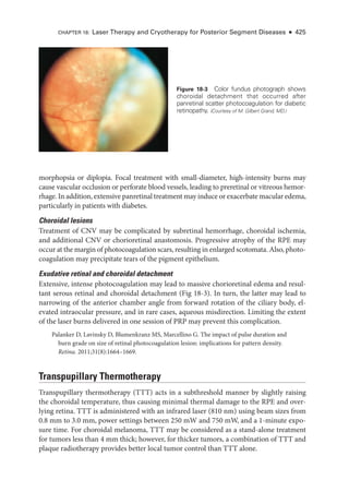
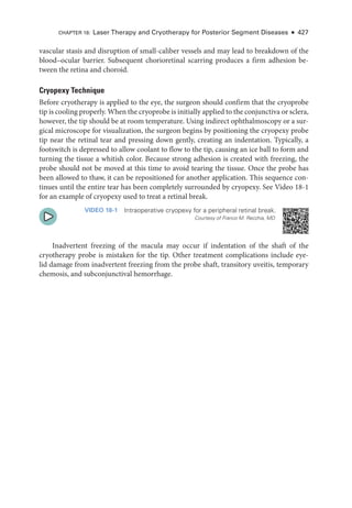
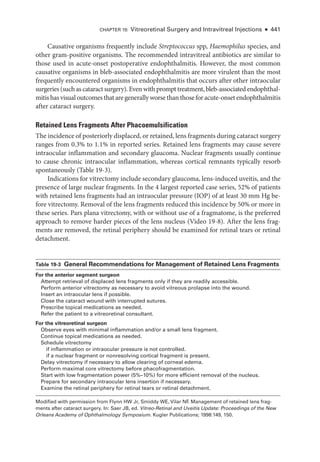
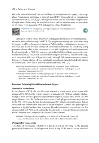
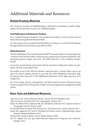
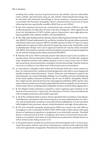
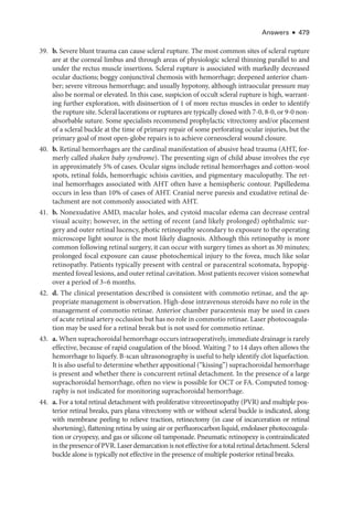
Figure 3-3 Multifocal ERG (mfERG), full-
field ERG (ffERG), and pattern ERG (PERG) from a pa-
tient with ABCA4 retinopathy (Stargardt disease; fundus flavimaculatus) show the importance
of fixation in mfERG recording and interpretation. A–C: Macular atrophy centralized on the
fovea. A, Fundus photograph. B, Fundus autofluorescence image. C, Near-
infrared and spectral-
domain optical coherence tomography (SD-
OCT) images. D, ffERG responses are normal (see
Fig 3-1 for explanation of abbreviations; x-
axis = ms; y-
axis = μV); PERG response to a 15° field
is undetectable; however, PERG response to a 30° field is pre
sent but subnormal. E, mfERG
shows an area of dysfunction that is localized but apparently not around the fovea, which sim-
ply reflects the eccentric fixation often pre
sent in a patient with a central scotoma. (Courtesy of
Graham E. Holder, PhD.)](https://image.slidesharecdn.com/12retinaandvitreous-250103182737-09041aee/85/Retina-and-Vitreous-69-320.jpg)
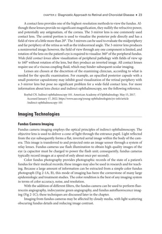
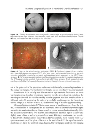
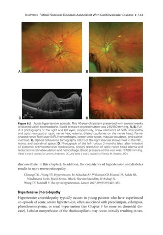
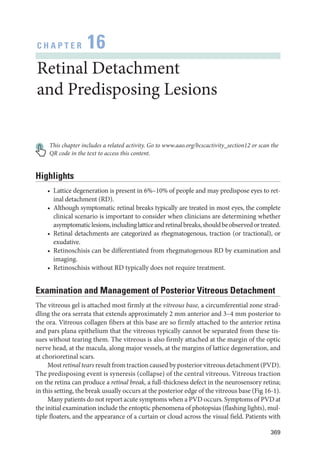
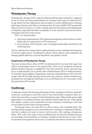
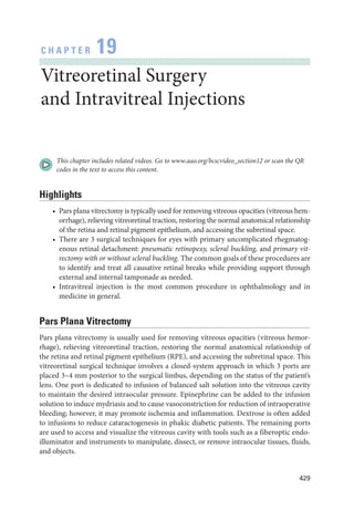
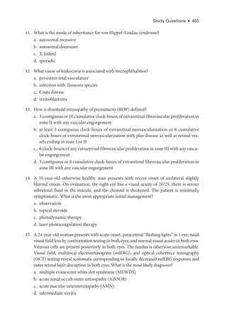
![52 ● Retina and Vitreous
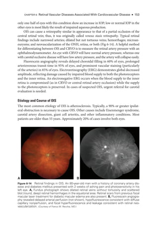
Clinical Considerations
The ERG provides objective data on ret
i
nal function and is therefore impor
tant in the diag
nosis, management, and follow-up of ret
i
nal disease. Symptomatic indications include nycta
lopia, which requires distinguishing between the potentially blinding rod–
cone dystrophies
and the relatively benign congenital stationary night blindness (CSNB). The dystrophies are
associated with markedly abnormal a-
waves in DA bright-
flash ERGs; CSNB is usually
associated with a normal a-
wave and a negative ERG waveform (see Fig 3 -2). Other symp
tomatic indications include photophobia, which indicates generalized cone dysfunction (as
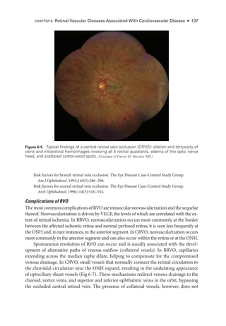
A B
C
D
200 μm
200 μm
2 μV
50 ms
Amp.P1 [nV/deg2
]
100 μV/Div
DA 0.01 DA 10.0
50 ms/Div
+
100 μV/Div
50 ms/Div
+
100 μV/Div
50 ms/Div
+
100 μV/Div
50 ms/Div
+
100 μV/Div
50 ms/Div
+
LA 3.0 30 Hz LA 3.0 PERG 15°
100 μV/Div
DA 10.0
50 ms/Div
+
100 μV/Div
50 ms/Div
+
100 μV/Div
50 ms/Div
+
100 μV/Div
50 ms/Div
+
LA 3.0 30 Hz LA 3.0 PERG 15°
100 μV/Div
DA 0.01 DA 10.0
50 ms/Div
+
100 μV/Div
50 ms/Div
+
100 μV/Div
50 ms/Div
+
100 μV/Div
50 ms/Div
+
LA 3.0 30 Hz LA 3.0
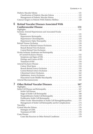
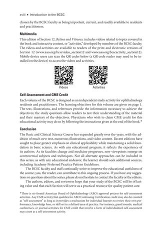
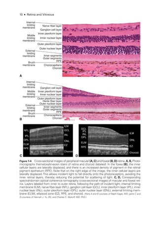
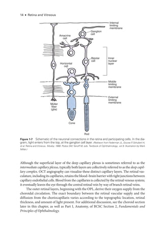
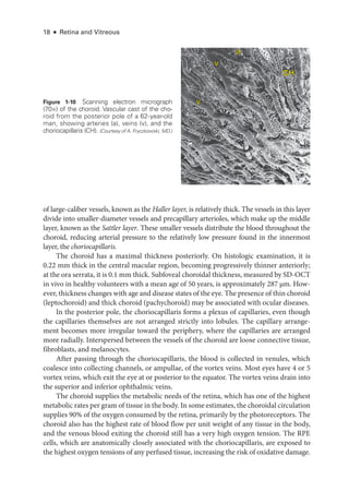
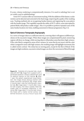
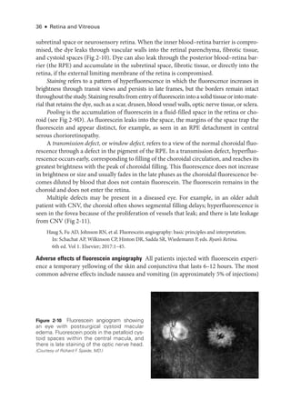
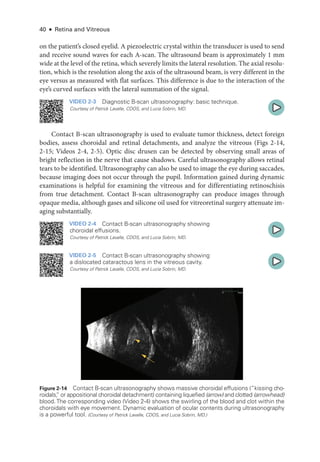
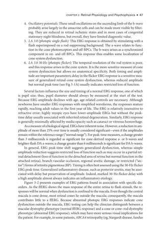
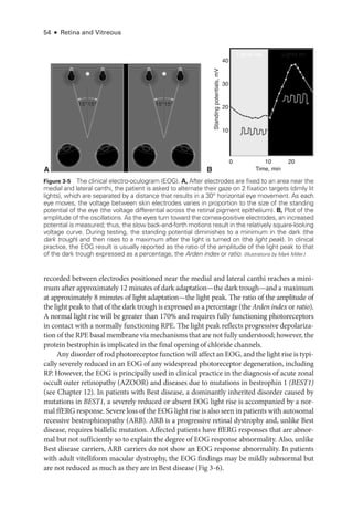
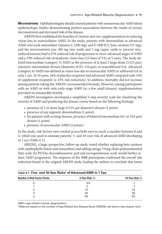
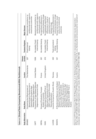
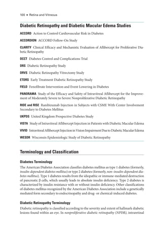
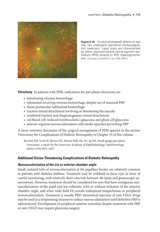
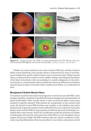
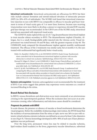
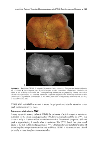
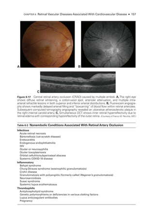
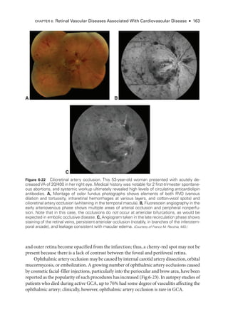
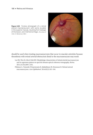
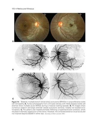
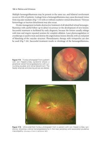
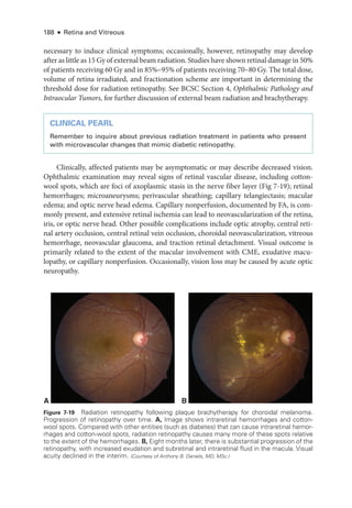
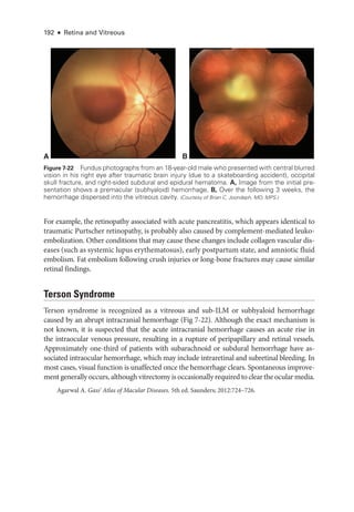
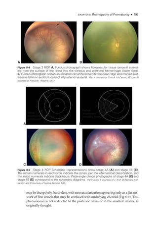
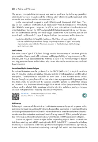
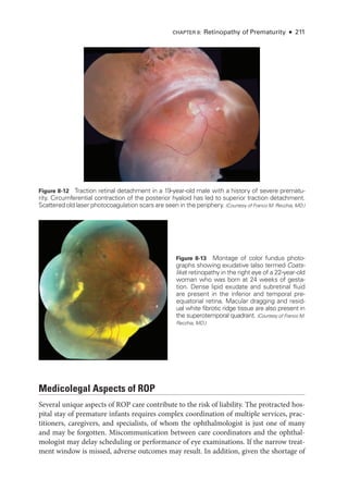
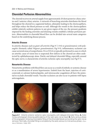
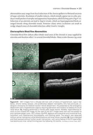
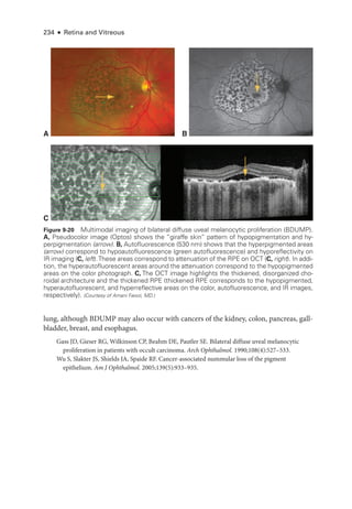
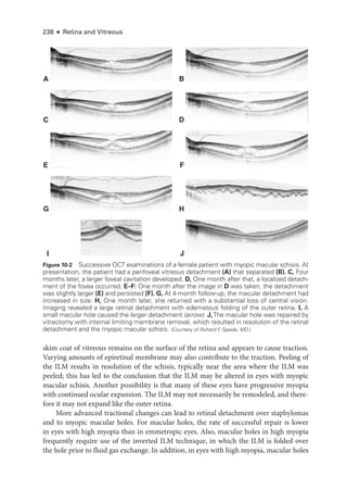
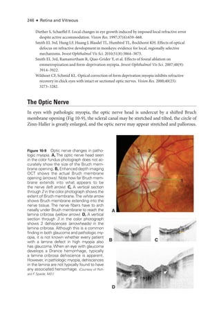
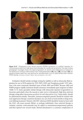
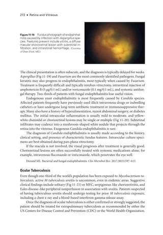
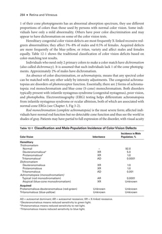
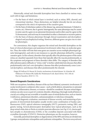
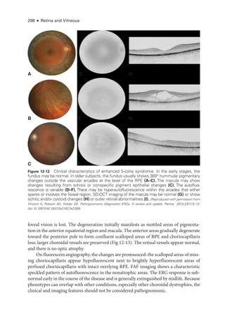
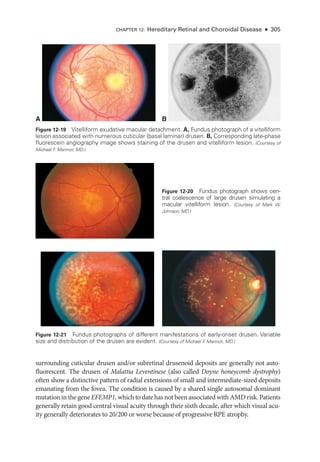
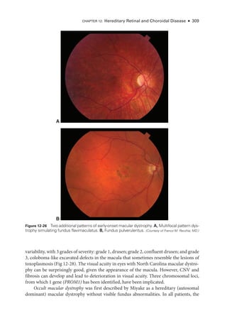
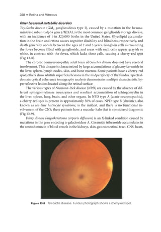
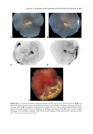
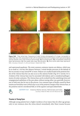
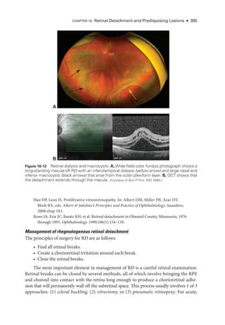
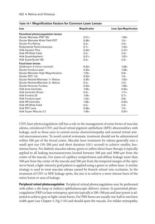
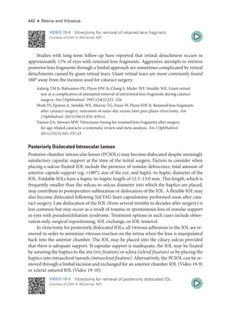
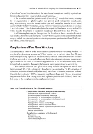
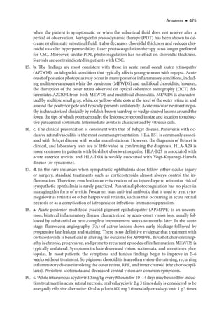
Figure 3-4 mfERG, ffERG, and PERG from a patient with hydroxychloroquine toxicity. A, Fun-
dus autofluorescence image. B, Near-
infrared and SD-
OCT images. The changes shown on
OCT are less marked, particularly temporal to the fovea, than may have been predicted by the
degree of functional vision loss. C, ffERG responses are normal (see Fig 3-1 for explanation of
abbreviations); PERG response to a 15° field is barely detectable. D,The mfERG shows marked
abnormality with some sparing of the response to the central foveal hexagon (inner circle) but
loss of parafoveal responses (outer circle). (Courtesy of Graham E. Holder, PhD.)](https://image.slidesharecdn.com/12retinaandvitreous-250103182737-09041aee/85/Retina-and-Vitreous-71-320.jpg)
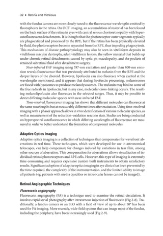
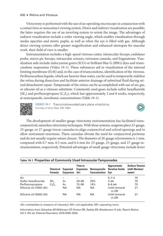
![Chapter 3: Ret
i
nal Physiology and Psychophysics ● 55
Arden GB, Constable PA. The electro-
oculogram. Prog Retin Eye Res. 2006;25(2):207–248.
Burgess R, Millar ID, Leroy BP, et al. Biallelic mutation of BEST1
causes a distinct retinopathy
in humans. Am J Hum Genet. 2008;82(1):19–31.
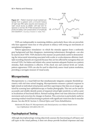
Visual Evoked Potentials
Visual evoked potential (VEP; also called visual evoked cortical potential [VECP] or re-
sponse [VER]) testing mea
sures electrical signals produced in the brain in response to
stimulation of the ret
ina by
either light flashes (flash VEP) or patterned stimuli, usually a
pattern-
reversing black-
and-
white checkerboard displayed on a monitor (pattern-reversal
VEP or pattern onset-
offset VEP). The signals are recorded via electrodes placed on the
occipital scalp. The VEP is extracted from the larger background electroencephalogram
by averaging the responses to multiple reversals or flashes. Pattern-
reversal VEPs have
a similar waveform across a population and a remarkably consistent timing; amplitudes
show greater variability. Flash VEPs are far more variable across a population but can be
useful when clinicians compare eyes or hemispheric responses in the same patient; hemi
spheric comparison requires multiple recording channels. A normal pattern-
reversal VEP
(Fig 3 -7) contains a major positive component at approximately 100 milliseconds, P100.
Measurement is usually taken of P100 amplitude and peak time (sometimes called latency).
In adults, VEPs are often used to demonstrate optic nerve conduction delay, particularly
in patients with suspected multiple sclerosis; patients with demyelinating optic neuritis
almost invariably show VEP delay even when vision recovers (see Fig 3 -7). However, there
can be subclinical delay in patients without any history or signs of optic neuropathy. In
most optic nerve diseases, VEP delay is pre
sent, but a VEP abnormality can be confined
to amplitude (interocular asymmetry), for example, as in patients with nonarteritic anterior
ischemic optic neuropathy. It is impor
tant to note that a delayed VEP is not diagnostic of
optic nerve disease. Macular dysfunction can cause similarly abnormal findings, and assess
ment of macular function with multifocal or pattern electroretinography enables improved
VEP interpretation. In patients with medically unexplained vision loss, VEPs are crucial
when the vision loss is suspected to be nonorganic. Though nonspecific, VEPs can objec
tively demonstrate normal function in the presence of symptoms that suggest other
wise.
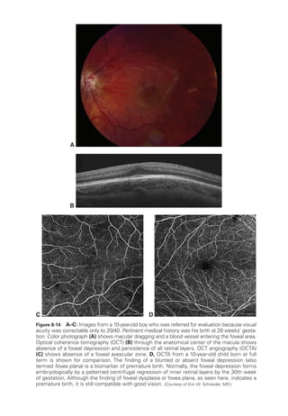
A B
Normal EOG Best disease
Dark
AR: 2.18 AR: 1.18
AR: 2.30
OD
OS
AR: 1.17
OS
Light Dark
OD Light
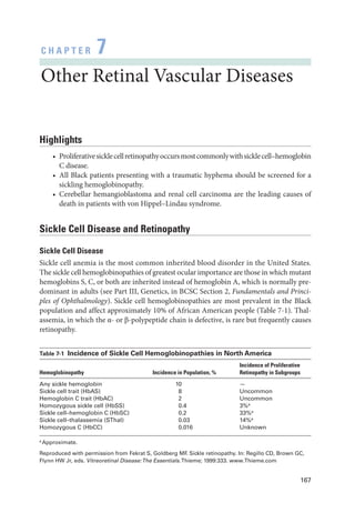
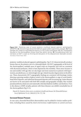
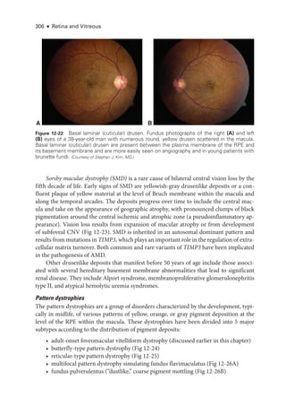
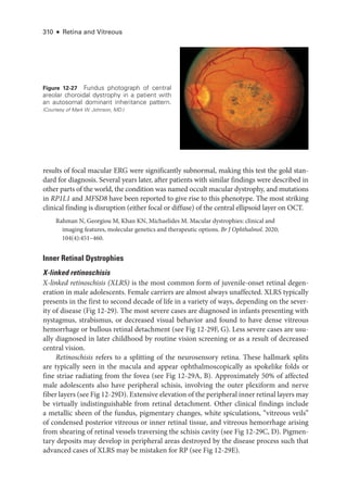
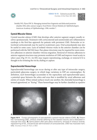
Figure 3-6 Best disease. A, Fundus photo
graph shows an egg yolk–
like vitelliform macular le-
sion. B,The EOG demonstrates a reduced light peak to dark trough (Arden) ratio compared with
a normal EOG. AR =Arden ratio. (From Gundogan FC, Yolcu U. Clinical ocular electrophysiology. In: Davey P
, ed.
Ophthalmology: Current Clinical and Research Updates. IntechOpen; 2014. doi:10.5772/57609)](https://image.slidesharecdn.com/12retinaandvitreous-250103182737-09041aee/85/Retina-and-Vitreous-74-320.jpg)
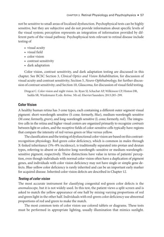
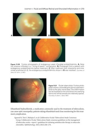
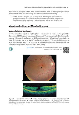
![58 ● Retina and Vitreous
Pseudoisochromatic plates, such as the Ishihara plates (which assess color discrimination
along protan [red] and deutan [green] axes only) and Hardy-Rand-Rittler plates (which
also assess the tritan [blue] axis), present colored numbers or figures against a background
of colored dots (Fig 3 -8). The colors of both figure and background are selected from hues
that are difficult for a person with abnormal color vision to distinguish. Individuals with
defective color vision see
either no pattern at all or an alternative pattern based on bright
ness rather than hue. These tests are quick to perform and sensitive for screening for color
vision abnormalities, but they are not effective in classifying the deficiency.
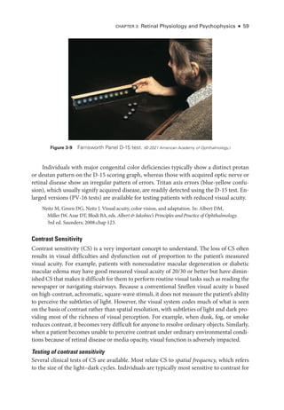
Panel tests, including the Farnsworth-
Munsell 100 and the Farnsworth Panel D-15
hue tests, are more accurate in classifying color deficiency. The Farnsworth-Munsell 100-hue
test is very sensitive
because the difference in hues between adjacent tablets approximates
the minimum that a typical observer can distinguish (1–4 nm). The spectrum is divided
into 4 parts of 25 colored tablets each, and the patient is asked to discriminate between
subtle shades of similar colors. Testing is tiring and time-
consuming but is more sensitive
in identifying difficulties with hue discrimination and color confusion.
Consisting of only 15 colored tablets, the Farnsworth Panel D-
15 test (Fig 3 -9) is
quicker than the Farnsworth-
Munsell 100 and more con
ve
nient for routine clinical use.
The hues are more saturated, and they cover the spectrum so that patients
will confuse
colors for which they have deficient perception (such as red and green). The patient is
asked to arrange the tablets in sequence, and errors can be quickly plotted to define the
color deficiency. The D-15 test may miss mildly affected individuals, but it is still deemed
useful
because of its speed. The relative insensitivity may also be an asset in judging the
practical significance of mild degrees of color deficiency. For example, individuals who
fail the Ishihara plates but pass the D-15 test
will prob
ably not have color discrimination
prob
lems
under most circumstances and in most occupations. Desaturated versions of
the D-15 test, such as the L’Anthony D-15, which recognize more subtle degrees of color
deficiency, are perhaps more clinically useful.
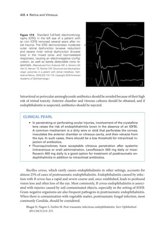
Figure 3-8 Pseudoisochromatic plates. (Courtesy of Carl Regillo, MD.)](https://image.slidesharecdn.com/12retinaandvitreous-250103182737-09041aee/85/Retina-and-Vitreous-77-320.jpg)


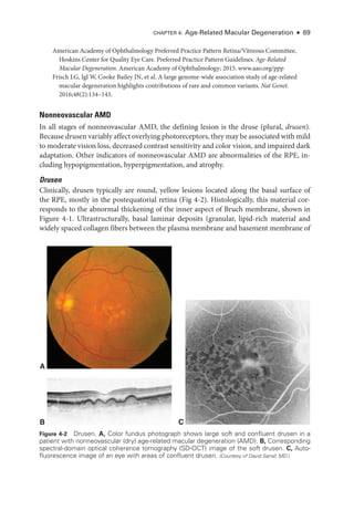
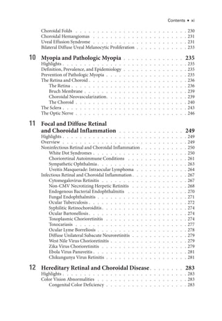
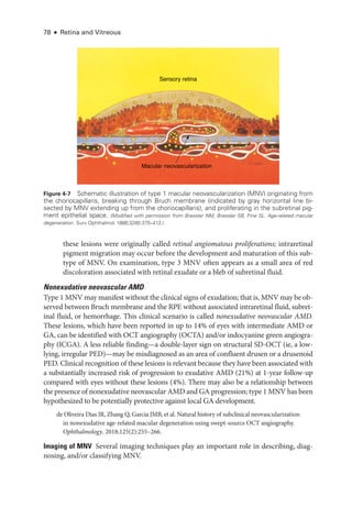
![Chapter 4: Age-
Related Macular Degeneration ● 77
patients with intermediate AMD, preferential hyperacuity perimetry (PHP), which has
been studied extensively, can detect recent-
onset MNV with high sensitivity (82%) and
high specificity (88%). The HOME study, a phase 3 randomized clinical trial with 1520
participants, demonstrated the efficacy and potential benefit of PHP in early detection
of MNV.
Shape-
discrimination hyperacuity uses a princi
ple similar to that of PHP but instead
tests for discrimination of shapes, such as the ability to discern a perfect circle from a
distorted contour.
Chew EY, Clemons TE, Harrington M, et al; AREDS2-
HOME Study Research Group.
Effectiveness of dif
fer
ent monitoring modalities in the detection of neovascular age-
related
macular degeneration: the Home Study, report number 3. Retina. 2016;36(8):1542–1547.
Neovascular AMD
The presence of MNV is the defining characteristic of neovascular AMD. Degenerative
changes in Bruch membrane (eg, the accumulation of drusen and progressive thickening
of the membrane that characterize nonneovascular AMD) and possibly the choriocapil-
laris may lead to a proangiogenic environment, with pathologic neovascularization devel-
oping from the choriocapillaris or from the neurosensory ret
ina itself.
These new vessels,
which may be accompanied by fibroblasts, may leak and bleed, disrupting the normal ret
i
nal architecture with a degenerate fibrovascular complex; when untreated, this complex
ultimately produces a hypertrophic, fibrotic, disciform scar.
Signs and symptoms of neovascular AMD
Patients with neovascular AMD may describe a sudden decrease in vision, metamorphop-
sia, and/or paracentral scotomata. Amsler grid self-testing by patients is highly effective in
detecting early exudative AMD. Clinical signs of MNV may include subretinal or intraret
i
nal fluid (eg, cystoid macular edema [CME]), exudate and/or blood, a pigment ring or
gray-
green membrane, irregular elevation of the RPE or a PED, an RPE tear, and/or a sea
fan pattern of subret
i
nal vessels.
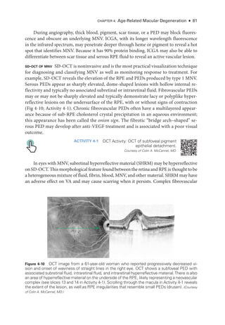
Anatomical classification of MNV
The 3 main subtypes of MNV are based on level of origin:
• In type 1 MNV, also called occult CNV, new vessels originating from the choriocap-
illaris grow through Bruch membrane into the sub-
RPE space (Fig 4-7). Fluid leak-
age and bleeding may produce a vascularized serous or fibrovascular PED.
These
fibrovascular PEDs typically have an irregular surface contour.
• In type 2 MNV, also called classic CNV, new vessels extend into the space between
the RPE and the neurosensory ret
ina. On examination, this may appear as a lacy
or gray-
green lesion. Neovascularization that exists beneath both the neurosensory
ret
ina and the RPE is designated as mixed type 1 and type 2 MNV, also termed
minimally classic CNV.
• In type 3 MNV, the pathologic vessels develop from the deep capillary plexus of the
ret
ina and grow downward
toward the RPE.
Because of their intraret
i
nal origin,](https://image.slidesharecdn.com/12retinaandvitreous-250103182737-09041aee/85/Retina-and-Vitreous-96-320.jpg)
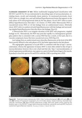
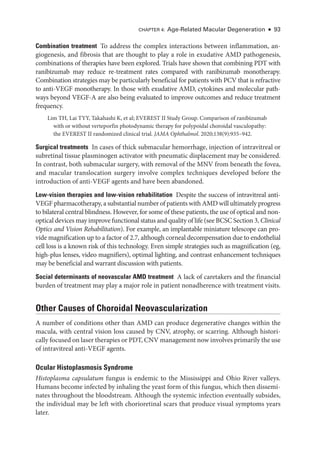

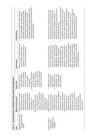
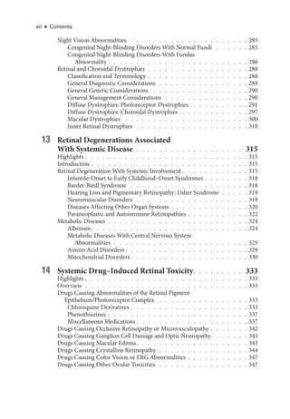
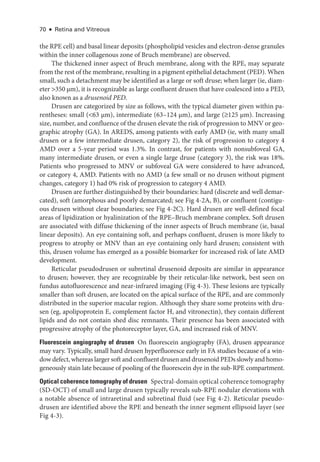
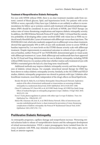
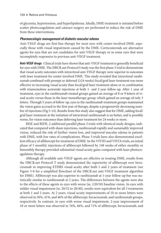
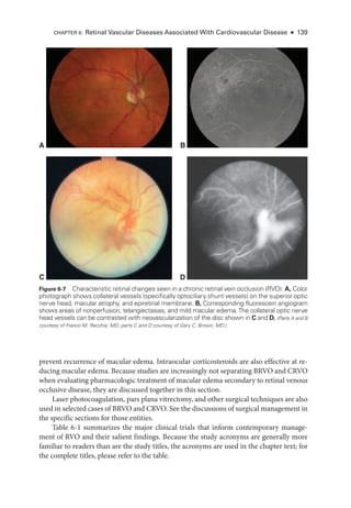
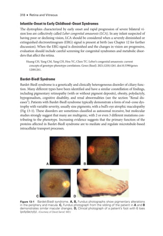
![Chapter 5: Diabetic Retinopathy ● 109
results from ETDRS Report Number 9 (see
Table 5-3). According to this rule, severe
NPDR manifests with any 1 of the following features:
• severe intraret
i
nal hemorrhages (typically estimated as 20 intraretinal hemor-
rhages) and microaneurysms in 4 quadrants (Fig 5-2)
• definite venous beading in 2 or more quadrants (Fig 5-3)
• moderate IRMA in 1 or more quadrants (Fig 5-4)
In the ETDRS, patients with severe NPDR had 15% and 60% chances of progression to
high-risk PDR within 1 and 3 years, respectively. In patients with very severe NPDR, which
was defined as having 2 or more of the features in the preceding list, the chance of progres-
sion to high-
risk PDR within 1 year increased to 45%.
The ETDRS severity scale has been the reference standard for classifying diabetic reti-
nopathy for several de
cades. To use it, clinicians need to acquire and interpret standardized
photographic fields that cover approximately 90° of the patient’s posterior ret
ina (eg, with
ultra-wide-field imaging, 80% of the ret
ina can now be visualized in a single 200° image).
Peripheral diabetic retinopathy lesions are often pre
sent outside the standard ETDRS fields;
in approximately 10% of eyes,
these lesions suggest more severe diabetic retinopathy. Pre-
liminary studies also suggest that a predominance of peripheral diabetic retinopathy lesions
increases the risk of diabetic retinopathy progression. This association is being evaluated in
the ongoing DRCR Ret
ina Network (DRCR.net) Protocol AA study.
In more advanced cases of NPDR, ret
i
nal capillary nonperfusion is a common finding.
Closure of ret
i
nal arterioles may expand areas of nonperfusion and progressive ischemia. In
addition, the foveal avascular zone may appear increasingly irregular on fluorescein angiog-
raphy (FA) or OCTA as well as enlarged when the innermost capillaries become nonperfused.
With increasing retinopathy severity, macular vessel density decreases in the superficial and
deep capillary plexuses. Peripheral nonperfusion is also frequently seen on ultra-
wide-
field
FA (Fig 5-5), even in eyes with mild NPDR.
When NPDR leads to loss of visual function, 1 of 2 mechanisms is typically impli-
cated: (1) increased intraret
i
nal vascular permeability, resulting in macular edema (see
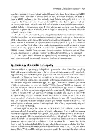
Figure 5-2 Fundus photo
graph shows diffuse
intraretinal hemorrhages (arrow) and microan-
eurysms in an eye with NPDR. (Standard photograph
2A, courtesy of the EarlyTreatment Diabetic Study [ETDRS].)
Figure 5-3 Fundus photo
graph shows venous
beading (arrows) in an eye with NPDR. (Standard
photo
graph 6B, courtesy of the ETDRS.)](https://image.slidesharecdn.com/12retinaandvitreous-250103182737-09041aee/85/Retina-and-Vitreous-128-320.jpg)
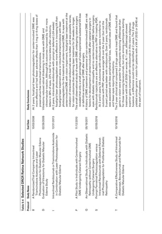
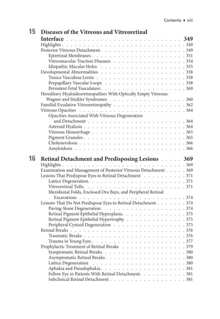
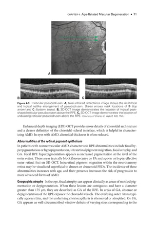
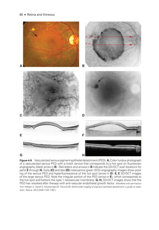
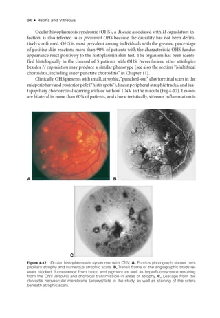
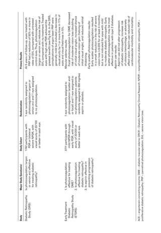
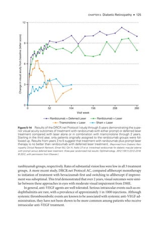
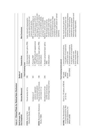
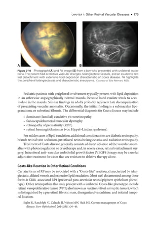
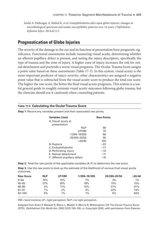
![114 ● Retina and Vitreous
Extraret
i
nal fibrovascular proliferation, which defines PDR, progresses through
3 stages:
1. Fine new vessels with minimal fibrous tissue cross and extend beyond the ILM,
often using the posterior hyaloid as a scaffold.
2. The new vessels grow in size and extent, developing an increased fibrous component.
3. The new vessels regress, leaving residual fibrovascular tissue that may be tethered
within the posterior hyaloid.
In PDR, neovascular proliferation is categorized by its location:
either on or within a
disc diameter of the optic nerve head (neovascularization of the disc [NVD]) or elsewhere
(neovascularization elsewhere [NVE]).
Patients may receive treatment at any stage of PDR. However, treatment is usually
considered mandatory once an eye has developed high-
risk characteristics, and it has been
shown to dramatically reduce rates of severe vision loss. PDR with high-
risk characteris-
tics is defined as the presence of any of the following findings:
• any NVD with vitreous or preret
i
nal hemorrhage
• extent of NVD greater than or equal to one-
fourth the disc area, with or without
vitreous or preret
i
nal hemorrhage (ie, greater than or equal to the extent shown in
ETDRS standard photo
graph 10A) (Fig 5-6)
• extent of NVE greater than or equal to one-
half the disc area, with vitreous or pre
retinal hemorrhage (Fig 5-7)
In eyes that have not developed high-
risk characteristics, treatment may be deferred.
Treatment may also be deferred in eyes that have peripheral neovascularization outside the
seven 30° photographic fields comprising the standard protocol for diabetic retinopathy
Figure 5-6 Fundus photo
graph of a left eye
shows neovascularization of the disc (NVD,
arrow) with a small amount of vitreous hemor-
rhage. Even without vitreous hemorrhage, this
degree of neovascularization is the lower limit
of moderate NVD and is considered high-
risk
proliferative diabetic retinopathy. (Standard photo
graph 10A, courtesy of the Diabetic Retinopathy Study.)
Figure 5-7 Fundus photo
graph of a right eye
shows cotton-
wool spots and moderate neo-
vascularization elsewhere with preret
i
nal hem-
orrhage. (Standard photo
graph 7
, courtesy of the Diabetic
Retinopathy Study.)](https://image.slidesharecdn.com/12retinaandvitreous-250103182737-09041aee/85/Retina-and-Vitreous-133-320.jpg)


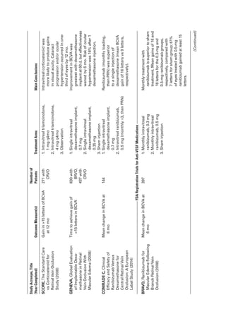

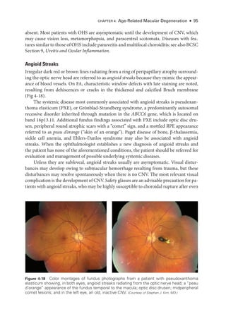
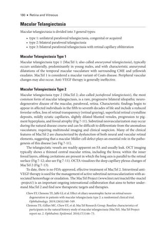
![At pre
sent,
there is no proven treatment for symptomatic RAO. Case reports and un-
controlled studies have suggested the utility of digital massage of the affected eye, anterior
chamberparacentesis,vasodilation,breathingintoapaperbag,carbogentherapy(amixtureof
95% oxygen and 5% carbon dioxide), topical IOP-
lowering medi
cations, hyperbaric oxygen,
and transvitreal Nd:YAG embolysis. However,
there are no level I data to support any specific
therapy. Treatment with antifibrinolytic agents (typically, tissue plasminogen activator [tPA])
A
C D
B
Figure 6-21 Acute ret
i
nal ischemia. This 64-
year-
old man with diabetes, hypertension, hyper-
lipidemia, and coronary artery disease presented with a 3-
day history of a central blind spot in
his left eye and VA of counting fin
gers. A, Fundus photo
graph shows parapapillary cotton-
wool
spots, macular whitening, and a cherry-
red spot. B, Fluorescein angiography shows arterial
filling beginning at 1 minute 19 seconds
after dye injection. Stroke workup was performed im-
mediately and revealed 90% carotid occlusion, which was treated with carotid endarterectomy
(CEA). Three weeks
after CEA, VA had improved to 20/40. C,
There was resolution of ret
i
nal
whitening and improvement in ret
i
nal perfusion. D, Dye appearance at 33 seconds
after infusion
demonstrates the improved perfusion. (Courtesy of Franco M. Recchia, MD.)
CHAPTER 6: Ret
i
nal Vascular Diseases Associated With Cardiovascular Disease ● 161](https://image.slidesharecdn.com/12retinaandvitreous-250103182737-09041aee/85/Retina-and-Vitreous-180-320.jpg)

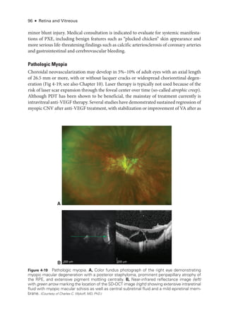
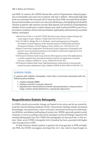
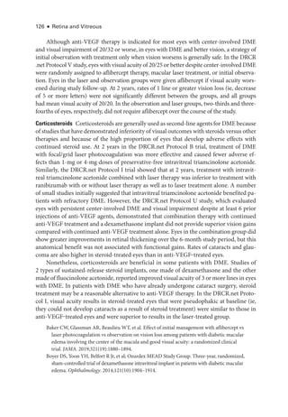
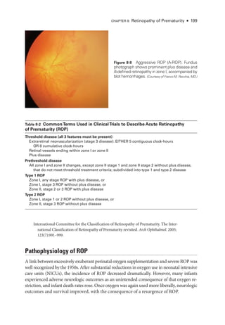
![196 ● Retina and Vitreous
Figure 8-2 Stage 1 ROP
. Fundus photo
graph reveals a faint white demarcation line temporally
(arrows). Arborizing, dilated vessels approach the line, and a choroidal vessel is seen extending
to the periphery (arrowheads). (Courtesy of Franco M. Recchia, MD.)
Figure 8-3 Stage 2 ROP
. Fundus photo
graph shows an elevated gray-
white ridge (arrows) at
the border of the vascularized (rosy) and avascular (grayish) retina. (Courtesy of Franco M. Recchia, MD.)
• Aggressive ROP (A-ROP; previously referred to as Rush disease and aggressive pos-
terior ROP [AP-
ROP]) is characterized by the rapid development of pathologic
neovascularization and severe plus disease without progression through the typical
stages of ROP. Hemorrhages at the junction of the vascular and avascular ret
ina,
as well as iris rubeosis, may be seen (see Fig 8-8). The vascular‒avascular junction](https://image.slidesharecdn.com/12retinaandvitreous-250103182737-09041aee/85/Retina-and-Vitreous-215-320.jpg)
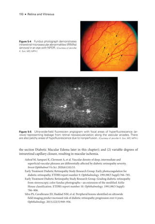
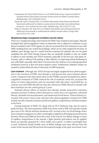
![198 ● Retina and Vitreous
According to the ICROP, an eye is classified on the basis of the most advanced disease
noted. However, documentation should reflect all affected zones and stages observed, in-
cluding their relative extent.
ACTIVITY 8-1 Interactive schematic for type 1 ROP
.
Developed by Franco M. Recchia, MD.
Chiang MF, Quinn GE, Fielder AR, et al. International Classification of Retinopathy of Pre
maturity, 3rd ed [epub ahead of print, July 8, 2021]. Ophthalmology. 2021;128(10):e51–e68.
doi:10.1016/j.ophtha.2021.05.031
Early Treatment for Retinopathy of Prematurity Cooperative Group. Revised indications for the
treatment of retinopathy of prematurity: results of the Early Treatment for Retinopathy of
Prematurity randomized trial. Arch Ophthalmol. 2003;121(12):1684–1694.
A B
Figure 8-7 Fundus photo
graphs show pronounced plus disease in eyes with ROP
. The ret
i
nal
arteries and veins are dilated and tortuous. A, The avascular ret
ina and preret
i
nal proliferations
can be seen inferiorly and inferotemporally (bottom right). B, Preret
i
nal hemorrhages are vis
i
ble, originating from the proliferative disease. (Part A courtesy of Colin A. McCannel, MD; part B courtesy
of Audina Berrocal, MD.)
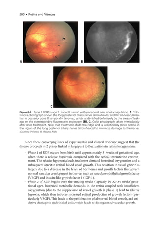
A B
Figure 8-6 Stage 5 ROP
. Wide-
angle fundus photo
graphs show total ROP ret
i
nal detachment
(RD). A, Total RD with vis
i
ble optic nerve head is now defined as stage 5A. Per
sis
tent vascular
activity accompanies the preret
i
nal fibrovascular ridge that contracts circumferentially, acting like
a purse string. B, Eventually, the vascular activity subsides, and the fibrosis starts to close the
funnel anteriorly (evolution into stage 5B). The arrow denotes the optic nerve head, which is al-
most completely obscured. (Part A courtesy of Audina Berrocal, MD; part B courtesy of Franco M. Recchia, MD.)](https://image.slidesharecdn.com/12retinaandvitreous-250103182737-09041aee/85/Retina-and-Vitreous-217-320.jpg)
![CHAPTER 8: Retinopathy of Prematurity ● 201
The onset of phase 2 corresponds to the clinical appearance of ROP on ophthal-
moscopy and is closely correlated with the infant’s gestational age, rather than his or
her postnatal age. Initially, this pro
cess
causes the formation of vis
i
ble flat or raised
whitish tissue (ROP stages 1 [flat line] and 2 [ridge]). As the disease progresses, vas-
cular growth proliferates into the vitreous cavity (ROP stage 3). In this vasoprolifera-
tive phase, new vessels varying widely in size and extent arise from ret
i
nal vessels just
posterior to the peripheral ridge.
These abnormal new vessels easily bleed, leading to
vitreous hemorrhage, and may induce contracture of the firmly attached vitreous gel.
Eventually, growth
factor and hormonal changes cause involution and fibrosis of the
blood vessels with cicatricial contraction that may lead to traction (also called tractional)
ret
i
nal detachment (ROP stages 4 and 5).
Hartnett ME, Penn JS. Mechanisms and management of retinopathy of prematurity. N Engl
J Med. 2012;367(26):2515–2526.
Natural Course
Although the precise systemic and/or local tissue
factors that influence progression and
regression of ROP are not known, the time course is predictable. ROP is typically a tran-
sient disease that regresses spontaneously in most infants, particularly
those with heavier
birth weights (closer to 1500 g) and older gestational age (closer to 32 weeks) at birth. In
general, eyes with ROP in zone III have a good visual prognosis. In contrast, the more
posterior the zone at the time of diagnosis, the greater the area of nonperfused ret
ina and
thus the more worrisome the prognosis.
Screening Recommendations
Screening for ROP consists of a dilated fundus examination using binocular indirect oph-
thalmoscopy. Use of a topical anesthetic and an eyelid speculum facilitates examination.
The pupils should be examined for failure to dilate and the iris inspected for vessel en-
gorgement or frank rubeosis, as
these may be signs of aggressive disease. In addition, the
optic nerve should be evaluated for plus disease and the periphery examined with scleral
depression to establish the extent of vessel growth.
Alternatively, telemedicine (ie, photographic) screening approaches may replace the
indirect ophthalmoscopic examination (see the section Fundus Photographic Screening
of ROP). However, clinicians should be aware that telemedicine screening criteria devel-
oped in one country may not apply in another country, especially when the level or quality
of available medical or perinatal care is not comparable.
Screening Criteria
All infants with a birth weight 1500 g or a gestational age of ≤30 weeks should be
screened for ROP. Infants with a birth weight between 1500 and 2000 g or a gestational
age 30 weeks who have an unstable clinical course and are considered by their attending
pediatrician or neonatologist to be at high risk for the disease should also be screened. The](https://image.slidesharecdn.com/12retinaandvitreous-250103182737-09041aee/85/Retina-and-Vitreous-220-320.jpg)
![CHAPTER 8: Retinopathy of Prematurity ● 203
• postmenstrual age of at least 65 weeks in patients for whom intravitreal anti-
VEGF
medi
cations caused regression of ROP (ie, this treatment alters the natu
ral history
of the disease);
because very late recurrences of proliferative ROP have been re-
ported, clinical judgment and caution should be used on a case-
by-
case basis to
determine when surveillance can be safely terminated in
these patients
• regression of ROP (care must be taken to confirm the absence of abnormal vascular
tissue that could reactivate)
Fierson WM; American Acad
emy of Pediatrics Section on Ophthalmology; American Acad
emy of Ophthalmology; American Association for Pediatric Ophthalmology and Strabismus;
American Association of Certified Orthoptists. Screening examination of premature infants
for retinopathy of prematurity. Pediatrics. 2018;142(6). doi:10.1542/peds.2018-3061 [Published
correction appears in Pediatrics. 2019;143(3):320183810.]
Fundus Photographic Screening of ROP
Ultra-
wide-
angle (120°) fundus photography is useful for documenting disease in pre-
mature infant eyes, for assessing disease progression, and for screening. Given short-
ages of willing examiners skilled in indirect ophthalmoscopy and ROP screening, remote
screening of photographic fundus images (ie, telemedicine) has efficiently and cost-
effectively improved access to eye care for premature infants at high risk for ROP, provid-
ing real-
time diagnosis and improving documentation. For detection of plus disease and
disease requiring treatment, photoscreening by experienced personnel is comparable
to binocular indirect ophthalmoscopy, which is considered the gold standard for ROP
screening examinations.
To help identify patients who require an in-
person examination
after photoscreening, the
Photographic Screening for Retinopathy of Prematurity (Photo-
ROP) Cooperative Group es-
tablished a definition of clinically significant ROP based on features detectable by photography:
• zone I, any ROP without plus disease
• zone II, stage 2 with no plus disease or up to 1 quadrant of plus disease
• zone II, stage 3 with no plus disease or up to 1 quadrant of plus disease
In addition, more advanced approaches to interpretation of fundus photo
graphs, such as
electronic image recognition and deep learning algorithms, are
under study.
Daniel E, Quinn GE, Hildebrand PL, et al; e-
ROP Cooperative Group. Validated system
for centralized grading of retinopathy of prematurity: telemedicine approaches to
Evaluating Acute-
Phase Retinopathy of Prematurity (e-
ROP) study. JAMA Ophthalmol.
2015;133(6):675–682.
Prevention and Risk
Factors
Preventing ROP begins with preventing prematurity through optimal prenatal, perinatal,
and postnatal care. During the postnatal clinical course,
factors that appear to increase
the risk of severe ROP include sepsis, blood transfusion, intraventricular hemorrhage,
necrotizing enterocolitis, and slow weight gain.](https://image.slidesharecdn.com/12retinaandvitreous-250103182737-09041aee/85/Retina-and-Vitreous-222-320.jpg)

![215
C H A P T E R 9
Choroidal Disease
Highlights
• Pachychoroid is an entity with an evolving definition but is generally thought of as
abnormally dilated choroidal vessels abutting the ret
i
nal pigment epithelium (with
or without choroidal thickening).
• Fluorescein angiography can contribute to the diagnosis of arteritic anterior is
chemic optic neuropathy.
• Hypertensive choroidopathy can be diagnosed by means of multimodal imaging
because of its unique characteristics on optical coherence tomography and fundus
autofluorescence.
Scope of Chapter
This chapter describes noninflammatory choroidal diseases that also involve the ret
ina.
Inflammatory disorders of the ret
ina and choroid are discussed in Chapter 11. See also
BCSC Section 9, Uveitis and Ocular Inflammation. Intraocular tumors such as melanoma
are covered in BCSC Section 4, Ophthalmic Pathology and Intraocular Tumors.
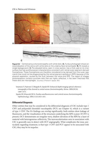
Central Serous Chorioretinopathy
Central serous chorioretinopathy (CSC; also called central serous retinopathy [CSR])
causes an
idiopathic serous detachment of the ret
ina related to leakage at the level of the ret
i
nal pigment
epithelium (RPE) secondary to hyperpermeability of the choriocapillaris, as seen on indocya
nine green angiography (ICGA). While the condition was originally described in 1866 by von
Graefe as recurrent central retinitis, it was Maumenee who first performed fluorescein angios
copy on patients with CSC and found a leak at the level of the RPE, not from retinal vessels (the
previously hypothesized source of leakage). Gass subsequently described the findings seen on
fluorescein angiography (FA) and suggested that
laser photocoagulation could be used to treat
affected patients. Gass also stated that the disease was secondary to hyperpermeability of the
choriocapillaris, a hypothesis that was confirmed de
cades
later via ICGA.
Demographics and Features
Central serous chorioretinopathy occurs primarily in persons between the ages of 35 and
55 years, with a male-to-female ratio of 3:1; at pre
sent,
there are no reliable statistics](https://image.slidesharecdn.com/12retinaandvitreous-250103182737-09041aee/85/Retina-and-Vitreous-234-320.jpg)


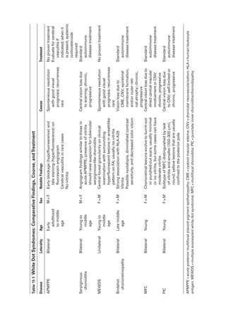
![252 ● Retina and Vitreous
ce
re
bral vasculitis. Severe APMPPE may be difficult to distinguish from serpiginous cho-
roiditis (discussed in the following section); this difficulty led to the introduction of the
term ampiginous to characterize the APMPPE–serpiginous choroiditis disease continuum.
Other entities in this placoid disorder continuum include persistent placoid maculopathy,
which is characterized by central macular involvement, a longer healing time, and a high
risk of choroidal neovascularization (CNV); and relentless placoid chorioretinitis, which
is characterized by the frequent occurrence of smaller, geo
graph
i
cally distributed lesions
that typically require immunosuppressive treatment.
Marchese A, Agarwal AK, Erba S, et al. Placoid lesions of the ret
i
na: pro
gress in multimodal
imaging and clinical perspective. Br J Ophthalmol. 2022;106(1):14–25.
B
A
C D
Figure 11-1 Acute posterior multifocal placoid pigment epitheliopathy (APMPPE). A, Color fundus
photo
graph of the left eye of a 23-
year-
old male patient shows confluent yellowish placoid le-
sions in the posterior pole.The right eye (not pictured) was also involved. Early (B) and late-
phase
(C) fluorescein angiography (FA) demonstrates hypofluorescence (due to decreased choriocap-
illaris perfusion and/or thickening of the ret
i
nal pigment epithelium [RPE]), and hyperfluorescence,
respectively. D, Indocyanine green angiography (ICGA; left) shows hypofluorescence of the
lesions, and the optical coherence tomography (OCT) scan (right) demonstrates outer ret
i
nal
involvement in the areas of lesions. (Courtesy of Lucia Sobrin, MD.)](https://image.slidesharecdn.com/12retinaandvitreous-250103182737-09041aee/85/Retina-and-Vitreous-271-320.jpg)
![274 ● Retina and Vitreous
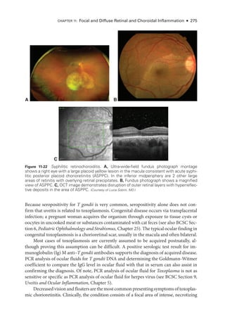
Syphilitic Retinochoroiditis
Uveitis is often the presenting sign of syphilis but can occur at any stage of the infection. Syphi-
litic uveitis is confirmed through serologic testing. The traditional screening algorithm for
syphilisusesanontreponemalassay(eg,thenonspecificbutquantitativeVDRLorrapidplasma
reagin[RPR]test)forprimaryevaluationandifthetestisreactive,atreponemalassay(eg,fluo
rescent treponemal antibody absorption [FTA-
ABS] test, Treponema pallidum particle agglu-
tination assay [TP-
PA], microhemagglutination assay for T pallidum antibodies [MHA-
TP])
for confirmation. More recently, laboratories have reversed the order in which treponemal and
nontreponemal tests are performed, leading to the development of the reverse sequence syph-
ilis screening (RSSS) algorithm. Although the CDC currently continues to recommend the
traditional testing approach, it provides a recommended algorithm for the use of RSSS. Briefly,
initial testing is done with a treponemal assay, followed by a quantitative nontreponemal test
for confirmation. Discordant samples are resolved on the basis of a second treponemal assay.
For patients with uveitis whose serologic test results are positive for syphilis, cerebro-
spinal fluid (CSF-) VDRL titers should be assessed before and, if the CSF-
VDRL results are
positive,
after completion of treatment in order to document a complete response.
Many patients with syphilitic uveitis present with a nondescript panuveitis, which sup-
ports the need for routine syphilis testing in all sexually active patients with uveitis. Spe-
cifically suggestive clinical findings include inflammatory ocular hypertensive syndrome,
iris roseola, and retinochoroiditis. The retinochoroiditis is often diaphanous—
appearing
less opaque than
either herpetic or toxoplasmic retinitis—
and is accompanied by overly-
ing inflammatory accumulations called retinal precipitates. A distinctive form of syphilitic
outer retinitis termed acute syphilitic posterior placoid chorioretinitis (ASPPC) is character-
ized by the presence of a placoid, round or oval, yellow lesion that involves or is near the
macula (Fig 11-22).
Because coinfection is common, all patients with syphilis should be
tested for HIV. Patients with syphilitic uveitis should be treated for neurosyphilis.
Begaj T, Sobrin L. Ophthalmic consequences of syphilis. Int Ophthalmol Clinics. 2022;62(2):251–268.
Ocular Bartonellosis
Cat-scratch disease, caused by Bartonella, is associated with 2 ocular syndromes: Parinaud
oculoglandular syndrome, which consists of conjunctival inflammation with preauricular
adenopathy; and Leber stellate neuroretinitis, which includes macular star formation and
optic nerve head swelling, often associated with a peripapillary serous macular detach-
ment (Fig 11-23). Cat-
scratch disease is the most common cause of neuroretinitis with
stellate maculopathy, but several other infectious diseases can have this pre
sen
ta
tion, in-
cluding toxoplasmosis, ehrlichiosis, and syphilis. Small, focal areas of retinitis or chorio-
retinitis are frequently noted in patients with Bartonella neuroretinitis. In rare cases, an
optic nerve head angiomatous lesion can develop. Treatment with antibiotics is necessary
in immunocompromised adults or in patients with per
sis
tent infection.
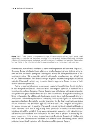
Toxoplasmic Chorioretinitis
Toxoplasmosis is the most common cause of posterior segment infectious disease world-
wide. The causative organism, Toxoplasma gondii, is an obligate intracellular parasite.](https://image.slidesharecdn.com/12retinaandvitreous-250103182737-09041aee/85/Retina-and-Vitreous-293-320.jpg)


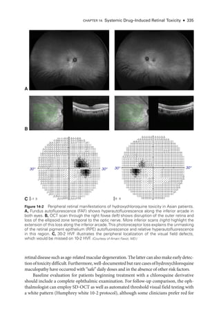
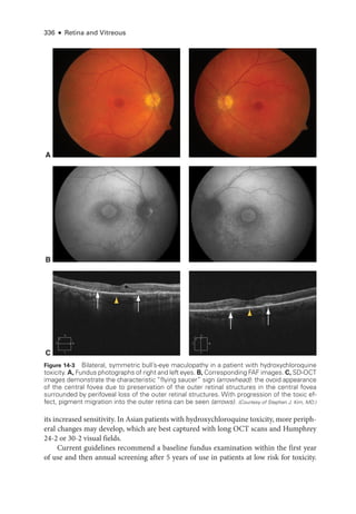
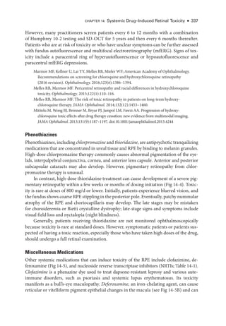
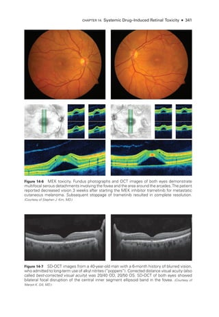
![333
C H A P T E R 14
Systemic Drug–Induced
Retinal Toxicity
Highlights
• Early detection of hydroxychloroquine toxicity is critical and requires the presence
of abnormalities on 2 modalities (spectral-
domain optical coherence tomography
[OCT] and automated threshold visual field testing).
• In Asian patients with hydroxychloroquine toxicity, more peripheral changes may
develop, which are best captured with long OCT scans and Humphrey 24-2 or 30-2
visual fields.
• Tamoxifen-
related maculopathy can have many of the same features as macular tel-
angiectasia type 2 (MacTel 2) and should be considered in the differential diagnosis
of MacTel 2.
Overview
Ret
i
nal toxicities caused by systemic therapeutic agents may be categorized according to
the ret
i
nal level affected and the pattern of toxicity. Broadly,
these toxicities manifest as
(1) abnormalities at the level of the retinal pigment epithelium (RPE)/photoreceptor com-
plex; (2) occlusive retinopathy or microvasculopathy; (3) ganglion cell and optic nerve dam-
age; and (4) other abnormalities, which include macular edema, crystalline retinopathy,
and alterations in color vision and electroretinogram (ERG) responses.
Drugs Causing Abnormalities of the Ret
i
nal Pigment
Epithelium/Photoreceptor Complex
Chloroquine Derivatives
Although ret
i
nal toxicity from chloroquine use remains a prob
lem in many parts of the
world, it is rare in the United States, where this medi
cation has largely been replaced by
the much safer, related drug hydroxychloroquine. These medications are used for the treat-
ment of malaria and rheumatologic and dermatologic diseases. Both medi
cations bind
to melanin in the RPE, which may concentrate or prolong their effects. Although the](https://image.slidesharecdn.com/12retinaandvitreous-250103182737-09041aee/85/Retina-and-Vitreous-352-320.jpg)
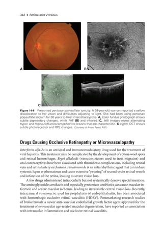
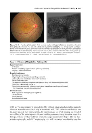


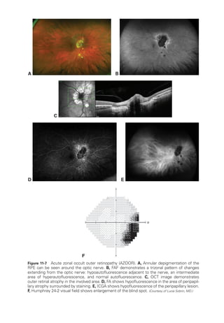
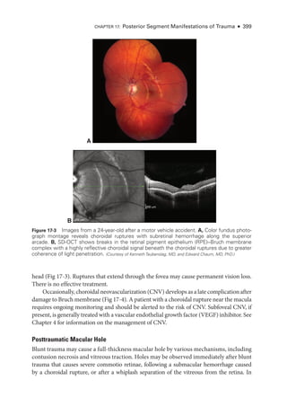
![398 ● Retina and Vitreous
• traumatic chorioret
i
nal disruption (chorioretinitis sclopetaria)
• vitreous base avulsion
• optic nerve avulsion
See Chapter 16 for discussion of traumatic retinal breaks and retinal detachment and Chap-
ter 19 for discussion of suprachoroidal hemorrhage. Sequelae of blunt trauma affecting the
posterior segment are discussed in the following sections.
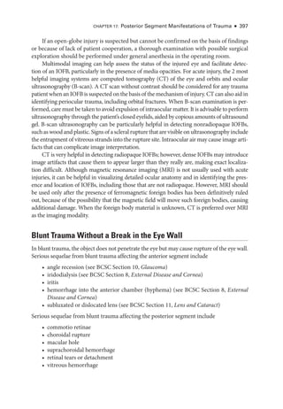
Commotio Retinae
The term commotio retinae refers to damage to the outer ret
i
nal layers caused by shock waves
that traverse the eye from the site of impact following blunt trauma. Ophthalmoscopic ex-
amination reveals a sheenlike ret
i
nal whitening that appears some hours
after the injury
(Fig 17-1). This ret
i
nal whitening occurs most commonly in the posterior pole but may also
be found peripherally. Spectral-
domain optical coherence tomography (SD-
OCT) findings
suggest that the major site of disruption is in the photoreceptor and ret
i
nal pigment epithelial
(RPE) layers, resulting in the observed ret
i
nal opacification (Fig 17-2). With foveal involve-
ment, a cherry-
red spot may appear
because the cells involved in the whitening are not pres
ent in the foveola. Commotio ret
i
nae in the posterior pole may reduce visual acuity to as
low as 20/200. Gradual visual recovery may occur if
there is no associated macular pigment
epitheliopathy, choroidal rupture, or macular hole formation. Disruption of the cone outer
segment tips, ellipsoid zone, and external limiting membrane is associated with poorer visual
and anatomical outcomes.
Choroidal Rupture
When the eye is compressed along its anteroposterior axis, tears may occur in Bruch mem-
brane, which has
little elasticity, as well as in the overlying RPE and fibrous tissue around
the choriocapillaris. Adjacent subret
i
nal hemorrhage is common. Choroidal ruptures may
be single or multiple and occur typically in the periphery and concentric to the optic nerve
*
Figure 17-1 Extensive commotio ret
i
nae with
sheenlike retinal whitening (arrow) and preret
i
nal hemorrhage (asterisk) following blunt ocular
trauma due to a rubber bullet. (Reproduced from
Barnes AC, Hudson LE, Jain N. Rubber bullet ocular trauma.
Ophthalmology. 2020;127(9):1190. Copyright 2020, with per-
mission from Elsevier.)
IR 30º ART [HS] OCT 30º (8.6 mm) ART (100) Q: 22 [HS]
200 μm
200 μm
Figure 17-2 Spectral-
domain optical coherence
tomography (SD-
OCT) image of the left eye of a
patient who experienced a rock injury at work.
Examination revealed macular ret
i
nal whiten
ing with visual acuity (VA) decreased to 20/100.
OCT image demonstrates increased reflectivity
of the parafoveal ellipsoid zone (arrows). (Courtesy
of Shriji Patel, MD, MBA.)](https://image.slidesharecdn.com/12retinaandvitreous-250103182737-09041aee/85/Retina-and-Vitreous-417-320.jpg)


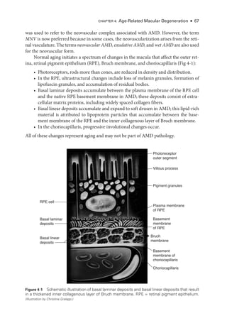
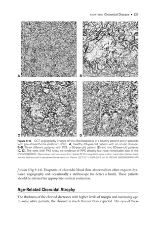
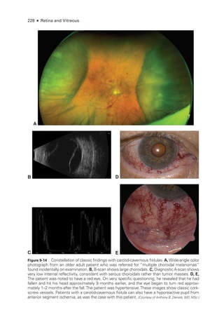
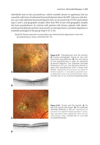
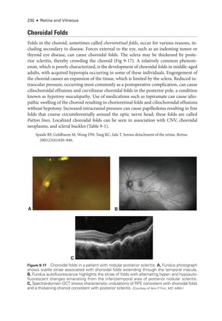
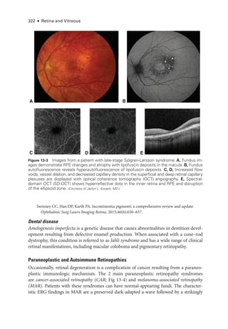
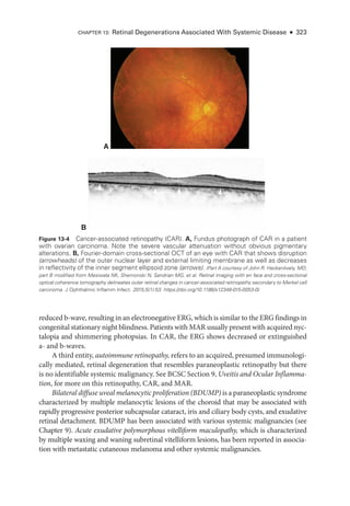
The document details the American Academy of Ophthalmology's 2023-2024 Basic and Clinical Science Course, specifically focusing on retina and vitreous education. It provides information on CME credits available for physicians, the editorial committee, and the acknowledgment of contributors and reviewers. The content aims to enhance ophthalmic knowledge and includes guidelines for claiming credits through assessments.


![Hypothalamus short ppt by Dr. Neha [PT].pptx](https://cdn.slidesharecdn.com/ss_thumbnails/hypothalamusbydr-260124145759-b9f94a93-thumbnail.jpg?width=640&height=640&fit=bounds)


