
Recommended
More Related Content
What's hot
What's hot (20)
Similar to Review of anatomy and physiology of respiratory system
Similar to Review of anatomy and physiology of respiratory system (20)
More from SanjaiKokila
More from SanjaiKokila (11)
Recently uploaded
Recently uploaded (20)
Review of anatomy and physiology of respiratory system
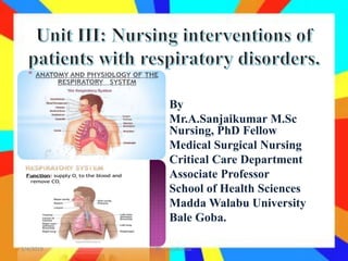
- 1. By Mr.A.Sanjaikumar M.Sc Nursing, PhD Fellow Medical Surgical Nursing Critical Care Department Associate Professor School of Health Sciences Madda Walabu University Bale Goba. 3/4/2019 BY: sanjaikumar
- 2. Review of anatomy and physiology of respiratory system Assessment of patient with respiratory disorder Upper respiratory disorders Tonsillitis Pharyngitis and adenoiditis Laryngitis Lower respiratory tract disorders and interventions for clients with LRT disorders Bronchitis (acute) Pneumonia COPD Chronic Bronchitis 3/4/2019 BY: sanjaikumar
- 3. Bronchiectasis Emphysema Asthma Lung abscess Pneumothorax TB Atelectasis Cor pulmonale Pulmonary embolism Pulmonary oedema Pluerisy Applying postural drainage Care of the pt with water-sealed drainage Care of the pt with tracheostomy Care of pt with Thoracentesis 3/4/2019 BY: sanjaikumar
- 5. Learning objectives Anatomic & physiologic overview of RS 3/4/2019 BY: sanjaikumar
- 6. At the end this presentation you will be able to: Identify the anatomy of RS structures Describe anatomical landmarks useful for respiratory system assessment 3/4/2019 BY: sanjaikumar
- 7. Function of the Respiratory System Major Functions of the Respiratory System Supply O2 for energy production Remove CO2 , waste product of energy reactions Homeostasis, acid-base balance of arterial blood Heat exchange 3/4/2019 BY: sanjaikumar
- 8. The respiratory system is composed of: Upper respiratory tract warms and filters inspired air Lower respiratory tracts. gas exchange. 3/4/2019 BY: sanjaikumar
- 9. Consist of the nose, sinuses and nasal passages, pharynx, tonsils and adenoids, larynx, and trachea. Nose Passageway for air to pass to and from the lungs. Filters impurities and humidifies and warms the air as it is inhaled. Is composed of an external and an internal portion. 3/4/2019 BY: sanjaikumar
- 10. External portion of nose: protrudes from the face and is supported by the nasal bones and cartilage. The anterior nares (nostrils) are the external openings of the nasal cavities. Internal portion of the nose o Hollow cavity separated into the right and left nasal cavities by nasal septum. o Each nasal cavity is divided into three passageways by the projection of the turbinates from the lateral walls. o The turbinate bones are also called conchae (the name suggested by their shell-like appearance). 3/4/2019 BY: sanjaikumar
- 12. Paranasal Sinuses Four pairs of bony cavities These air spaces are connected by a series of ducts that drain into the nasal cavity. Named by their location: frontal, ethmoidal, sphenoidal, and maxillary . A prominent function of the sinuses is to serve as a resonating chamber in speech. The sinuses are a common site of infection. 3/4/2019 BY: sanjaikumar
- 14. Pharynx, tonsils, and adenoids Pharynx (throat): Is a tubelike structure that connects the nasal and oral cavities to the larynx. Has three regions: nasal, oral, and laryngeal. Nasopharynx: is located posterior to the nose and above the soft palate. Oropharynx: houses the faucial, or palatine, tonsils. Laryngopharynx: extends from the hyoid bone to the cricoid cartilage. The epiglottis forms the entrance to the 3/4/2019 BY: sanjaikumar
- 16. Adenoids(pharyngeal tonsils) Are located in the roof of the nasopharynx. The tonsils, the adenoids, and other lymphoid tissue encircle the throat. These structures are important links in the chain of lymph nodes guarding the body from invasion by organisms entering the nose and the throat. The pharynx functions as a passageway for the respiratory and digestive tracts. 3/4/2019 BY: sanjaikumar
- 17. Larynx Is a cartilaginous epithelium lined structure that connects the pharynx and the trachea. Its major function is vocalization. It also protects the lower airway from foreign substances and facilitates coughing. It is frequently referred to as the voice box. 3/4/2019 BY: sanjaikumar
- 18. Trachea (windpipe) Is composed of smooth muscle with C-shaped rings of cartilage at regular intervals. Trachea bifurcates into its main stem bronchi at the levels of the sternal angle anteriorly and the T4 spinous process posteriorly. The trachea serves as the passage between the larynx and the bronchi. 3/4/2019 BY: sanjaikumar
- 20. B. LOWER RESPIRATORY TRACT • Consists of the lungs, which contain the bronchial and alveolar structures 3/4/2019 BY: sanjaikumar
- 21. Lungs Paired elastic structures enclosed in the thoracic cage When the capacity of the chest is increased, air enters through the trachea (inspiration) because of the lowered pressure within and inflates the lungs. When the chest wall and diaphragm return to their previous positions (expiration), the lungs recoil and force the air out through the bronchi and trachea. 3/4/2019 BY: sanjaikumar
- 23. Pleura: A serous membrane that lines the lungs and wall of the thorax Visceral pleura: covers the lungs Parietal pleura : lines the thorax. Small amount of pleural fluid between these two membranes 3/4/2019 BY: sanjaikumar
- 24. Mediastinum. • Middle of the thorax, between the pleural sacs that contain the two lungs. • Extends from the sternum to the vertebral column and contains all the thoracic tissue outside the lungs (heart, thymus, certain large blood vessels [ie, aorta, vena cava], and esophagus). 3/4/2019 BY: sanjaikumar
- 25. Bronchi and Bronchioles. There are several divisions of the bronchi within each lobe of the lung. First are the lobar bronchi (3 in the right lung and 2 in the left lung). Lobar bronchi divide into segmental bronchi (10 on the right and 8 on the left), which are the structures identified when choosing the most effective postural drainage position for a given patient. 3/4/2019 BY: sanjaikumar
- 26. Alveoli. About 300 million alveoli, which are arranged in clusters of 15 to 20. Three types of alveolar cells. • Type i alveolar cells: – Are epithelial cells - form the alveolar walls. • Type ii alveolar cells: – metabolically active. – Secrete surfactant, a phospholipid • Type III alveolar cell : – Macrophages are large phagocytic – Act as an important defense mechanism. 3/4/2019 BY: sanjaikumar
- 28. Breathing Terms for Various Breathing Activities • Hyperpnea- increased breathing movement • Eupnea- normal breathing movements • Hypopnea - decreased breathing movements • Apnea- arrested breathing • Bradypnea - decreased rate of breathing • Tachypnea- increased rate of breathing • Dyspnea- labored breathing (subjective feeling) • Asphyxia- inability to breathe • Orthopnea- labored breathing, except in the sitting or upright position 3/4/2019 BY: sanjaikumar
- 29. Breathing. • Automatic act & controlled in the brainstem and mediated by the muscles of respiration. Dome-shaped diaphragm ( primary muscle of inspiration. Contracts- descends & enlarges thoracic cavity, compresses the abdominal contents, pushing abdominal wall outward. Rib cage & neck expand thorax during inspiration 3/4/2019 BY: sanjaikumar
- 30. • During inspiration: Muscles contract Thorax expands Intrathoracic pressure decreases, drawing air through the tracheobronchial tree into the alveoli, or distal air sacs, and expanding the lungs.3/4/2019 BY: sanjaikumar
- 31. Expiratory phase: • Begins after inspiratory effort stops • Chest wall and lungs recoil, diaphragm relaxes and rises passively, Air flows outward, and Chest and abdomen return to their resting positions. 3/4/2019 BY: sanjaikumar
- 32. Cont’d... • Normal breathing is quiet and easy—barely audible near the open mouth as a faint whish. • When a healthy person lies supine, the breathing movements of the thorax are relatively slight. In contrast, the abdominal movements are usually easy to see. • In the sitting position, movements of the thorax become more prominent. 3/4/2019 BY: sanjaikumar
- 33. Cont’d... • During exercise and in certain diseases, extra work is required to breathe, and accessory muscles join the inspiratory effort. • The sternomastoids are the most important of these, and the scalenes may become visible. • Abdominal muscles assist in expiration. 3/4/2019 BY: sanjaikumar
- 34. Real anatomical land marks and imaginary lines on chest wall. Help in description of chest examination findings. Grouped in to two:- I. Used to describe findings along vertical axis. II. Used to describe findings along the circumference of the chest. 3/4/2019 BY: sanjaikumar
- 35. Anatomic & physiologic overview.... Land mark of anterior chest: :- - “U” shaped depression at the top of sternum, b/n clavicles. adjacent to 2nd rib & just below it is 2nd ICS. - Site of tracheal bifurcation( Rt & Lt main bronchi. - 2.5 cm below sternal notch. - Reference to count ICS by walking down with 2 fingers. 3/4/2019 BY: sanjaikumar
- 37. Anatomic & physiologic overview.... Land mark of posterior chest • Spinous process of C7 vertebrae. - Prominent bone at the back of flexed neck. - At same level with the 1st rib . • Inferior tip of the scapula - Lies at the level of 7th rib/ICS. 3/4/2019 BY: sanjaikumar
- 38. • Vertical imaginary lines drawn on anterior & posterior chest walls. 3/4/2019 BY: sanjaikumar
- 39. Anatomic & physiologic overview.... . • Vertebral line- overlies the spinous processes of the vertebrae. • Right and left Scapular lines:- drops from the inferior angle of the scapula. 3/4/2019 BY: sanjaikumar
- 40. Anatomic & physiologic overview.... Anteriorly Apex of lung : 2 - 4 cm above the inner third of the clavicle. Lower border lung: crosses the 6th rib at the MCL & the 8th rib at the MAL. Posteriorly Lower border: at T10 spinous process (on inspiration, it descends further). 3/4/2019 BY: sanjaikumar
- 42. Anatomic & physiologic overview.... Each lung is divided roughly in half by an oblique (major) fissure: Runs from T3 spinous process obliquely down to 6th rib at MCL. Rt lung - divided by horizontal (minor) fissure Runs close to 4th rib & meets oblique fissure in MAL near the 5th rib. 3/4/2019 BY: sanjaikumar
- 44. • Right Lung – Divided into 3 lobes, upper, middle , lower lobes. – Shorter due to liver • Left Lung – Has only two lobes = Left Upper and Lower – Narrower due to heart 3/4/2019 BY: sanjaikumar
- 49. Used to locate chest findings, such as:- • Supraclavicular—above the clavicles • Infraclavicular—below the clavicles • Interscapular—between the scapulae • Infrascapular—below the scapula • Bases of the lungs —the lower most portions. • Upper, middle, and lower lung fields 3/4/2019 BY: sanjaikumar
Editor's Notes
- The cartilaginous rings are incomplete on the posterior surface and give firmness to the wall of the trachea, preventing it from collapsing.