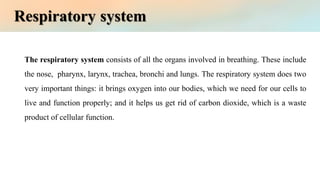
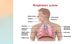
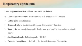
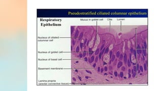
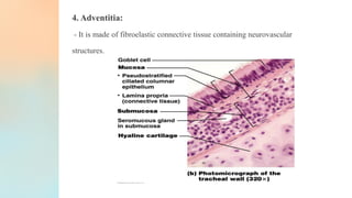
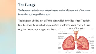
Respiratory system
Practical Histology
First stage
Lab 4
‫م‬
.
‫م‬
.
‫جواد‬ ‫محسن‬ ‫نور‬
The respiratory system consists of all the organs involved in breathing. These include
the nose, pharynx, larynx, trachea, bronchi and lungs. The respiratory system does two
very important things: it brings oxygen into our bodies, which we need for our cells to
live and function properly; and it helps us get rid of carbon dioxide, which is a waste
product of cellular function.
Respiratory system
Respiratory epithelium
Lined by pseudostratified ciliated columnar epithelium
➢ Ciliated columnar cells: most common, each cell has about 300 cilia
➢ Goblet cells: secret mucous
➢ Brush cells: have short microvilli, nerve fibers, sensory function
➢ Basal cells: are rounded stem cells that located near basal lamina and show mitotic
figures
➢ Small granule cells (kulchitsky cells = DNEs)
➢ Exocrine bronchiolar cells (club cells, formerly known as Clara cells)
Trachea
❑ Extends through the mediastinum anterior to the esophagus.
❑ It’s made up of 15 - 20 C-shaped tracheal cartilages that keep the tracheal
lumen open and numerous seromucous glands that produce a more fluid mucus
❑ Trachea is lined by pseudostratified columnar
epithelium.
General features:
Structure:
The trachea is made of four coats, similar to the general structure of the
respiratory tract. From inner to outer they are:
1. Mucosa:
❑ It is composed of epithelium and lamina propria.
❑ The epithelium is thick and is formed of pseudostratified ciliated column
ar variety with goblet cells (respiratory epithelium).
❑ The lamina propria consists of fibroelastic vascular connective tissue in
which the elastic fibers are longitudinally oriented.
❑ Lymphocytes and mast cells are abundant.
2. Submucosa:
- It is made of loose connective tissue found deep to lamina propria, containing mixed
glands. (There is no clear demarcation between lamina propria and submucosa.)
3. Cartilage and smooth muscle layer:
- It is formed by C-shaped hyaline cartilaginous rings. The posterior free ends of the
cartilage ring is bridged by smooth muscle and fibroelastic ligament.
4. Adventitia:
- It is made of fibroelastic connective tissue containing neurovascular
structures.
The Lungs
The lungs are paired, cone-shaped organs which take up most of the space
in our chests, along with the heart.
The lungs are divided into different parts which are called lobes. The right
lung has three lobes called upper, middle and lower lobes. The left lung
only has two lobes, the upper and lower.
Structure:
❑ The lining epithelium of bronchial tree gradually decreases in thickness as it is traced
distally.
❑ Similarly, glands and goblet cells also gradually decrease and disappear completely
at the distal part.
❑ The cartilaginous support decreases while the number of elastic fibers increase as the
bronchial tree is traced distally.
1. Intrapulmonary bronchus: (i.e., Secondary and tertiary bronchi)
- In the lung each principal bronchus divides into secondary or lobar
bronchi. Following layers are observed in these bronchi from inner to
outer:
(a) Mucosa:
– It consists of epithelium and lamina propria.
– The epithelium is pseudostratified ciliated columnar variety with few
goblet cells.
– The lamina propria is rich in elastic fibers.
– Mucosa is thrown into folds by the contraction of underlying smooth
muscle.
(b) Smooth muscle layer:
– This layer consists of spirally running criss-cross bundles of smooth muscle.
(c) Submucosa:
– It contains few seromucous glands.
(d) Cartilage layer and adventitia: The intrapulmonary bronchus contains isolated
plates of hyaline cartilage.
Lab 4 The Respiratory system.pdf

Lab 4 The Respiratory system.pdf

  • 1.
    Respiratory system Practical Histology Firststage Lab 4 ‫م‬ . ‫م‬ . ‫جواد‬ ‫محسن‬ ‫نور‬
  • 2.
    The respiratory systemconsists of all the organs involved in breathing. These include the nose, pharynx, larynx, trachea, bronchi and lungs. The respiratory system does two very important things: it brings oxygen into our bodies, which we need for our cells to live and function properly; and it helps us get rid of carbon dioxide, which is a waste product of cellular function. Respiratory system
  • 4.
    Respiratory epithelium Lined bypseudostratified ciliated columnar epithelium ➢ Ciliated columnar cells: most common, each cell has about 300 cilia ➢ Goblet cells: secret mucous ➢ Brush cells: have short microvilli, nerve fibers, sensory function ➢ Basal cells: are rounded stem cells that located near basal lamina and show mitotic figures ➢ Small granule cells (kulchitsky cells = DNEs) ➢ Exocrine bronchiolar cells (club cells, formerly known as Clara cells)
  • 6.
    Trachea ❑ Extends throughthe mediastinum anterior to the esophagus. ❑ It’s made up of 15 - 20 C-shaped tracheal cartilages that keep the tracheal lumen open and numerous seromucous glands that produce a more fluid mucus ❑ Trachea is lined by pseudostratified columnar epithelium.
  • 7.
    General features: Structure: The tracheais made of four coats, similar to the general structure of the respiratory tract. From inner to outer they are: 1. Mucosa: ❑ It is composed of epithelium and lamina propria. ❑ The epithelium is thick and is formed of pseudostratified ciliated column ar variety with goblet cells (respiratory epithelium). ❑ The lamina propria consists of fibroelastic vascular connective tissue in which the elastic fibers are longitudinally oriented. ❑ Lymphocytes and mast cells are abundant.
  • 8.
    2. Submucosa: - Itis made of loose connective tissue found deep to lamina propria, containing mixed glands. (There is no clear demarcation between lamina propria and submucosa.) 3. Cartilage and smooth muscle layer: - It is formed by C-shaped hyaline cartilaginous rings. The posterior free ends of the cartilage ring is bridged by smooth muscle and fibroelastic ligament.
  • 9.
    4. Adventitia: - Itis made of fibroelastic connective tissue containing neurovascular structures.
  • 10.
    The Lungs The lungsare paired, cone-shaped organs which take up most of the space in our chests, along with the heart. The lungs are divided into different parts which are called lobes. The right lung has three lobes called upper, middle and lower lobes. The left lung only has two lobes, the upper and lower.
  • 11.
    Structure: ❑ The liningepithelium of bronchial tree gradually decreases in thickness as it is traced distally. ❑ Similarly, glands and goblet cells also gradually decrease and disappear completely at the distal part. ❑ The cartilaginous support decreases while the number of elastic fibers increase as the bronchial tree is traced distally.
  • 12.
    1. Intrapulmonary bronchus:(i.e., Secondary and tertiary bronchi) - In the lung each principal bronchus divides into secondary or lobar bronchi. Following layers are observed in these bronchi from inner to outer: (a) Mucosa: – It consists of epithelium and lamina propria. – The epithelium is pseudostratified ciliated columnar variety with few goblet cells. – The lamina propria is rich in elastic fibers. – Mucosa is thrown into folds by the contraction of underlying smooth muscle.
  • 13.
    (b) Smooth musclelayer: – This layer consists of spirally running criss-cross bundles of smooth muscle. (c) Submucosa: – It contains few seromucous glands. (d) Cartilage layer and adventitia: The intrapulmonary bronchus contains isolated plates of hyaline cartilage.