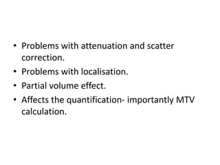
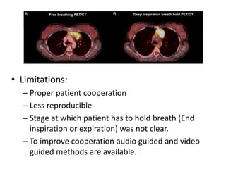
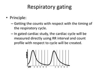
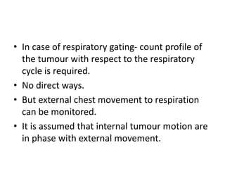
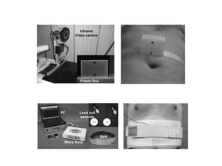
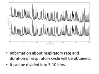
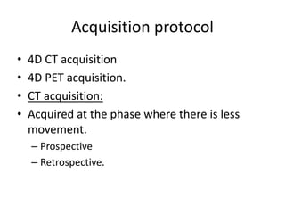
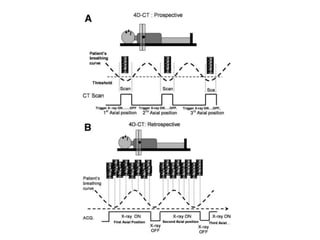
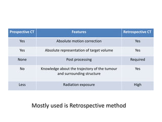
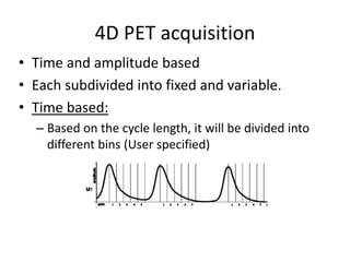
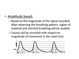
Call Girls Visakhapatnam Just Call 9907093804 Top Class Call Girl Service Ava...
Respiratory gating
1. Respiratory gating
Dr.Vishnukumar R, MD Nuclear Medicine
Jawaharlal Institute of Post graduate Medical Education and Research (JIPMER),
Puducherry, India.
2. Learning objectives
• Need for gating
• Types of respiratory gating.
• Acquisition protocol: CT and PET.
• Data processing
• Limitations
3. Need for gating
• CT acquisition will be fast. Cycle that will be
captured depends on the respiratory cycle.
• PET CT is acquired in free breathing.
• Normal cycle: Expiration > Inspiration.
• PET images of thorax mostly represents data
of end expiration.
4.
5.
6. • Problems with attenuation and scatter
correction.
• Problems with localisation.
• Partial volume effect.
• Affects the quantification- importantly MTV
calculation.
7. Available solutions
• Respiratory gating
• Deep inspiratory breath hold (DIBH).
• DIBH:
– Both PET and CT studies will acquired at breath
hold.
– Duration of holding the breath might vary (10-40s)
8. • Limitations:
– Proper patient cooperation
– Less reproducible
– Stage at which patient has to hold breath (End
inspiration or expiration) was not clear.
– To improve cooperation audio guided and video
guided methods are available.
9. Respiratory gating
• Principle:
– Getting the counts with respect with the timing of
the respiratory cycle.
– In gated cardiac study, the cardiac cycle will be
measured directly using RR interval and count
profile with respect to cycle will be created.
10. • In case of respiratory gating- count profile of
the tumour with respect to the respiratory
cycle is required.
• No direct ways.
• But external chest movement to respiration
can be monitored.
• It is assumed that internal tumour motion are
in phase with external movement.
13. • Information about respiratory rate and
duration of respiratory cycle will be obtained.
• It can be divided into 5-10 bins.
14. Acquisition protocol
• 4D CT acquisition
• 4D PET acquisition.
• CT acquisition:
• Acquired at the phase where there is less
movement.
– Prospective
– Retrospective.
15.
16. Prospective CT Features Retrospective CT
Yes Absolute motion correction Yes
Yes Absolute representation of target volume Yes
None Post processing Required
No Knowledge about the trajectory of the tumour
and surrounding structure
Yes
Less Radiation exposure High
Mostly used is Retrospective method
17. 4D PET acquisition
• Time and amplitude based
• Each subdivided into fixed and variable.
• Time based:
– Based on the cycle length, it will be divided into
different bins (User specified)
18. • Amplitude based:
– Based on the magnitude of the signal recorded.
After observing the breathing pattern, region of
maximal and minimal breathing will be studied.
– Counts will be recorded with respect to
magnitude of movement in the voxel (cm).
19. • Fixed cycle:
– User specified number of gates and length will be
fixed.
– If combined with time based model, excess part of
the lengthier cycle will be excluded.
– Problems with amplitude gating is limited.
• Variable cycle:
– Number is constant but length will be optimised.
20. • If 8 gates are used and length of a cycle is
16ms, length will be 2 ms.
• If the cycle length is 24 ms, the new optimised
length would be 3 ms.
21. • Problem will be counts between the gates will
not be equal.
• This leads to blurring effect.
• In order to minimize it, number of gates has to
be increased.
• Amplitude based methods yields good result.
22. Data processing
• Goal of data processing
– To represent the volume of the tumour
encompassing its motion during the respiration.
• For delineating GTV in RT planning.
– To accurately define the target with respect to its
physical characteristics.
• For absolute quantification.
23. • 4D CT data are expressed as Maximum
Intensity Projection (MIP) and Average CT
image (AVE).
• MIP:
– Image formed with pixels (out of 8 set of data, if 8
gates are used) recording maximum CT number
(HU).
• AVE:
– Image formed with average CT numbers recorded
in each pixel throughout the acquisition.
24.
25.
26. • 4D PET:
– MIP images
– Summed images: Summing all the available data.
– Summed images are better, due to its less
susceptibility to noise.
27. Limitations
• Longer acquisition period: 7-8 minutes/ bed,
compared to 2-3 minutes/bed
• Patient discomfort causing additional motion
artefact.
• Problematic in patients with irregular
breathing cycles.
• Requiring separate algorithm for acquisition
and processing.
• High cost.
28. Summary
• Need for gating: Accurate tumour volume
delineation.
• Respiratory gating and DIBH
• Optoelectric and Pressure methods are
commonly used.
• 4D CT: Retrospective and prospective.
Retrospective is preferred.
• 4D PET: Time and Amplitude based. Amplitude
based is preferred.
29. • Processed as MIP and AVE for 4D CT; MIP and
Summed image for 4D PET.
• Summed preferred over MIP in PET and MIP
over AVE in CT.
• Limitations are time consuming, patient
discomfort, presence of irregular breathing
cycles and high cost.