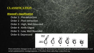
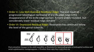
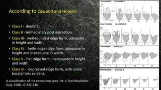
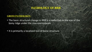
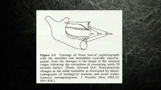
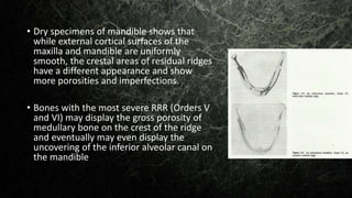
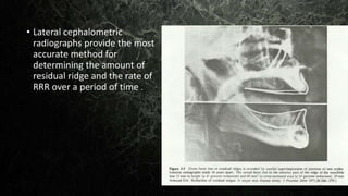
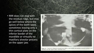
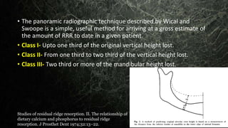
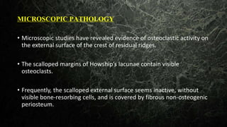
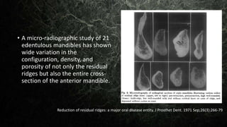
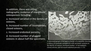
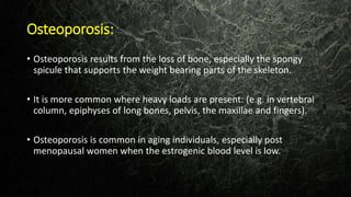
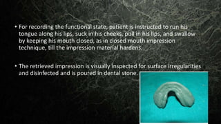
RESIDUAL RIDGE
RESORPTION
CONTENTS
• INTRODUCTION
• DEFINITION
• CLASSIFICATION OF RRR
• PATHOLOGY OF RRR
• PATHOPHYSIOLOGY OF RRR
• PATHOGENESIS OF RRR
• EPIDEMIOLOGY OF RRR
• ETIOLOGY OF RRR
• CLINICAL FEATURES OF RRR
• MANAGEMENT
• CONCLUSION
• REFERENCES
INTRODUCTION
• Residual ridge is the portion of the residual bone and its soft tissue
covering that remains after the removal of teeth.
• It consists of the denture-bearing mucosa, submucosa and
periosteum, and the underlying residual alveolar bone.
• alveolar bone.
•After tooth extraction, a cascade of inflammatory reactions is
immediately activated, and the extraction socket is temporarily closed
by the blood clot.
• Epithelial tissue begins its proliferation and migration within the first
week and the disrupted tissue integrity is quickly restored.
• The most striking feature of the extraction wound healing is that even
after the healing of wounds, the residual alveolar ridge bone
undergoes a life-long catabolic remodeling.
• The size of the residual ridge is reduced most rapidly in the first 6
months, but the bone resorption activity continues throughout life at
a slower rate, resulting in removal of a large amount of jaw structure.
• This unique phenomenon has been described as RESIDUAL RIDGE
RESORPTION (RRR).
Definition: The diminishing quantity and quality of the residual ridge
after teeth are removed (GPT8).
• In the maxillae, the labial and buccal alveolar plates resorb much
faster than the palatal plates, while, in the mandible, the amounts of
bone resorbed at the lingual and labial plates are approximately the
same.
• Swenson stated that after tooth extraction the alveolar process of the
maxillae resorbs upward and inward to become progressively smaller
because of the direction and inclination of the roots of the teeth.
Alveolar ridge resorption following tooth extraction: J Prosthet Dent January 1967
• Consequently, the older the edentulous maxillae, the smaller is the
potential tooth-bearing area. He felt the opposite to be true in the
mandible, which inclines outward and becomes progressively wider
with edentulous age.
Alveolar ridge resorption following tooth extraction: J Prosthet Dent January 1967
CLASSIFICATION
Atwood’s classification
Order 1 : Pre-extraction
Order 2 : Post-extraction
Order 3 : High, Well Rounded
Order 4 : Knife Edged
Order 5 : Low, Well Rounded
Order 6: Depressed
Post-extraction changes in the adult mandible as illustrated by microradiographs of midsagittal sections and
serial cephalometric roentgenograms. J Prosthet Dent.1963 Sep 1;13(5):810-24.
• Order I: Pre-extraction: The lower central incisor is in its socket with
very thin labial and lingual cortical plates merged with the lamina
dura
• Order II: Post-extraction: The healing period includes clot formation,
clot organization, filling of the socket to the height of the cortical
plates with new trabecular bone, and epithelization over the socket
site. The edges of the residual ridge are still sharp
Post-extraction changes in the adult mandible as illustrated by microradiographs of midsagittal sections and
serial cephalometric roentgenograms. J Prosthet Dent.1963 Sep 1;13(5):810-24.
• Order III: High, Well-Rounded Residual Ridge: The cortical plates are
rounded off, narrowing of the crest of the ridge has begun, and
remodeling of the internal trabecular structure has taken place .
• Order IV: Knife-Edge Residual Ridge: There is marked narrowing of the
labiolingual diameter of the crest of the ridge with a compensatory
internal remodeling which sometimes leads to an incredibly sharp
crest of the ridge
Post-extraction changes in the adult mandible as illustrated by microradiographs of midsagittal sections and
serial cephalometric roentgenograms. J Prosthet Dent.1963 Sep 1;13(5):810-24.
• Order V.: Low Well-Rounded Residudal Ridge: The end result of
progressive labiolingual narrowing of a knife-edge ridge is the
disappearance of the knife-edge portion. A more widely rounded, but
considerably lower residual ridge remains
• Order VI: Depressed Residual Ridge: Resorption has continued below
the level of the genial tubercle.
Post-extraction changes in the adult mandible as illustrated by microradiographs of midsagittal sections and
serial cephalometric roentgenograms. J Prosthet Dent.1963 Sep 1;13(5):810-24.
• Class I - dentate.
• Class II - immediately post extraction.
• Class III- well-rounded ridge form, adequate
in height and width.
• Class IV - knife-edge ridge form, adequate in
height and inadequate in width.
• Class V - flat ridge form, inadequate in height
and width.
• Class VI - depressed ridge form, with some
basalar loss evident.
A classification of the edentulous jaws. Int. J. Oral Maxillofac.
Surg. 1988; 17:232-236
According to Cawood and Howell.
PATHOLOGY OF RRR
GROSS PATHOLOGY:
• The basic structural change in RRR is a reduction in the size of the
bony ridge under the mucoperiosteum.
• It is primarily a localized loss of bone structure.
• Longitudinal radiographic cephalometric studies have provided
excellent visualization of the gross patterns of this bone loss from a
lateral viewpoint.
• The careful superimposition of portions of tracings of these lateral
cephalograms has clearly shown the gross reduction of bone in size
and shape that occurs on the external surface on the labial, crestal,
and lingual aspects of the residual ridge.
• Dry specimens of mandible shows that
while external cortical surfaces of the
maxilla and mandible are uniformly
smooth, the crestal areas of residual ridges
have a different appearance and show
more porosities and imperfections.
• Bones with the most severe RRR (Orders V
and VI) may display the gross porosity of
medullary bone on the crest of the ridge
and eventually may even display the
uncovering of the inferior alveolar canal on
the mandible
• Lateral cephalometric
radiographs provide the most
accurate method for
determining the amount of
residual ridge and the rate of
RRR over a period of time .
• RRR does not stop with
the residual ridge, but may
go well below where the
apices of the teeth were,
sometimes leaving only a
thin cortical plate on the
inferior border of the
mandible or virtually no
maxillary alveolar process
on the upper jaw.
• The panoramic radiographic technique described by Wical and
Swoope is a simple, useful method for arriving at a gross estimate of
the amount of RRR to date in a given patient.
• Class I- Upto one third of the original vertical height lost.
• Class II- From one third to two third of the vertical height lost.
• Class III- Two third or more of the mandibular height lost.
Studies of residual ridge resorption. II. The relationship of
dietary calcium and phosphorus to residual ridge
resorption. J Prosthet Dent 1974;32:13–22.
MICROSCOPIC PATHOLOGY
• Microscopic studies have revealed evidence of osteoclastic activity on
the external surface of the crest of residual ridges.
• The scalloped margins of Howship’s lacunae contain visible
osteoclasts.
• Frequently, the scalloped external surface seems inactive, without
visible bone-resorbing cells, and is covered by fibrous non-osteogenic
periosteum.
• A micro-radiographic study of 21
edentulous mandibles has shown
wide variation in the
configuration, density, and
porosity of not only the residual
ridges but also the entire cross-
section of the anterior mandible.
Reduction of residual ridges: a major oral disease entity. J Prosthet Dent. 1971 Sep;26(3):266-79
In addition, there was micro-
radiographic evidence of mandibular
osteoporosis including:
a. Increased variation in the density of
osteons,
b. Increased number of incompletely
closed osteons,
c. Increased endosteal porosity,
d. Increased number of plugged
osteons in about half the specimens.
A microradiograph of the inferior border of a mandible showing
evidence of moderate osteoporosis with increased variation in
the density of osteons, increased number of incompletely
closed osteons, and increased endosteal porosity ( x 15)
PATHOPHYSIOLOGY OF RRR
• It is a normal function of bone to undergo constant remodeling
throughout life through the processes of bone resorption and bone
formation.
• Except during growth, when bone formation exceeds bone
resorption, bone resorption and bone formation are normally in
equilibrium.
• Osteoporosis is a generalized disease of bone in which bone is in
negative balance, because bone resorption exceeds bone formation.
• In periodontal disease, there is a localized destruction of the bone
around teeth, perhaps due to certain local pathologic processes.
• In both generalized osteoporosis and localized periodontal disease,
when bone matrix is lost it does not ordinarily return.
• Physiologic process of internal bone remodeling goes on even in the
presence of this pathologic external osteoclastic activity.
• Remodeling must take place in three dimensions such that certain
portions of bone become narrower to the extent that all existing
cortical bone in that area is removed by external osteoclastic activity.
• And it should be replaced by a new cortical layer that is formed by
simultaneous endosteal bone formation.
• If endosteal bone growth fails to keep pace with the external
osteoclastic activity, there will be absence of a cortical layer and
exposure of the medullary layer to the external surface of the bone,
resulting in defects on the crest of the ridge.
PATHOGENESIS OF RRR
• Immediately following the extraction (Order II), any sharp edges
remaining are rounded off by external osteoclastic resorption, leaving
a high, well-rounded residual ridge (Order III).
• As resorption continues from the labial and lingual aspects, the crest
of the ridge becomes increasingly narrow, ultimately becoming knife
edged (Order IV).
• As the process continues, the knife edge becomes shorter and
eventually disappears, leaving a low well-rounded or flat ridge (Order
V).
• Eventually, this too resorbs, leaving a depressed ridge (Order VI).
• RRR is chronic, progressive, irreversible, and cumulative.
EPIDEMIOLOGY
• There have been no large-scale studies of RRR in man.
• RRR is worldwide, occurs in males and females, young and old, in
sickness and in health, with and without dentures.
• It is unrelated to the primary reason for the extraction of the teeth
(caries or periodontal disease).
ETIOLOGY
• It is postulated that RRR is a multifactorial disease and that the rate of
RRR depends not on one single factor but on the concurrence of two
or more factors, which may be called cofactors.
• For convenience, possible factors could be divided into four
categories : Anatomic, Metabolic, Functional, And Prosthetic.
• f
• Since all of these factors may vary from one patient to the next, these
different co-factors may combine in an infinite variety of ways, thus
explaining the variations in RRR between patients.
• The anatomic factors include things such as the size and shape of the
ridge, the type of bone, and the type of mucoperiosteum.
• The metabolic factors include age, sex, hormonal balance, osteoporosis,
etc.
• The functional factors include the frequency, direction, and amount of
force applied to the ridge.
• The prosthetic factors include the type of denture base, the form and type
of teeth, the interocclusal distance etc
• For further convenience, since the
functional factors must function
through the prosthetic factors,
they may be grouped together as
mechanical factors.
• This gives us three groups of
cofactors-anatomic, biologic, and
mechanical
Some clinical factors related to rate of resorption of
residual ridges .J Prosthet Dent. 1962;441-50.
Anatomic Factors
• It is postulated that RRR varies with the quantity and quality of the
bone of the residual ridges.
RRR ∝ anatomic factors
Amount of Bone
• More bone there is, the more RRR there will ultimately be.
• Broad, high ridge may have a greater potential bone loss, the rate of
vertical bone loss may actually be slower than that of a small ridge
because there is more bone to be resorbed per unit of time and
because the rate of resorption also depends on the density of the
bone.
Quality of Bone.
• On theoretic grounds, the denser the bone, the slower the rate of
resorption because there is more bone to be resorbed per unit of
time.
• Another way to evaluate the anatomic factors is to consider the
mechanical factors that would be favorable to stability and retention
of a denture.
• Thus, large well-rounded ridges and broad palates would seem to be
favourable anatomic factors.
Metabolic Factors
• RRR varies directly with certain systemic or localized bone resorptive
factors and inversely with certain bone formation factors:
RRR ∝
𝑩𝑶𝑵𝑬 𝑹𝑬𝑺𝑶𝑹𝑷𝑻𝑰𝑶𝑵 𝑭𝑨𝑪𝑻𝑶𝑹𝑺
𝑩𝑶𝑵𝑬 𝑭𝑶𝑹𝑴𝑨𝑻𝑰𝑶𝑵 𝑭𝑨𝑪𝑻𝑶𝑹𝑺
• RRR is a localized loss of bone on the crest of the residual ridge.
• Local biochemical factors in relation to periodontal disease play an
important role in RRR.
• These factors include:
a) Endotoxins from dental plaque,
b) Osteoclast-activating-factor (OAF),
c) Prostaglandins,
d) Human gingival bone-resorption stimulating factor, and others.
• Heparin, a cofactor in bone resorption, is associated with mast cells, and
can be observed in microscopic sections of residual ridges close to the
bone margin.
• Other local bone resorption factors are related to trauma, which leads to
increased or decreased vascularity and changes in oxygen tension.
• Local bone resorbing factors must be considered in the environment
of the systemic factors that influence the balance between normal
bone formation and bone resorption .
• Some patients have a natural resistance to unfavorable local factors
whether it be
i. Calculus or bacteria;
ii. Occlusal force in patients with natural teeth;
iii. Vertical dimension,
iv. Cusp form,
v. Other prosthetic factors in denture wearers.
• Perhaps such individuals have the correct amounts of circulating:
• Estrogen,
• Thyroxine,
• Growth hormone,
• Androgens,
• Calcium,
• Phosphorus,
• Vitamin D,
• Protein,
• Fluoride, and so on to
compensate for poor local
factors
• Bone has its own specific metabolism and undergoes equivalent
changes.
• The four main levels of bone activity are
(1) equilibrium,
(2) growth,
(3) atrophy, resulting from decreased osteoblastic activity, as in
osteoporosis and in disuse atrophy, and
(4) resorption, caused by increased osteoclastic activity, as in
hyperparathyroidism and in pressure resorption.
• In equilibrium the two antagonistic actions (of osteoblasts and
osteoclasts) are in balance.
• Whereas in osteoporosis, osteoblasts are hypoactive, and, in the
resorption related to hyperparathyroidism, increased osteoblastic
activity is unable to keep up with the increased osteoclastic activity.
• Since bone metabolism is dependent on cell metabolism, anything
that influences cell metabolism of osteoblasts and osteoclasts is
important.
• The thyroid hormone affects the rate of metabolism of cells in general
and hence the activity of both, the osteoblasts and osteoclasts.
• Parathyroid hormone influences the excretion of phosphorous in the
kidney and also directly influences osteoclasts.
• The degree of absorption of Ca, P and proteins determines the
amount of building blocks available for the growth and maintenance
of bone.
• Vit C aids in bone matrix formation.
• Vit D acts through its influence on the rate of absorption of calcium in
the intestines and on the citric acid content of bone.
• Various members of Vit B complex are necessary for bone cell
metabolism.
Some clinical factors related to rate of resorption of residual ridges .J Prosthet
Dent. 1962;441-50.
Mechanical Factors
• Bone that is “used” as by regular physical activity, will tend to
strengthen within certain limits, while bone that is in “disuse” will
tend to atrophy.
• Masticatory and non-masticatory force is ordinarily transmitted to the
dento-alveolar bone through the periodontal ligament.
• Some postulate that RRR is an inevitable “ disuse atrophy .”
• Others postulate that RRR is an “ abuse” bone resorption due to
excessive forces transmitted through dentures.
• Hence both hypothesis are true which states with or without
dentures some patients have little or no RRR and some have severe
RRR.
• It is not the amount of force that is taken into concern, but also
The frequency of force,
The duration of force,
The direction of force,
The area over which force is distributed and
The damping effect of the underlying tissue.
• Abnormal parafunctional forces from clenching and grinding of teeth
may last up to several hours per day which likely leads to pathologic
loads on the residual ridges.
• Force is a cofactor in RRR that can be expressed as:
• There is a tendency for more RRR in the mandible than in the maxilla.
RRR ∝ FORCE
• The amount of force applied to the bone may be affected inversely by
the “damping effect,” or energy absorption.
• This cofactor may be expressed as follows:
• The “damping effect” may take place in the mucoperiosteum which
can be considered a viscoelastic material.
RRR ∝ 1
DAMPING EFFECT
• Muco-periosteum varies in its viscoelastic properties from patient to
Patient and from maxilla to mandible, its energy absorption qualities
may influence the rate of RRR.
• Maxillary residual ridge is frequently broader, flatter, and more
cancellous than its mandibular counterpart and may be a factor in the
frequently observed differences in the RRR of the two jaws.
• The traditional design of dentures includes many features whose goal
is to reduce the amount of force to the ridge and thereby to reduce
RRR.
• These prosthetic factors include:
• Broad-area coverage (to reduce the force per unit area);
• Decreased number of dental units,
• Decreased bucco-lingual width of teeth,
• Improved tooth form(to decrease the amount of force required to penetrate a bolus
of food);
• Avoidance of inclined planes(to minimize dislodgement of dentures and shear
forces);
• Centralization of occlusal contacts(to increase stability of dentures and to
maximize compressive forces)
• Provision of adequate tongue room(to improve stability of denture in speech and
mastication)
• Adequate inter-occlusal distance during rest jaw (to decrease the frequency
and duration of tooth-contacts)
• In addition to the three major categories of factors (anatomic,
metabolic, and mechanical), the importance of the time since
extraction to the bone-loss curves, should be emphasized by adding
an inverse relation.
RRR ∝
1
𝑡𝑖𝑚ⅇ
Osteoporosis:
• Osteoporosis results from the loss of bone, especially the spongy
spicule that supports the weight bearing parts of the skeleton.
• It is more common where heavy loads are present: (e.g. in vertebral
column, epiphyses of long bones, pelvis, the maxillae and fingers).
• Osteoporosis is common in aging individuals, especially post
menopausal women when the estrogenic blood level is low.
• Osteoporosis is caused by a variety of factors such as calcium loss,
calcium deficiency, hormonal deficiency, change in protein nutrition
and decreased physical activity.
• Progressive loss of alveolar bone may be a manifestation of
osteoporosis.
• In patients whose residual ridges are low, osteoporosis should be
considered, especially in women.
• Massler had reported that Prosthodontists are in a strategic position
to intercept early evidence of osteoporosis and educate the geriatric
patient towards good nutrition.
CLINICAL FEATURES OF RRR
• The depth and width of the sulcus is reduced due to the resorption of
the ridge till the level of the muscle attachment.
• Decreased vertical dimension at occlusion (VDO).
• Reduction of the lower facial height (due to decreased VDO).
• Anterior rotation of the mandible
• Increase in relative prognathism.
• Resorption is centripetal (towards the centre) in the maxilla, and
centrifugal (away from the centre) in the mandible.
• Sharp, spiny and uneven ridge appear in the crest due to difference in
rate of resorption from one place to another.
• Long-term resorption affects support stability and retention of
dentures.
The mandible's rotation may produce the following consequences:
• Loss of centric occlusion in the dentures.
• Changes in the structures that support the upper denture.
• Movement of the lower denture in a backward direction. This may
lead to traumatic changes in the supporting structures of the
mandible.
• Movement of the lower jaw anteriorly, with an ensuing prognathic
appearance.
MANAGEMENT OF RRR
• The best treatment is to avoid total tooth extraction, preserve a few teeth, and
make overdentures, which are associated with much lower rates of bone
resorption.
• The placement of dental implants and the insertion of an implant-supported
prosthesis have been shown to reduce bone loss in the edentulous jaw.
Clinical morbidity and sequelae of treatment with complete dentures. J Prosthet Dent 1997;79:17-23.
MOUTH PREPARARTION
1. Physical health.
• Any systemic illness that is contributing to the degenerative bone
condition must be corrected or stabilized.
• In cases where only limited help can be rendered, the patient must be
carefully counseled as to the effect this will have on dental health.
2. Diet.
• The patients need a diet high in protein, vitamin, and mineral content.
• They should totally eliminate refined carbohydrates, white flour, and
white sugar or, specifically, sucrose and glucose.
3. Pre-prosthetic surgery.
• Its role may be considered where the following procedures are
necessary:
(1) removal of local prosthetic problems such as high frenal
attachments,
(2) increase in the height of the alveolus,
(3) repositioning of attachments of the soft tissues to the jaws, so
increasing the denture-bearing area, and
(4) insertion of subperiosteal implant dentures.
It includes the following surgical procedures:
•Ridge correction.
•Ridge extension/vestibuloplasty.
•Ridge augmentation
•Surgical correction of maxillomandibular relation.
4. Tissue treatment therapy.
• The use of soft conditioning material to rejuvenate the tissue-bearing
area has been well established.
Soft liners.
• Used routinely for patients with severe alveolar resorption, in two
clear instances:
(1) Where non-surgically removable alveolar irregularities are
traumatizing the denture-bearing mucosa and
(2) where maximum stability and minimum soft-tissue loading have
been incorporated into a prosthesis and the denture-bearing mucosa is
still unable to accept the load.
Disadvantages:
• A soft liner will not aid denture stability, and care must be taken to
ensure that the prosthesis to which it is fitted has good balanced
occlusion.
• The soft liner should be used where there is adequate bulk is and
must be carefully processed if its properties are to be retained and
separation from the denture base avoided.
Accessory aids to retention
Magnets:
• Magnets have been employed embedded either in the alveolar
process and denture, so as to attract the denture to the alveolus, or in
the dentures alone, normally in the molar region, with the poles
placed so that they repel each other.
Springs
• Used as an aid to retention although they tend to stabilize the
denture antero-posteriorly.
• Also, the constant pressure they produce on the bone may be a factor
in further alveolar resorption.
• The length and site of insertion of the springs must be very carefully
chosen, for if too short they tend to expel the dentures from the
mouth when opened, and if too long they impinge upon the cheeks.
Prosthetic management:
• Impression techniques.
• Denture base selection.
• Teeth selection and arrangement.
• Implant supported prosthesis.
1.Impression techniques
•
• In patients with severely resorbed ridges, lack of ideal amount of
supporting structures decreases support and the encroachment of
the surrounding mobile tissues onto the denture border reduces both
stability and retention.
• Thus the main aim of the impression procedure is to gain maximum
area of coverage.
Mc-Cord and Tyson’s admixed technique
• Impression compound and green tracing stick compound in the ratio
of 3 : 7 parts by weight are placed in a bowl of water at 60 C and
kneaded to a homogenous mass that provides a working time of
about 90 seconds.
• Wax spacer is removed; this homogenous mass is loaded and patient
is made to do various tongue movements.
All Green Technique
• Green stick compound is kneaded to a homogenous mass and is
loaded on the special tray and border movements are done.
• Final impression is made using zinc oxide eugenol.
Winkler’s technique (Closed mouth functional impression)
• In this technique, denture bases with occlusal
rim are fabricated on primary cast.
• Jaw relations are done to record appropriate
horizontal and vertical dimensions.
• Three applications of tissue conditioner
material are done at an interval of 8–10
minutes and functional movements are made
by the patients.
• Tissue conditioning material is applied on the tissue surface of
mandibular denture base and patient is asked to close the mouth in
the prerecorded vertical dimension and do various functional
movements such as puffing, blowing, whistling, and smiling.
• Final impression is made with light body addition silicone material
with closed mouth technique.
Cocktail Impression Technique
• In this technique customized tray is fabricated with autopolymerizing
acrylic resin according to Dynamic Impression Technique.
• A tray with 1 mm wax spacer and cylindrical mandibular rests in the
posterior region is made at increased vertical height.
• Patient is advised to close his mouth so that the mandibular rests fit
against the maxillary alveolar ridge.
• This helps to stabilize the tray in position by preventing
anteroposterior and mediolateral displacement of the tray during
definitive impression.
• Lingual surfaces of mandibular rests are made concave to provide
space for the tongue to move freely during functional movements.
• McCord and Tyson’s technique for flat mandibular ridges is followed
for definitive impression.
• For recording the functional state, patient is instructed to run his
tongue along his lips, suck in his cheeks, pull in his lips, and swallow
by keeping his mouth closed, as in closed mouth impression
technique, till the impression material hardens.
• The retrieved impression is visually inspected for surface irregularities
and disinfected and is poured in dental stone.
Elastomeric Technique
• Tray adhesive is applied over the border, internal and external surface
of the acrylic custom tray, to facilitate the retention of the silicone
border moulding material.
• An addition silicon putty with an extended working time is loaded
along the borders of special tray.
• The special tray is placed in the mouth and its border is molded; the
patient is asked to move the tongue according to standard impression
procedures.
• The tray is removed from the mouth, and the impression is examined.
• Light-body addition of silicon impression material is loaded in the
impression and inserted in the mouth.
• The patient is instructed to repeat the tongue movements, more
vigorously, while the light-body impression material is border molded
along the buccal and labial flange areas.
• After the material has set, the impression was removed from the
mouth and examined for any discrepancy
2. Selection of denture base
For degenerative ridge patients there are three types of denture bases:
• Methyl methacrylate resin denture bases
• Cast metal bases
• Processed resilient , lined denture bases
The degenerative denture ridge—Care and treatment. J Prosthet Dent. 1974 Nov;32(5):477-92
Methyl methacrylate resin denture bases
• These are the standard bases
normally used.
• These bases are quickly and easily
processed.
• Dimensionally stable.
• But in a short time the base appears
to soften and change color, and is not
strong.
The degenerative denture ridge—Care and treatment. J Prosthet Dent. 1974 Nov;32(5):477-92
Cast metal bases
Main• Main advantage is the great accuracy of fit to the tissues by surface
tension, than acrylic denture bases.
• They maybe of gold, chromium cobalt or aluminium.
Processed resilient , lined denture bases
• Its greatest advantage is its cushioning effect on the mucosa and its
ability to distort and spring back.
Indications:
• Patients with severely undercut ridges, but for whom surgery is
contraindicated.
• Patients with parafunctional mandibular movement habits.
• Patients with flat ridge and delicate tissues.
Teeth selection and arrangement
Teeth can be selected acc. to their form and size:
• Anatomic or cuspal teeth
• Semi anatomic teeth
• Non anatomic or zero degree teeth.
The following requirements have to be met during teeth arrangement:
• Stability of occlusion in centric relation.
• Balanced occlusion for eccentric contacts.
• Control of horizontal force by buccolingual cusp height reduction acc.
to residual ridge shape and inter arch space.
• Functional balance by favorable tooth to ridge crest position.
• Cutting and shearing efficiency.
• Anterior clearance of teeth during mastication.
• Minimal occlusal stop areas for reduced pressure during function.
• Teeth should be placed in neutral zone to create co ordination
between the primary and secondary masticatory organs.
Implant Supported Prosthesis
The
• The various problems associated with RRR and stability of removable
soft tissue borne dentures have aroused interest in dental
implantology to provide stable mechanical support to the dental
prosthesis.
• This is because of the following advantages offered by implant
supported prosthesis:
• Maintenance of alveolar bone.
• Maintenance of occlusal vertical dimension.
• Height of alveolar bone is found to be maintained as long as the
implant remains healthy.
• Improved psychological health.
• Overall volume of bone is maintained.
• Efficiency to take up stress and strain.
• There is 20 fold decrease in the loss of structure with implants when
compared with resorption that occurs with removable prosthesis.
SUMMARY
1. Reduction of residual ridges (RRR) needs to be recognized for what
it is a major unsolved oral disease which causes physical,
psychologic, and economic problems.
2. RRR is a chronic, progressive, irreversible, and disabling disease,
probably of multifactorial origin.
3. Much is known about the pathology and the pathophysiology of
this oral disease, but we need to know much more about its
pathogenesis, epidemiology, and etiology.
4. The ultimate goal of research of RRR is to find better methods of
prevention or control of the disease.
5. More research in RRR with new methods and new thinking are badly
needed in order to provide the best possible oral health care for
millions of edentulous patients.
REFERENCES
• Sheldon Winkler. Essentials of complete denture prosthodontics. 2nd edition
• A local pathophysiologic mechanism of the resorption of residual ridges:
Prostaglandin as a mediator of bone resorption.
• Current perspectives in residual ridge remodeling and its clinical implications: A
review. (J Prosthet Dent 1998;80:224-37.)
• Post-extraction changes in the adult mandible as illustrated by
microradiographs of midsagittal sections and serial cephalometric
roentgenograms. J Prosthet Dent.1963 Sep 1;13(5):810-24.
• Studies of residual ridge resorption. II. The relationship of dietary calcium
and phosphorus to residual ridge resorption. J Prosthet Dent 1974;32:13–
22.
• Reduction of residual ridges: A maior oral disease entity. J Prosthet Dent.
1971 Sep;26(3):266-79
• Some clinical factors related to rate of resorption of residual ridges .J Prosthet
Dent. 1962;441-50.
• The management of gross alveolar resorption. J Prosthet Dent. 1973
Apr;29(4):397-404.
• Factors of bone resorption of the residual ridge .J Prosthet Dent. 1960; 10: 605–
611.
• A classification of the edentulous jaws. Int. J. Oral Maxillofac. Surg.
1988; 17:232-236
• Clinical morbidity and sequelae of treatment with complete dentures.
J Prosthet Dent 1997;79:17-23.
• The degenerative denture ridge—Care and treatment. J Prosthet
Dent. 1974 Nov;32(5):477-92
• Changes caused by a mandibular removable partial denture opposing
a maxillary complete denture. J Prosthet Dent 1972;27:140–50.
• Dynamic impression methods.Tryde, G., Olsson, K., Jensen, S. A., Cantor, R.,
Tarsetano, J. J., & Brill, N. (1965). The Journal of Prosthetic Dentistry, 15(6), 1023–
1034.
• Modified fluid wax impression for a severely resorbed edentulous mandibular
ridge. J Prosthet Dent 2009;101:279-282
• Cocktail Impression Technique: A New Approach to Atwood’s Order VI
Mandibular Ridge Deformity. J Indian Prosthodont Soc (Jan-Mar 2011) 11(1):32–
35.
• Comparison of Different Final Impression Techniques for Management of
Resorbed Mandibular Ridge: A Case Report. Yadav, B., Jayna, M., Yadav, H., Suri,
S., Phogat, S., & Madan, R. (2014).Case Reports in Dentistry, 2014, 1–6.

residual ridge resorption

  • 1.
  • 2.
    CONTENTS • INTRODUCTION • DEFINITION •CLASSIFICATION OF RRR • PATHOLOGY OF RRR • PATHOPHYSIOLOGY OF RRR • PATHOGENESIS OF RRR • EPIDEMIOLOGY OF RRR • ETIOLOGY OF RRR • CLINICAL FEATURES OF RRR • MANAGEMENT • CONCLUSION • REFERENCES
  • 3.
    INTRODUCTION • Residual ridgeis the portion of the residual bone and its soft tissue covering that remains after the removal of teeth. • It consists of the denture-bearing mucosa, submucosa and periosteum, and the underlying residual alveolar bone. • alveolar bone.
  • 4.
    •After tooth extraction,a cascade of inflammatory reactions is immediately activated, and the extraction socket is temporarily closed by the blood clot. • Epithelial tissue begins its proliferation and migration within the first week and the disrupted tissue integrity is quickly restored. • The most striking feature of the extraction wound healing is that even after the healing of wounds, the residual alveolar ridge bone undergoes a life-long catabolic remodeling.
  • 5.
    • The sizeof the residual ridge is reduced most rapidly in the first 6 months, but the bone resorption activity continues throughout life at a slower rate, resulting in removal of a large amount of jaw structure. • This unique phenomenon has been described as RESIDUAL RIDGE RESORPTION (RRR).
  • 6.
    Definition: The diminishingquantity and quality of the residual ridge after teeth are removed (GPT8).
  • 7.
    • In themaxillae, the labial and buccal alveolar plates resorb much faster than the palatal plates, while, in the mandible, the amounts of bone resorbed at the lingual and labial plates are approximately the same. • Swenson stated that after tooth extraction the alveolar process of the maxillae resorbs upward and inward to become progressively smaller because of the direction and inclination of the roots of the teeth. Alveolar ridge resorption following tooth extraction: J Prosthet Dent January 1967
  • 8.
    • Consequently, theolder the edentulous maxillae, the smaller is the potential tooth-bearing area. He felt the opposite to be true in the mandible, which inclines outward and becomes progressively wider with edentulous age. Alveolar ridge resorption following tooth extraction: J Prosthet Dent January 1967
  • 9.
    CLASSIFICATION Atwood’s classification Order 1: Pre-extraction Order 2 : Post-extraction Order 3 : High, Well Rounded Order 4 : Knife Edged Order 5 : Low, Well Rounded Order 6: Depressed Post-extraction changes in the adult mandible as illustrated by microradiographs of midsagittal sections and serial cephalometric roentgenograms. J Prosthet Dent.1963 Sep 1;13(5):810-24.
  • 10.
    • Order I:Pre-extraction: The lower central incisor is in its socket with very thin labial and lingual cortical plates merged with the lamina dura • Order II: Post-extraction: The healing period includes clot formation, clot organization, filling of the socket to the height of the cortical plates with new trabecular bone, and epithelization over the socket site. The edges of the residual ridge are still sharp Post-extraction changes in the adult mandible as illustrated by microradiographs of midsagittal sections and serial cephalometric roentgenograms. J Prosthet Dent.1963 Sep 1;13(5):810-24.
  • 11.
    • Order III:High, Well-Rounded Residual Ridge: The cortical plates are rounded off, narrowing of the crest of the ridge has begun, and remodeling of the internal trabecular structure has taken place . • Order IV: Knife-Edge Residual Ridge: There is marked narrowing of the labiolingual diameter of the crest of the ridge with a compensatory internal remodeling which sometimes leads to an incredibly sharp crest of the ridge Post-extraction changes in the adult mandible as illustrated by microradiographs of midsagittal sections and serial cephalometric roentgenograms. J Prosthet Dent.1963 Sep 1;13(5):810-24.
  • 12.
    • Order V.:Low Well-Rounded Residudal Ridge: The end result of progressive labiolingual narrowing of a knife-edge ridge is the disappearance of the knife-edge portion. A more widely rounded, but considerably lower residual ridge remains • Order VI: Depressed Residual Ridge: Resorption has continued below the level of the genial tubercle. Post-extraction changes in the adult mandible as illustrated by microradiographs of midsagittal sections and serial cephalometric roentgenograms. J Prosthet Dent.1963 Sep 1;13(5):810-24.
  • 13.
    • Class I- dentate. • Class II - immediately post extraction. • Class III- well-rounded ridge form, adequate in height and width. • Class IV - knife-edge ridge form, adequate in height and inadequate in width. • Class V - flat ridge form, inadequate in height and width. • Class VI - depressed ridge form, with some basalar loss evident. A classification of the edentulous jaws. Int. J. Oral Maxillofac. Surg. 1988; 17:232-236 According to Cawood and Howell.
  • 14.
    PATHOLOGY OF RRR GROSSPATHOLOGY: • The basic structural change in RRR is a reduction in the size of the bony ridge under the mucoperiosteum. • It is primarily a localized loss of bone structure.
  • 15.
    • Longitudinal radiographiccephalometric studies have provided excellent visualization of the gross patterns of this bone loss from a lateral viewpoint. • The careful superimposition of portions of tracings of these lateral cephalograms has clearly shown the gross reduction of bone in size and shape that occurs on the external surface on the labial, crestal, and lingual aspects of the residual ridge.
  • 17.
    • Dry specimensof mandible shows that while external cortical surfaces of the maxilla and mandible are uniformly smooth, the crestal areas of residual ridges have a different appearance and show more porosities and imperfections. • Bones with the most severe RRR (Orders V and VI) may display the gross porosity of medullary bone on the crest of the ridge and eventually may even display the uncovering of the inferior alveolar canal on the mandible
  • 18.
    • Lateral cephalometric radiographsprovide the most accurate method for determining the amount of residual ridge and the rate of RRR over a period of time .
  • 19.
    • RRR doesnot stop with the residual ridge, but may go well below where the apices of the teeth were, sometimes leaving only a thin cortical plate on the inferior border of the mandible or virtually no maxillary alveolar process on the upper jaw.
  • 20.
    • The panoramicradiographic technique described by Wical and Swoope is a simple, useful method for arriving at a gross estimate of the amount of RRR to date in a given patient. • Class I- Upto one third of the original vertical height lost. • Class II- From one third to two third of the vertical height lost. • Class III- Two third or more of the mandibular height lost. Studies of residual ridge resorption. II. The relationship of dietary calcium and phosphorus to residual ridge resorption. J Prosthet Dent 1974;32:13–22.
  • 21.
    MICROSCOPIC PATHOLOGY • Microscopicstudies have revealed evidence of osteoclastic activity on the external surface of the crest of residual ridges. • The scalloped margins of Howship’s lacunae contain visible osteoclasts. • Frequently, the scalloped external surface seems inactive, without visible bone-resorbing cells, and is covered by fibrous non-osteogenic periosteum.
  • 22.
    • A micro-radiographicstudy of 21 edentulous mandibles has shown wide variation in the configuration, density, and porosity of not only the residual ridges but also the entire cross- section of the anterior mandible. Reduction of residual ridges: a major oral disease entity. J Prosthet Dent. 1971 Sep;26(3):266-79
  • 23.
    In addition, therewas micro- radiographic evidence of mandibular osteoporosis including: a. Increased variation in the density of osteons, b. Increased number of incompletely closed osteons, c. Increased endosteal porosity, d. Increased number of plugged osteons in about half the specimens. A microradiograph of the inferior border of a mandible showing evidence of moderate osteoporosis with increased variation in the density of osteons, increased number of incompletely closed osteons, and increased endosteal porosity ( x 15)
  • 24.
    PATHOPHYSIOLOGY OF RRR •It is a normal function of bone to undergo constant remodeling throughout life through the processes of bone resorption and bone formation. • Except during growth, when bone formation exceeds bone resorption, bone resorption and bone formation are normally in equilibrium.
  • 25.
    • Osteoporosis isa generalized disease of bone in which bone is in negative balance, because bone resorption exceeds bone formation. • In periodontal disease, there is a localized destruction of the bone around teeth, perhaps due to certain local pathologic processes. • In both generalized osteoporosis and localized periodontal disease, when bone matrix is lost it does not ordinarily return.
  • 26.
    • Physiologic processof internal bone remodeling goes on even in the presence of this pathologic external osteoclastic activity. • Remodeling must take place in three dimensions such that certain portions of bone become narrower to the extent that all existing cortical bone in that area is removed by external osteoclastic activity. • And it should be replaced by a new cortical layer that is formed by simultaneous endosteal bone formation.
  • 27.
    • If endostealbone growth fails to keep pace with the external osteoclastic activity, there will be absence of a cortical layer and exposure of the medullary layer to the external surface of the bone, resulting in defects on the crest of the ridge.
  • 28.
    PATHOGENESIS OF RRR •Immediately following the extraction (Order II), any sharp edges remaining are rounded off by external osteoclastic resorption, leaving a high, well-rounded residual ridge (Order III). • As resorption continues from the labial and lingual aspects, the crest of the ridge becomes increasingly narrow, ultimately becoming knife edged (Order IV).
  • 29.
    • As theprocess continues, the knife edge becomes shorter and eventually disappears, leaving a low well-rounded or flat ridge (Order V). • Eventually, this too resorbs, leaving a depressed ridge (Order VI). • RRR is chronic, progressive, irreversible, and cumulative.
  • 30.
    EPIDEMIOLOGY • There havebeen no large-scale studies of RRR in man. • RRR is worldwide, occurs in males and females, young and old, in sickness and in health, with and without dentures. • It is unrelated to the primary reason for the extraction of the teeth (caries or periodontal disease).
  • 31.
    ETIOLOGY • It ispostulated that RRR is a multifactorial disease and that the rate of RRR depends not on one single factor but on the concurrence of two or more factors, which may be called cofactors. • For convenience, possible factors could be divided into four categories : Anatomic, Metabolic, Functional, And Prosthetic. • f • Since all of these factors may vary from one patient to the next, these different co-factors may combine in an infinite variety of ways, thus explaining the variations in RRR between patients.
  • 32.
    • The anatomicfactors include things such as the size and shape of the ridge, the type of bone, and the type of mucoperiosteum. • The metabolic factors include age, sex, hormonal balance, osteoporosis, etc. • The functional factors include the frequency, direction, and amount of force applied to the ridge. • The prosthetic factors include the type of denture base, the form and type of teeth, the interocclusal distance etc
  • 33.
    • For furtherconvenience, since the functional factors must function through the prosthetic factors, they may be grouped together as mechanical factors. • This gives us three groups of cofactors-anatomic, biologic, and mechanical Some clinical factors related to rate of resorption of residual ridges .J Prosthet Dent. 1962;441-50.
  • 34.
    Anatomic Factors • Itis postulated that RRR varies with the quantity and quality of the bone of the residual ridges. RRR ∝ anatomic factors
  • 35.
    Amount of Bone •More bone there is, the more RRR there will ultimately be. • Broad, high ridge may have a greater potential bone loss, the rate of vertical bone loss may actually be slower than that of a small ridge because there is more bone to be resorbed per unit of time and because the rate of resorption also depends on the density of the bone.
  • 36.
    Quality of Bone. •On theoretic grounds, the denser the bone, the slower the rate of resorption because there is more bone to be resorbed per unit of time.
  • 37.
    • Another wayto evaluate the anatomic factors is to consider the mechanical factors that would be favorable to stability and retention of a denture. • Thus, large well-rounded ridges and broad palates would seem to be favourable anatomic factors.
  • 38.
    Metabolic Factors • RRRvaries directly with certain systemic or localized bone resorptive factors and inversely with certain bone formation factors: RRR ∝ 𝑩𝑶𝑵𝑬 𝑹𝑬𝑺𝑶𝑹𝑷𝑻𝑰𝑶𝑵 𝑭𝑨𝑪𝑻𝑶𝑹𝑺 𝑩𝑶𝑵𝑬 𝑭𝑶𝑹𝑴𝑨𝑻𝑰𝑶𝑵 𝑭𝑨𝑪𝑻𝑶𝑹𝑺 • RRR is a localized loss of bone on the crest of the residual ridge. • Local biochemical factors in relation to periodontal disease play an important role in RRR.
  • 39.
    • These factorsinclude: a) Endotoxins from dental plaque, b) Osteoclast-activating-factor (OAF), c) Prostaglandins, d) Human gingival bone-resorption stimulating factor, and others. • Heparin, a cofactor in bone resorption, is associated with mast cells, and can be observed in microscopic sections of residual ridges close to the bone margin. • Other local bone resorption factors are related to trauma, which leads to increased or decreased vascularity and changes in oxygen tension.
  • 40.
    • Local boneresorbing factors must be considered in the environment of the systemic factors that influence the balance between normal bone formation and bone resorption . • Some patients have a natural resistance to unfavorable local factors whether it be i. Calculus or bacteria; ii. Occlusal force in patients with natural teeth; iii. Vertical dimension, iv. Cusp form, v. Other prosthetic factors in denture wearers.
  • 41.
    • Perhaps suchindividuals have the correct amounts of circulating: • Estrogen, • Thyroxine, • Growth hormone, • Androgens, • Calcium, • Phosphorus, • Vitamin D, • Protein, • Fluoride, and so on to compensate for poor local factors
  • 42.
    • Bone hasits own specific metabolism and undergoes equivalent changes. • The four main levels of bone activity are (1) equilibrium, (2) growth, (3) atrophy, resulting from decreased osteoblastic activity, as in osteoporosis and in disuse atrophy, and (4) resorption, caused by increased osteoclastic activity, as in hyperparathyroidism and in pressure resorption.
  • 43.
    • In equilibriumthe two antagonistic actions (of osteoblasts and osteoclasts) are in balance. • Whereas in osteoporosis, osteoblasts are hypoactive, and, in the resorption related to hyperparathyroidism, increased osteoblastic activity is unable to keep up with the increased osteoclastic activity.
  • 44.
    • Since bonemetabolism is dependent on cell metabolism, anything that influences cell metabolism of osteoblasts and osteoclasts is important. • The thyroid hormone affects the rate of metabolism of cells in general and hence the activity of both, the osteoblasts and osteoclasts. • Parathyroid hormone influences the excretion of phosphorous in the kidney and also directly influences osteoclasts.
  • 45.
    • The degreeof absorption of Ca, P and proteins determines the amount of building blocks available for the growth and maintenance of bone. • Vit C aids in bone matrix formation. • Vit D acts through its influence on the rate of absorption of calcium in the intestines and on the citric acid content of bone. • Various members of Vit B complex are necessary for bone cell metabolism.
  • 46.
    Some clinical factorsrelated to rate of resorption of residual ridges .J Prosthet Dent. 1962;441-50.
  • 47.
    Mechanical Factors • Bonethat is “used” as by regular physical activity, will tend to strengthen within certain limits, while bone that is in “disuse” will tend to atrophy. • Masticatory and non-masticatory force is ordinarily transmitted to the dento-alveolar bone through the periodontal ligament. • Some postulate that RRR is an inevitable “ disuse atrophy .”
  • 48.
    • Others postulatethat RRR is an “ abuse” bone resorption due to excessive forces transmitted through dentures. • Hence both hypothesis are true which states with or without dentures some patients have little or no RRR and some have severe RRR.
  • 49.
    • It isnot the amount of force that is taken into concern, but also The frequency of force, The duration of force, The direction of force, The area over which force is distributed and The damping effect of the underlying tissue.
  • 50.
    • Abnormal parafunctionalforces from clenching and grinding of teeth may last up to several hours per day which likely leads to pathologic loads on the residual ridges. • Force is a cofactor in RRR that can be expressed as: • There is a tendency for more RRR in the mandible than in the maxilla. RRR ∝ FORCE
  • 51.
    • The amountof force applied to the bone may be affected inversely by the “damping effect,” or energy absorption. • This cofactor may be expressed as follows: • The “damping effect” may take place in the mucoperiosteum which can be considered a viscoelastic material. RRR ∝ 1 DAMPING EFFECT
  • 52.
    • Muco-periosteum variesin its viscoelastic properties from patient to Patient and from maxilla to mandible, its energy absorption qualities may influence the rate of RRR. • Maxillary residual ridge is frequently broader, flatter, and more cancellous than its mandibular counterpart and may be a factor in the frequently observed differences in the RRR of the two jaws.
  • 53.
    • The traditionaldesign of dentures includes many features whose goal is to reduce the amount of force to the ridge and thereby to reduce RRR. • These prosthetic factors include: • Broad-area coverage (to reduce the force per unit area); • Decreased number of dental units, • Decreased bucco-lingual width of teeth, • Improved tooth form(to decrease the amount of force required to penetrate a bolus of food); • Avoidance of inclined planes(to minimize dislodgement of dentures and shear forces);
  • 54.
    • Centralization ofocclusal contacts(to increase stability of dentures and to maximize compressive forces) • Provision of adequate tongue room(to improve stability of denture in speech and mastication) • Adequate inter-occlusal distance during rest jaw (to decrease the frequency and duration of tooth-contacts)
  • 55.
    • In additionto the three major categories of factors (anatomic, metabolic, and mechanical), the importance of the time since extraction to the bone-loss curves, should be emphasized by adding an inverse relation. RRR ∝ 1 𝑡𝑖𝑚ⅇ
  • 56.
    Osteoporosis: • Osteoporosis resultsfrom the loss of bone, especially the spongy spicule that supports the weight bearing parts of the skeleton. • It is more common where heavy loads are present: (e.g. in vertebral column, epiphyses of long bones, pelvis, the maxillae and fingers). • Osteoporosis is common in aging individuals, especially post menopausal women when the estrogenic blood level is low.
  • 57.
    • Osteoporosis iscaused by a variety of factors such as calcium loss, calcium deficiency, hormonal deficiency, change in protein nutrition and decreased physical activity. • Progressive loss of alveolar bone may be a manifestation of osteoporosis.
  • 58.
    • In patientswhose residual ridges are low, osteoporosis should be considered, especially in women. • Massler had reported that Prosthodontists are in a strategic position to intercept early evidence of osteoporosis and educate the geriatric patient towards good nutrition.
  • 59.
    CLINICAL FEATURES OFRRR • The depth and width of the sulcus is reduced due to the resorption of the ridge till the level of the muscle attachment. • Decreased vertical dimension at occlusion (VDO). • Reduction of the lower facial height (due to decreased VDO). • Anterior rotation of the mandible
  • 60.
    • Increase inrelative prognathism. • Resorption is centripetal (towards the centre) in the maxilla, and centrifugal (away from the centre) in the mandible. • Sharp, spiny and uneven ridge appear in the crest due to difference in rate of resorption from one place to another. • Long-term resorption affects support stability and retention of dentures.
  • 61.
    The mandible's rotationmay produce the following consequences: • Loss of centric occlusion in the dentures. • Changes in the structures that support the upper denture. • Movement of the lower denture in a backward direction. This may lead to traumatic changes in the supporting structures of the mandible. • Movement of the lower jaw anteriorly, with an ensuing prognathic appearance.
  • 62.
    MANAGEMENT OF RRR •The best treatment is to avoid total tooth extraction, preserve a few teeth, and make overdentures, which are associated with much lower rates of bone resorption. • The placement of dental implants and the insertion of an implant-supported prosthesis have been shown to reduce bone loss in the edentulous jaw. Clinical morbidity and sequelae of treatment with complete dentures. J Prosthet Dent 1997;79:17-23.
  • 63.
    MOUTH PREPARARTION 1. Physicalhealth. • Any systemic illness that is contributing to the degenerative bone condition must be corrected or stabilized. • In cases where only limited help can be rendered, the patient must be carefully counseled as to the effect this will have on dental health.
  • 64.
    2. Diet. • Thepatients need a diet high in protein, vitamin, and mineral content. • They should totally eliminate refined carbohydrates, white flour, and white sugar or, specifically, sucrose and glucose.
  • 65.
    3. Pre-prosthetic surgery. •Its role may be considered where the following procedures are necessary: (1) removal of local prosthetic problems such as high frenal attachments, (2) increase in the height of the alveolus, (3) repositioning of attachments of the soft tissues to the jaws, so increasing the denture-bearing area, and (4) insertion of subperiosteal implant dentures.
  • 66.
    It includes thefollowing surgical procedures: •Ridge correction. •Ridge extension/vestibuloplasty. •Ridge augmentation •Surgical correction of maxillomandibular relation.
  • 67.
    4. Tissue treatmenttherapy. • The use of soft conditioning material to rejuvenate the tissue-bearing area has been well established.
  • 68.
    Soft liners. • Usedroutinely for patients with severe alveolar resorption, in two clear instances: (1) Where non-surgically removable alveolar irregularities are traumatizing the denture-bearing mucosa and (2) where maximum stability and minimum soft-tissue loading have been incorporated into a prosthesis and the denture-bearing mucosa is still unable to accept the load.
  • 69.
    Disadvantages: • A softliner will not aid denture stability, and care must be taken to ensure that the prosthesis to which it is fitted has good balanced occlusion. • The soft liner should be used where there is adequate bulk is and must be carefully processed if its properties are to be retained and separation from the denture base avoided.
  • 70.
    Accessory aids toretention Magnets: • Magnets have been employed embedded either in the alveolar process and denture, so as to attract the denture to the alveolus, or in the dentures alone, normally in the molar region, with the poles placed so that they repel each other.
  • 71.
    Springs • Used asan aid to retention although they tend to stabilize the denture antero-posteriorly. • Also, the constant pressure they produce on the bone may be a factor in further alveolar resorption. • The length and site of insertion of the springs must be very carefully chosen, for if too short they tend to expel the dentures from the mouth when opened, and if too long they impinge upon the cheeks.
  • 72.
    Prosthetic management: • Impressiontechniques. • Denture base selection. • Teeth selection and arrangement. • Implant supported prosthesis.
  • 73.
    1.Impression techniques • • Inpatients with severely resorbed ridges, lack of ideal amount of supporting structures decreases support and the encroachment of the surrounding mobile tissues onto the denture border reduces both stability and retention. • Thus the main aim of the impression procedure is to gain maximum area of coverage.
  • 74.
    Mc-Cord and Tyson’sadmixed technique • Impression compound and green tracing stick compound in the ratio of 3 : 7 parts by weight are placed in a bowl of water at 60 C and kneaded to a homogenous mass that provides a working time of about 90 seconds. • Wax spacer is removed; this homogenous mass is loaded and patient is made to do various tongue movements.
  • 75.
    All Green Technique •Green stick compound is kneaded to a homogenous mass and is loaded on the special tray and border movements are done. • Final impression is made using zinc oxide eugenol.
  • 76.
    Winkler’s technique (Closedmouth functional impression) • In this technique, denture bases with occlusal rim are fabricated on primary cast. • Jaw relations are done to record appropriate horizontal and vertical dimensions. • Three applications of tissue conditioner material are done at an interval of 8–10 minutes and functional movements are made by the patients.
  • 77.
    • Tissue conditioningmaterial is applied on the tissue surface of mandibular denture base and patient is asked to close the mouth in the prerecorded vertical dimension and do various functional movements such as puffing, blowing, whistling, and smiling. • Final impression is made with light body addition silicone material with closed mouth technique.
  • 78.
    Cocktail Impression Technique •In this technique customized tray is fabricated with autopolymerizing acrylic resin according to Dynamic Impression Technique. • A tray with 1 mm wax spacer and cylindrical mandibular rests in the posterior region is made at increased vertical height. • Patient is advised to close his mouth so that the mandibular rests fit against the maxillary alveolar ridge. • This helps to stabilize the tray in position by preventing anteroposterior and mediolateral displacement of the tray during definitive impression.
  • 79.
    • Lingual surfacesof mandibular rests are made concave to provide space for the tongue to move freely during functional movements. • McCord and Tyson’s technique for flat mandibular ridges is followed for definitive impression.
  • 80.
    • For recordingthe functional state, patient is instructed to run his tongue along his lips, suck in his cheeks, pull in his lips, and swallow by keeping his mouth closed, as in closed mouth impression technique, till the impression material hardens. • The retrieved impression is visually inspected for surface irregularities and disinfected and is poured in dental stone.
  • 81.
    Elastomeric Technique • Trayadhesive is applied over the border, internal and external surface of the acrylic custom tray, to facilitate the retention of the silicone border moulding material. • An addition silicon putty with an extended working time is loaded along the borders of special tray. • The special tray is placed in the mouth and its border is molded; the patient is asked to move the tongue according to standard impression procedures.
  • 82.
    • The trayis removed from the mouth, and the impression is examined. • Light-body addition of silicon impression material is loaded in the impression and inserted in the mouth.
  • 83.
    • The patientis instructed to repeat the tongue movements, more vigorously, while the light-body impression material is border molded along the buccal and labial flange areas. • After the material has set, the impression was removed from the mouth and examined for any discrepancy
  • 84.
    2. Selection ofdenture base For degenerative ridge patients there are three types of denture bases: • Methyl methacrylate resin denture bases • Cast metal bases • Processed resilient , lined denture bases The degenerative denture ridge—Care and treatment. J Prosthet Dent. 1974 Nov;32(5):477-92
  • 85.
    Methyl methacrylate resindenture bases • These are the standard bases normally used. • These bases are quickly and easily processed. • Dimensionally stable. • But in a short time the base appears to soften and change color, and is not strong. The degenerative denture ridge—Care and treatment. J Prosthet Dent. 1974 Nov;32(5):477-92
  • 86.
    Cast metal bases Main•Main advantage is the great accuracy of fit to the tissues by surface tension, than acrylic denture bases. • They maybe of gold, chromium cobalt or aluminium.
  • 87.
    Processed resilient ,lined denture bases • Its greatest advantage is its cushioning effect on the mucosa and its ability to distort and spring back. Indications: • Patients with severely undercut ridges, but for whom surgery is contraindicated. • Patients with parafunctional mandibular movement habits. • Patients with flat ridge and delicate tissues.
  • 88.
    Teeth selection andarrangement Teeth can be selected acc. to their form and size: • Anatomic or cuspal teeth • Semi anatomic teeth • Non anatomic or zero degree teeth.
  • 89.
    The following requirementshave to be met during teeth arrangement: • Stability of occlusion in centric relation. • Balanced occlusion for eccentric contacts. • Control of horizontal force by buccolingual cusp height reduction acc. to residual ridge shape and inter arch space. • Functional balance by favorable tooth to ridge crest position.
  • 90.
    • Cutting andshearing efficiency. • Anterior clearance of teeth during mastication. • Minimal occlusal stop areas for reduced pressure during function. • Teeth should be placed in neutral zone to create co ordination between the primary and secondary masticatory organs.
  • 91.
    Implant Supported Prosthesis The •The various problems associated with RRR and stability of removable soft tissue borne dentures have aroused interest in dental implantology to provide stable mechanical support to the dental prosthesis.
  • 92.
    • This isbecause of the following advantages offered by implant supported prosthesis: • Maintenance of alveolar bone. • Maintenance of occlusal vertical dimension. • Height of alveolar bone is found to be maintained as long as the implant remains healthy. • Improved psychological health.
  • 93.
    • Overall volumeof bone is maintained. • Efficiency to take up stress and strain. • There is 20 fold decrease in the loss of structure with implants when compared with resorption that occurs with removable prosthesis.
  • 94.
    SUMMARY 1. Reduction ofresidual ridges (RRR) needs to be recognized for what it is a major unsolved oral disease which causes physical, psychologic, and economic problems. 2. RRR is a chronic, progressive, irreversible, and disabling disease, probably of multifactorial origin. 3. Much is known about the pathology and the pathophysiology of this oral disease, but we need to know much more about its pathogenesis, epidemiology, and etiology.
  • 95.
    4. The ultimategoal of research of RRR is to find better methods of prevention or control of the disease. 5. More research in RRR with new methods and new thinking are badly needed in order to provide the best possible oral health care for millions of edentulous patients.
  • 96.
    REFERENCES • Sheldon Winkler.Essentials of complete denture prosthodontics. 2nd edition • A local pathophysiologic mechanism of the resorption of residual ridges: Prostaglandin as a mediator of bone resorption. • Current perspectives in residual ridge remodeling and its clinical implications: A review. (J Prosthet Dent 1998;80:224-37.) • Post-extraction changes in the adult mandible as illustrated by microradiographs of midsagittal sections and serial cephalometric roentgenograms. J Prosthet Dent.1963 Sep 1;13(5):810-24. • Studies of residual ridge resorption. II. The relationship of dietary calcium and phosphorus to residual ridge resorption. J Prosthet Dent 1974;32:13– 22.
  • 97.
    • Reduction ofresidual ridges: A maior oral disease entity. J Prosthet Dent. 1971 Sep;26(3):266-79 • Some clinical factors related to rate of resorption of residual ridges .J Prosthet Dent. 1962;441-50. • The management of gross alveolar resorption. J Prosthet Dent. 1973 Apr;29(4):397-404. • Factors of bone resorption of the residual ridge .J Prosthet Dent. 1960; 10: 605– 611.
  • 98.
    • A classificationof the edentulous jaws. Int. J. Oral Maxillofac. Surg. 1988; 17:232-236 • Clinical morbidity and sequelae of treatment with complete dentures. J Prosthet Dent 1997;79:17-23. • The degenerative denture ridge—Care and treatment. J Prosthet Dent. 1974 Nov;32(5):477-92 • Changes caused by a mandibular removable partial denture opposing a maxillary complete denture. J Prosthet Dent 1972;27:140–50.
  • 99.
    • Dynamic impressionmethods.Tryde, G., Olsson, K., Jensen, S. A., Cantor, R., Tarsetano, J. J., & Brill, N. (1965). The Journal of Prosthetic Dentistry, 15(6), 1023– 1034. • Modified fluid wax impression for a severely resorbed edentulous mandibular ridge. J Prosthet Dent 2009;101:279-282 • Cocktail Impression Technique: A New Approach to Atwood’s Order VI Mandibular Ridge Deformity. J Indian Prosthodont Soc (Jan-Mar 2011) 11(1):32– 35.
  • 100.
    • Comparison ofDifferent Final Impression Techniques for Management of Resorbed Mandibular Ridge: A Case Report. Yadav, B., Jayna, M., Yadav, H., Suri, S., Phogat, S., & Madan, R. (2014).Case Reports in Dentistry, 2014, 1–6.