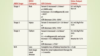
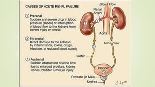
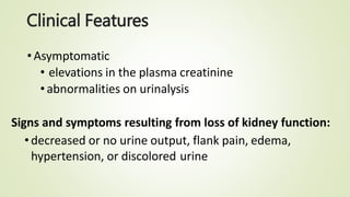
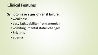
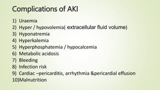
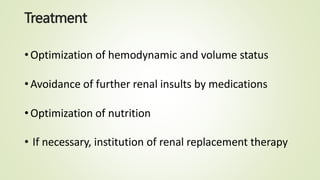
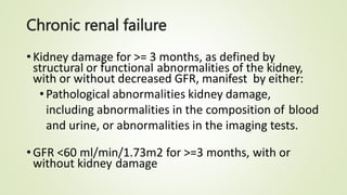
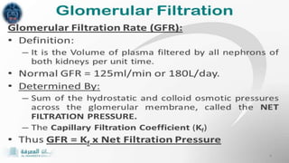
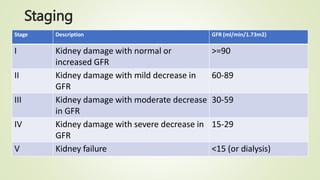
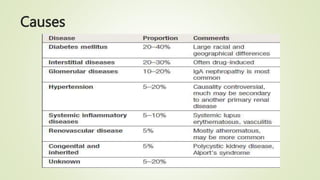
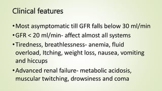
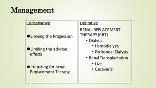
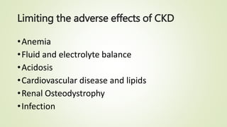
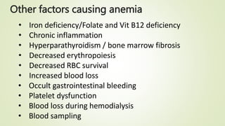
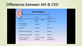
Renal Failure
By ,
DARSHAN
RENAL FAILURE
• Renal failure Results when the kidneys cannot
remove the body’s metabolic wastes or perform
their regulatory functions.
• The substances normally eliminated in the urine
accumulate in the body fluids as a result of
impaired renal excretion, leading to a disruption in
endocrine and metabolic functions as well as fluid,
electrolyte, and acid-base disturbances.
• It is a systemic disease and is a final common
pathway of many different kidney and urinary tract
diseases
ACUTE RENAL FAILURE
It is a reversible clinical syndrome where there
is a sudden and almost complete loss of kidney
function (decreased GFR) over a period of hours to
days with failure to excrete nitrogenous waste
products and to maintain fluid and electrolyte
homeostasis.
ARF manifests as an increase in serum creatinine
and BUN, Urine volume may be normal, or
changes may occur. Possible changes include
oliguria(<400ml/day), nonoliguria (>400ml/ day), or
anuria (<50 ml/day)
Categories of Acute Renal Failure
1. Prerenal:
occurs in 60%-70% of cases, it is the
result of impaired blood flow to kidney that
leads to hypoperfusion of the kidney and a
decrease in the GFR.
Common clinical conditions are volume-
depletion states (hemorrhage or GI losses),
impaired cardiac performance (cardiogenic
shock), and vasodilation (sepsis, infection)
2. Intrarenal:
Conditions such as burns, infections,
crush injuries, and severe blood transfusion
reactions can lead to intrarenal ARF.
With burns and crush injuries,
myoglobin and hemoglobin are liberated,
causing obstruction, renal toxicity, and
ischemia
Medications such as NSAIDs interfere
with the normal auto regulatory mechanisms
of the kidneys and may cause hypoperfusion
and eventual ischemia.
3. Postrenal :
it is usually the result of an
obstruction somewhere distal to the
kidneys. Pressure rises in the kidney
tubules and eventually, the GFR
decreases
Clinical Features
•Asymptomatic
• elevations in the plasma creatinine
• abnormalities on urinalysis
Signs and symptoms resulting from loss of kidney function:
•decreased or no urine output, flank pain, edema,
hypertension, or discolored urine
Clinical Features
Symptoms or signs of renal failure:
•weakness
•easy fatiguability (from anemia)
•vomiting, mental status changes
•Seizures
•edema
Diagnosis
•Detailed history
•Blood urea nitrogen and serum creatinine
•CBC, peripheral smear, and serology
•Urinalysis
•Urine electrolytes
•Ultrasonography, CT
•Serology: Anti DNA, HBV, HCV, cryoglobulin, urinary
Myoglobulin, HBsAG
Complications of AKI
1) Uraemia
2) Hyper / hypovolemia( extracellular fluid volume)
3) Hyponatremia
4) Hyperkalemia
5) Hyperphosphatemia / hypocalcemia
6) Metabolic acidosis
7) Bleeding
8) Infection risk
9) Cardiac –pericarditis, arrhythmia &pericardial effusion
10)Malnutrition
Treatment
•Optimization of hemodynamic and volume status
•Avoidance of further renal insults by medications
•Optimization of nutrition
• If necessary, institution of renal replacement therapy
Chronic renal failure
•Kidney damage for >= 3 months, as defined by
structural or functional abnormalities of the kidney,
with or without decreased GFR, manifest by either:
•Pathological abnormalities kidney damage,
including abnormalities in the composition of blood
and urine, or abnormalities in the imaging tests.
•GFR <60 ml/min/1.73m2 for >=3 months, with or
without kidney damage
Staging
Stage Description GFR (ml/min/1.73m2)
I Kidney damage with normal or
increased GFR
>=90
II Kidney damage with mild decrease in
GFR
60-89
III Kidney damage with moderate decrease
in GFR
30-59
IV Kidney damage with severe decrease in
GFR
15-29
V Kidney failure <15 (or dialysis)
Causes
Clinical features
•Most asymptomatic till GFR falls below 30 ml/min
•GFR < 20 ml/min- affect almost all systems
•Tiredness, breathlessness- anemia, fluid
overload, Itching, weight loss, nausea, vomiting
and hiccups
•Advanced renal failure- metabolic acidosis,
muscular twitching, drowsiness and coma
Investigations
19
Management
•High blood pressure medications.
•Medications to relieve swelling.
•Medications to treat anemia.
•Medications to lower cholesterol levels.
•Medications to protect your bones.
•A lower protein diet to minimize waste products in
your blood.
Management
Conservative
⚫Slowing the Progession
⚫Limiting the adverse
effects
⚫Preparing for Renal
Replacement Therapy
Definitive
RENAL REPLACEMENT
THERAPY (RRT)
• Dialysis:
• Hemodialysis
• Peritoneal Dialysis
• Renal Transplantation
• Live
• Cadaveric
Limiting the adverse effects of CKD
•Anemia
•Fluid and electrolyte balance
•Acidosis
•Cardiovascular disease and lipids
•Renal Osteodystrophy
•Infection
Anaemia
• Defined as Hemoglobin < 13.5 g/dl in males
< 12 g/dl in females
• Anemia is a condition in which you lack enough
healthy red blood cells to carry adequate oxygen to
your body's tissues.
• Primary cause : insufficient production of
Erythropoetin
Other factors causing anemia
• Iron deficiency/Folate and Vit B12 deficiency
• Chronic inflammation
• Hyperparathyroidism / bone marrow fibrosis
• Decreased erythropoiesis
• Decreased RBC survival
• Increased blood loss
• Occult gastrointestinal bleeding
• Platelet dysfunction
• Blood loss during hemodialysis
• Blood sampling
Anemia - goals
⚫Target Hb : not less than 11.5g/dl
⚫Check Hb monthly while on ESAs (Erythropoeisis
stimulating agents)
⚫Iron studies monthly when started on ESA
⚫On stable ESA Therapy : Iron studies can be done 3times in
a month
Anemia – treatment options
⚫Oral iron
⚫IV Iron Dextran
⚫IV Iron Sucrose
⚫IV Sodium Ferric Gluconate Complex
⚫Folic acid and Vitamin B 12 supplements
⚫Erythropoetin Stimulating Agents : Epoetin alfa*
Epoetin beta, Darbepoetin alfa
⚫Epoetin alfa / beta : 50 -100 IU / Kg SC per week
⚫Darbepoetin alfa : 40 mcg SC every 2 weeks
Bone disorder
•Renal bone disease- There are changes in the
concentrations of calcium, phosphate, vitamin d and
parathyroid hormone.
• Systemic complications include renal osteodystrophy
and soft tissue calcification, which contribute to morbidity
and mortality.
Preparation for Renal Replacement
Therapy
⚫Patients of CKD Stage IV approaching Stage V should be referred for
Vascular access ifhemodialysis is preferred
Peritoneal dialysis catheter placement if peritoneal dialysis is
preferred
⚫AVF is most preferred access for HD patients
Ideally created 6 months prior to start of HD
Non dominant upper extremity
And that arm is to be preserved – no iv lines
⚫AVG : 3-6 weeks prior to start of HD
⚫PD Catheter : 2 weeks prior to start of HD
Differences between AKI & CKD
Thank You

Renal Failure.pptx

  • 1.
  • 2.
    RENAL FAILURE • Renalfailure Results when the kidneys cannot remove the body’s metabolic wastes or perform their regulatory functions. • The substances normally eliminated in the urine accumulate in the body fluids as a result of impaired renal excretion, leading to a disruption in endocrine and metabolic functions as well as fluid, electrolyte, and acid-base disturbances. • It is a systemic disease and is a final common pathway of many different kidney and urinary tract diseases
  • 3.
    ACUTE RENAL FAILURE Itis a reversible clinical syndrome where there is a sudden and almost complete loss of kidney function (decreased GFR) over a period of hours to days with failure to excrete nitrogenous waste products and to maintain fluid and electrolyte homeostasis. ARF manifests as an increase in serum creatinine and BUN, Urine volume may be normal, or changes may occur. Possible changes include oliguria(<400ml/day), nonoliguria (>400ml/ day), or anuria (<50 ml/day)
  • 4.
    Categories of AcuteRenal Failure 1. Prerenal: occurs in 60%-70% of cases, it is the result of impaired blood flow to kidney that leads to hypoperfusion of the kidney and a decrease in the GFR. Common clinical conditions are volume- depletion states (hemorrhage or GI losses), impaired cardiac performance (cardiogenic shock), and vasodilation (sepsis, infection)
  • 5.
    2. Intrarenal: Conditions suchas burns, infections, crush injuries, and severe blood transfusion reactions can lead to intrarenal ARF. With burns and crush injuries, myoglobin and hemoglobin are liberated, causing obstruction, renal toxicity, and ischemia Medications such as NSAIDs interfere with the normal auto regulatory mechanisms of the kidneys and may cause hypoperfusion and eventual ischemia.
  • 6.
    3. Postrenal : itis usually the result of an obstruction somewhere distal to the kidneys. Pressure rises in the kidney tubules and eventually, the GFR decreases
  • 9.
    Clinical Features •Asymptomatic • elevationsin the plasma creatinine • abnormalities on urinalysis Signs and symptoms resulting from loss of kidney function: •decreased or no urine output, flank pain, edema, hypertension, or discolored urine
  • 10.
    Clinical Features Symptoms orsigns of renal failure: •weakness •easy fatiguability (from anemia) •vomiting, mental status changes •Seizures •edema
  • 11.
    Diagnosis •Detailed history •Blood ureanitrogen and serum creatinine •CBC, peripheral smear, and serology •Urinalysis •Urine electrolytes •Ultrasonography, CT •Serology: Anti DNA, HBV, HCV, cryoglobulin, urinary Myoglobulin, HBsAG
  • 12.
    Complications of AKI 1)Uraemia 2) Hyper / hypovolemia( extracellular fluid volume) 3) Hyponatremia 4) Hyperkalemia 5) Hyperphosphatemia / hypocalcemia 6) Metabolic acidosis 7) Bleeding 8) Infection risk 9) Cardiac –pericarditis, arrhythmia &pericardial effusion 10)Malnutrition
  • 13.
    Treatment •Optimization of hemodynamicand volume status •Avoidance of further renal insults by medications •Optimization of nutrition • If necessary, institution of renal replacement therapy
  • 14.
    Chronic renal failure •Kidneydamage for >= 3 months, as defined by structural or functional abnormalities of the kidney, with or without decreased GFR, manifest by either: •Pathological abnormalities kidney damage, including abnormalities in the composition of blood and urine, or abnormalities in the imaging tests. •GFR <60 ml/min/1.73m2 for >=3 months, with or without kidney damage
  • 16.
    Staging Stage Description GFR(ml/min/1.73m2) I Kidney damage with normal or increased GFR >=90 II Kidney damage with mild decrease in GFR 60-89 III Kidney damage with moderate decrease in GFR 30-59 IV Kidney damage with severe decrease in GFR 15-29 V Kidney failure <15 (or dialysis)
  • 17.
  • 18.
    Clinical features •Most asymptomatictill GFR falls below 30 ml/min •GFR < 20 ml/min- affect almost all systems •Tiredness, breathlessness- anemia, fluid overload, Itching, weight loss, nausea, vomiting and hiccups •Advanced renal failure- metabolic acidosis, muscular twitching, drowsiness and coma
  • 19.
  • 20.
    Management •High blood pressuremedications. •Medications to relieve swelling. •Medications to treat anemia. •Medications to lower cholesterol levels. •Medications to protect your bones. •A lower protein diet to minimize waste products in your blood.
  • 21.
    Management Conservative ⚫Slowing the Progession ⚫Limitingthe adverse effects ⚫Preparing for Renal Replacement Therapy Definitive RENAL REPLACEMENT THERAPY (RRT) • Dialysis: • Hemodialysis • Peritoneal Dialysis • Renal Transplantation • Live • Cadaveric
  • 22.
    Limiting the adverseeffects of CKD •Anemia •Fluid and electrolyte balance •Acidosis •Cardiovascular disease and lipids •Renal Osteodystrophy •Infection
  • 23.
    Anaemia • Defined asHemoglobin < 13.5 g/dl in males < 12 g/dl in females • Anemia is a condition in which you lack enough healthy red blood cells to carry adequate oxygen to your body's tissues. • Primary cause : insufficient production of Erythropoetin
  • 24.
    Other factors causinganemia • Iron deficiency/Folate and Vit B12 deficiency • Chronic inflammation • Hyperparathyroidism / bone marrow fibrosis • Decreased erythropoiesis • Decreased RBC survival • Increased blood loss • Occult gastrointestinal bleeding • Platelet dysfunction • Blood loss during hemodialysis • Blood sampling
  • 25.
    Anemia - goals ⚫TargetHb : not less than 11.5g/dl ⚫Check Hb monthly while on ESAs (Erythropoeisis stimulating agents) ⚫Iron studies monthly when started on ESA ⚫On stable ESA Therapy : Iron studies can be done 3times in a month
  • 26.
    Anemia – treatmentoptions ⚫Oral iron ⚫IV Iron Dextran ⚫IV Iron Sucrose ⚫IV Sodium Ferric Gluconate Complex ⚫Folic acid and Vitamin B 12 supplements ⚫Erythropoetin Stimulating Agents : Epoetin alfa* Epoetin beta, Darbepoetin alfa ⚫Epoetin alfa / beta : 50 -100 IU / Kg SC per week ⚫Darbepoetin alfa : 40 mcg SC every 2 weeks
  • 27.
    Bone disorder •Renal bonedisease- There are changes in the concentrations of calcium, phosphate, vitamin d and parathyroid hormone. • Systemic complications include renal osteodystrophy and soft tissue calcification, which contribute to morbidity and mortality.
  • 28.
    Preparation for RenalReplacement Therapy ⚫Patients of CKD Stage IV approaching Stage V should be referred for Vascular access ifhemodialysis is preferred Peritoneal dialysis catheter placement if peritoneal dialysis is preferred ⚫AVF is most preferred access for HD patients Ideally created 6 months prior to start of HD Non dominant upper extremity And that arm is to be preserved – no iv lines ⚫AVG : 3-6 weeks prior to start of HD ⚫PD Catheter : 2 weeks prior to start of HD
  • 29.
  • 30.