
Recommended
More Related Content
What's hot
What's hot (20)
Similar to Registration of Mandibular movements .pdf
Similar to Registration of Mandibular movements .pdf (20)
Recently uploaded
Recently uploaded (20)
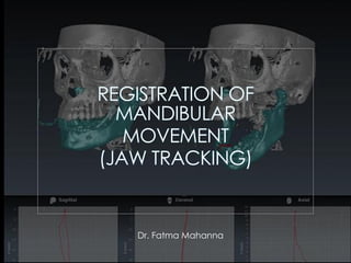
Registration of Mandibular movements .pdf
- 2. • The masticatory system is a functional unit composed of the teeth; their supporting structures, the jaws; the temporomandibular joints; the muscles involved directly or indirectly in mastication (including the muscles of the lips and tongue); and the vascular and nervous systems supplying these tissues.
- 3. • This system has been analyzed for many years, but the methods used record statistic points or single positions of the mandible (e.g., protrusion, excursion, etc.). The masticatory system is, however, dynamic, and the main component lies in the mandible. • Developments in digital dentistry have resulted in mechanical articulators that simulate mandibular movements being replaced and/or supplemented with virtual articulators in dental computer-aided design/computer-aided manufacturing (CAD/CAM) systems
- 4. In contrast to conventional mechanical procedures, the virtual articulator enables the visualization of three-dimensional (3D) calculated jaw movements for specific TMJ parameters or based on patient-specific dynamic motion data obtained using special devices. The main indications for the VA that have been proposed include individualized diagnostics and avoidance of the common problems encountered with MAs, such as creation of new occlusal contacts, material deformation, errors during orientation and positioning of dental casts, and difficulties simulating patient data in three dimensions (3D).
- 5. Types of Virtual articulator: There are currently two major types of VAs: 1. Completely adjustable (CA) 2. Mathematically simulated (MS). Completely adjustable (CA) Mathematically simulated (MS). The CA type reproduces exact movement paths of the mandible through the use of digital device accessories. Indication: involves complex cases where the morphology of the occlusal plane needs to be assessed during mandibular movements to avoid interferences in excursions. In order to capture the dynamic elements of occlusion and masticatory function, it was necessary to use a specific device called a Jaw Motion Analyzer (JMA) The MS type is an average value articulator which requires adjustment of additional settings in order to reproduce mandibular movements. Indication: involves cases where reproducing the relationship between the arches is sufficient for planning the occlusal morphology of the prosthesis. Disadvantage: • Couldn’t obtain individualized movements • must repeat the procedures for using a mechanical articulator, such as making impressions, performing facebow transfer, obtaining interocclusal records, and mounting dental casts (limitation)
- 6. ❑ Based on the chosen method of data acquisition and transfer, techniques for assembling a VA can be classified as : direct or indirect workflows. - The first step in a direct workflow involves digital scanning of the arches by means of an intraoral scanner (IOS), and then subsequently transferring this data to the VA without the use of analogue steps. - The indirect workflow involves taking analogue impressions of the arches, digitally scanning the casts mounted in a MA by means of a desktop laboratory scanner (DLS), and then transferring this data to the VA.
- 7. Lepidi et al, 2020
- 8. Importance of registration of jaw movements
- 9. Understanding of mandibular movement(electromyography coupled with jaw -tracking devices has provided much more information of the correlation between jaw movements and muscle activity) was considered important in: 1. This information was used in the design and setting of articulators. 2. The design of the dentures and denture teeth . 3. Importance of jaw movements has become apparent in fixed prosthodontics,( maintain and replace missing teeth should be to provide patients with good masticatory abilities)periodontics, orthodontics. 4. The diagnosis and treatment of disorders of the masticatory system . 5. Follow up after treatment of TMJ disorders
- 10. Review of Jaw Tracking methods: The extent to which jaw tracking provides a useful research tool, a diagnostic aid, or a therapeutic monitor clearly depends on what is being measured, how the process is carried out, and why the information is important To better understand differences between various systems to record mandibular motion a review of some recording methods presented over the years was made.
- 11. 1. Methods Using Mechanical Devices (Graphical) 2. Photographic Methods 3. Roentgenographic Methods 4. Electronic And Telemetric Methods 5. Magnetometry 6. Opto-electronic Methods 7. Axiography 8. Cadiax Copmact 9. Computerized Analysis Of Mandibular Movements 10. Electromagnetic Articulography(EMA) 11. Computer-monitored Radionuclide Tracking
- 12. Axiography Computerized Axiography Axiography By locating the condylar axis, the mandibular function can be analysed in relation to both condylar hinge axis and occlusal relationships. Axiographic recording procedure includes the following: • Clutch fixation • Analyzer bow preparation and placement • Placement of recording arm bow • Hinge axis location • Recording of movements
- 13. Cadiax Compact • The Cadiax compact axiographic device was designed to produce a fast joint analysis for articulator programming and also to aid in diagnosing the functional mandibular disorders. • It allows computerized recording of the opening, protrusion, and lateral tracings, and it calculates the sagittal and transversal condylar inclination angles for the adjustment of articulators
- 14. Electromagnetic Articulography (EMA) • This device measures displacements of the structure in real time as well as the acoustics and mechanics of speech using a microphone connected to the measurement system. • It has transmitter coils that determine magnetic fields to collect information about movements from sensors located on various structures (tongue, palate, mouth, incisors, skin, etc.). • After measurement, the information is passed on to a computer and read to visualize the recording of the mandibular movements registered by the EMA.
- 15. • Computer-monitored Radionuclide Tracking • A three-dimensional recording of mandibular movements . • A small and harmless radioactive source is fixed on the patient’s skin at the point of interest or sealed in a tooth cavity. • Using the proper collimation, the motion of the point source is recorded through a gamma camera and minicomputer. • Long-term storage on a magnetic device offers playback, slow-motion facilities, and data analysis.
- 16. Radiograph and TMJ: The hard and soft tissue structures of TMJ have been reconstructed by spiral and helical CT and magnetic resonance imaging (MRI). There are previous studies that have merged these data with jaw movement recordings by ultrafast MRI, electromagnetic tracking device, or optoelectric measuring systems. (Terajima et al.,2008) (Baltali., 2008)
- 17. MRIs disadvantages are related to 1-The high cost 2-The need for the patient to lie down during MRI imaging, which might alter normal jaw movements. 3-It is also contraindicated to patients with pacemakers and metallic heart valves. (Terajima et al.,2008) (Ferreira et al.,2016)
- 18. The disadvantage of the conventional CT is that it shows higher exposure values than CBCT. CBCTs main advantage is the observation of bony joint structures in all three planes in addition to the possible image manipulation at different depths and three-dimensional (3D) reconstruction. (Ferreira et al.,2016)
- 19. C
- 22. Motion Capture System Using Infrared Cameras: (Furtado et al.,2013) Requirements: • 3 infrared cameras These cameras are natively capable to find out white points in the images, which correspond to reflective objects (usually markers) in the scene. All cameras synchronously take images of the scene and reduce the image data to a set of 2D image coordinate representing the detections of the markers. Each camera is able to capture up to 100 frames per second, which is sufficiently high to guarantee detailed register of lower jaw movements.
- 23. • 9 Markers: A set of nine retro-reflective markers is proposed to allow mandibular movement analysis. Eight of them are called the secondary tracking markers and their purpose includes estimating some morphological parameters of the mandible. The primary tracking marker is the one primarily employed to track the movement of the jawbone.
- 24. Secondary tracking markers can be fixed on skin by using adhesive tape and a plastic support. They must be positioned on the following regions of the face: (1) TMJ external surface (left and right) (2) mandible angle region (left and right) (3) middle region between the chin and the mandible angle (left and right) (4) above upper lip (5) on the forehead.
- 25. • A prototype soft is implemented to communicate with the cameras and perform: 2D point identification and tracking 3D data processing
- 27. Top view of the proposed camera setup. The left and right cameras are placed at 1 meter distance from the subject forming an angle of 120 degrees. The central camera is positioned at 1.3 meters distance.
- 28. Advantages: 1- Good precision and accuracy 2- Minimum obstruction. 3- Real- time 3D reconstruction and analysis. 4- Moderate cost 5- It can give parameters of facial morphology and automatically recognize markers.
- 30. Magnetic motion capture Jaw tracking system: The jaw tracking system is based on the magnetic motion capture system with wireless resonated marker. The resonated markers are composed of an inductor and a capacitor, neither electric wires nor batteries inside the marker are necessary. Therefore, the markers can be easily attached to a tooth without disturbing the physiology. The system can be used for highly accurate jaw tracking without magnetic shielding because it is free of earth field noise, thus making it superior to conventional jaw movement tracking devices.
- 32. • 1- MODJAW • 2- ARCUS DIGMA • 3- FREECORDER BlueFox • 4- JMA Zebris • 5- Planmeca 4D • 6- SICAT JMT
- 33. Movement registration systems 1. MODJAW • The MODJAW system uses optical scanning solution to dental surgeons at the diagnostic and therapeutic stage. • By simply capturing jaw motion, MODJAW models the kinematics of patients. • The software, designed with an ergonomic interface and touch- screen diagnostic functionalities can provide dynamic visuals of models in 2D and 4D, patient’s occlusion plans, dynamic mapping of teeth contacts and automated calculation of posterior determinant parameters (Bennett Angles, condylar slopes, Spee curve…).
- 35. • 2- ARCUS digma • The ARCUS digma system uses ultrasound transmission to measure and reproduce the jaw movements. • Arcus Digma systems consists of a self-contained computer with a colour touch screen (tabletop unit) or a two- color screen (handheld), a head bow, a pair of ultrasonic transmitter-sensors that attach to the mandible with a small device called a paraocclussial clutch and to the maxilla with a bite fork. • KaVo’s system is an articulator- regulated registration technique and requires the use of a KaVo PROTAR articulator. During the measuring time, this articulator is “virtually” projected into the mouth of the patient.
- 36. 3- Freecorder BlueFox • This contactless system tracks a series of encoded visual patterns. First, to measure the position of the skull, a bow with references is placed on the ears and then, it is attached to the nose’s bridge. Another light modular arch is attached to the jaw to capture its movement. • Using special cameras, them patterns are captured 100 times per second, thus achieving very high resolutions (1/1000 mm.)
- 37. 4- JMA Zebris • JMA Zebris system has a customized jaw’s anchor that joins the lower arch by means of magnets. • Another upper arch is placed on the skull and the nose’s bridge. Both of them have electronic sensors that measure relative distances. • The system determines the jaw’s relative position by calculating the flight times of ultrasonic pulses.
- 38. 5- Planmeca 4D jaw motion • The system tracks and visualizes jaw movements with the Planmeca ProFace camera feature of Planmeca ProMax 3D Mid and Max X-ray units, showing the movements in real-time in a 3D CBCT image. • The camera tracks the position and movement of eight spheres. Half of them are fixed to a bow and the others to glasses. • Glasses position defines skull’s movement and the bow is fixed to the lower arch determining relative distances.
- 41. 6- SICAT JMT: The integration of CBCT diagnostics with CAD/CAM and other objective data from biometric instrumentation, like the SICAT JMT (Jaw Motion Tracker), gives us an opportunity to formulate a definitive treatment plan with a common goal: optimal oral health.
- 42. Advantages: • Real condyle-fossa relationship during jaw movement can be reproduced for any point on the mandible • In combination with CEREC SW 4.4, accurate articulation and functional prosthetics are now possible for the first time • SICAT OPTIMOTION all-digital workflow, which is based on the movement of the individual patient, reduces the amount of adjustment and adaptation effort required for a treatment appliance • Completely integrated workflow allows for diagnosis and treatment planning in a single patient appointment
- 43. Software SICAT Function showing the combined CBCT image with CAD/CAM image and jaw motion tracking data
- 44. Combining CAD/CAM and CBCT in a single practice with complete integration creates a magical experience for the clinician and the patient alike. CBCT provides an opportunity to provide an objective assessment based upon results of the 3-D image, proper diagnosis, and higher treatment plan acceptance and increased precision in dental therapy.
- 45. The introduction of digital intra oral impression in combination with Jaw tracking/CBCT, adds the advantages of • Time saving • The reproducibility of the method (Ender et al., 2015)
- 47. The jaw tracking system in this study consisted of a camera which can obtain depth data and RGB data of the object simultaneously, a laptop computer and Face shift software for data analysis and reconstruction of the participant face.
- 50. 1- Jaw tracking and TMD: The study of mandibular motion is essential to the management of TMDs. The need to duplicate the mandibular movements extra-orally led to the employment of various methods to record and analyze them. Patient information can be transferred to an articulator with mounted casts and thus, mandibular movements can be evaluated. Questionnaires, clinical and radiographic examinations, computed tomography and magnetic resonance imaging are some of the instruments used to assess TMD. However, there is a scarcity of studies on mandibular dynamics in TMD in the literature.
- 51. There are several limitations concerning bite registration and articulators. • Bite registrations are static recordings; thus, articulators are unable to record real life dynamics of occlusion during mandibular movements. • Another basic limitation is the lack of visualization of condyle position, which is essential to the management of TMDs. • There are also various difficulties with transferring the registration onto the articulator and mounting the casts with accuracy. (Kandasamy.,2015)
- 52. • Cone beam computed tomography (CBCT) in conjunction with jaw tracking devices enabled the virtual evaluation of the occlusion and the TMJs and helped substantially in overcoming these problems. • These system presenting a real, 3D simulation of mandibular movements relative to the patient-specific anatomy of the jaw. In addition, changes in the joint space during resting or other positions can be recorded. Thus, the system can be used as a useful supporting tool in the diagnosis, treatment, and management of TMDs.
- 53. TMD Diagnosis: Examples: • Detection of narrowing of intra articular distance during condyle sliding movement along the articular eminence, (especially in patients with severe facial asymmetry) • The reduced intra-articular distance by physical loading may be physiological. • However, under repeated harmful activities like biting with wide mouth opening and para-functions such as clenching and bruxism, the physiological threshold can be crossed, and overloading and subsequently TMDS may result. Chang AR, Han JJ, Kim DS, Yi WJ, Hwang SJ. Evaluation of intra-articular distance narrowing during temporomandibular joint movement in patients with facial asymmetry using 3-dimensional computed tomography image and tracking camera system. J Craniomaxillofac Surg. 2015 Apr;43(3):342-8. doi: 10.1016/j.jcms.2014.12.015. Epub 2015 Jan 7. PMID: 25648068.
- 54. Treatment of TMD: The SICAT Function software can combine cone bean CT, electronic JMT data, and digital impressions; thus, it is capable of presenting a real, 3D simulation of mandibular movements relative to the patient-specific anatomy of the jaw. In addition, changes in the joint space during resting or other positions can be recorded. Thus, the system can be used as a useful supporting tool in the diagnosis, treatment, and management of TMDs.
- 57. Other devices may be combined with jaw tracker during recording of mandibular movements: e.g., Deconditioning or relaxing the mandibular musculature by the application of TENS. With the help of TENS, complete relaxation of the muscles was achieved, and the mandibular musculature was induced to guide the mandible in the physiologic position.
- 58. • In maxillofacial prosthetic dentistry, the use of tracking system with 3D facial scanner can help to confirm that a prosthesis is harmonized with a particular patient’s aesthetics through superposition with facial scan data, with respect to the occlusal plane, smile line, and midline. • In addition, loss of TMJ function and muscle contraction can occur due to the development of scar tissue after resective surgery in patients with cancer. A symmetric articulator cannot reproduce or mimic asymmetric and individualized jaw movements that are characterized by severe deflection. 2- Maxillofacial prosthodontics: J.-E. Kim, et al., Complete assessment of occlusal dynamics and establishment of a digital workflow by using target tracking with a three-dimensional facial scanner, J Prosthodont Res (2018), https://doi.org/10.1016/j.jpor.2018.10.003
- 59. 3- Detection of Phasic Sleep Bruxism in OSA Patients: • Repetitive jaw muscle activity may lead to development of sleep bruxism in some individuals, which is characterized by tooth clenching or grinding and/or mandible bracing or thrusting. • Until now, the size and discomfort imposed by jaw tracking recording devices have been a major limitation for human sleep studies in a natural home environment. • Recent non-invasive and easy to use technological innovation has made possible the continuous data collection during sleep of mandibular jaw movements.(Martinot et al, 2021)
- 60. 4- Studies of speech difficulties: Jaw movement patterns were examined in subjects with childhood apraxia of speech (CAS) . A movement tracking system was used to study jaw duration, displacement, velocity, and stability.
- 61. A Comprehensive Treatment Approach For Idiopathic Condylar Resorption And AnteriorOpen Bite With 3D Virtual Surgical Planning And Self-ligated Customized Lingual Appliance. (Rahman Et Al, 2019) 5- Perform proper diagnosis for comprehensive treatment:
- 62. Studies suggest that patients with ICR remain undiagnosed and unrecognized in the orthodontic clinic owing to the poorly understood aetiology of the disease and lack of diagnostic tools. ICR often causes occlusal and skeletal changes, TMJ dysfunction and pain, and maxillofacial deformities. The treatment plan included: (1) presurgical alignment and levelling of the teeth in both arches (2) jaw motion tracking (JMT) to detect mandibular movement (3) 3-piece maxillary osteotomies with mandibular reconstruction and bilateral coronoidectomies (4) postsurgical correction of the malocclusion.