
Recommended
More Related Content
Similar to Computers in ortodontics.ppt
Similar to Computers in ortodontics.ppt (20)
Recently uploaded
Recently uploaded (20)
Computers in ortodontics.ppt
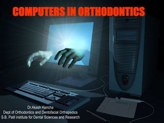
- 1. COMPUTERS IN ORTHODONTICS Dr.Akash Kencha Dept of Orthodontics and Dentofacial Orthopedics S.B. Patil institute for Dental Sciences and Research
- 2. Contents 1. Introduction 2. Use of computers in dental practice 3. Classification of appliance a)Administrative b)Clinical c)miscellaneous 5. Computed Tomography 6. Digigraph 7. VTO 8. Computerised tooth width analysis 8. Cephalometric applications 9. Dento facial planner plus 10. Vistadent image management system 11. Video imaging 12. MRI 13. Invisalign Application of computers in orthodontics – Contents sem1/sb/09-04
- 3. INTRODUCTION When the word "technology" is mentioned, most people think about computers. Virtually every facet of our lives has some computerized component. A few years ago, it was considered a specialized component, which has now been converted to an everyday appliance. The computer is basically an automatic electronic machine that performs calculations or derives results based on the data fed into it and the software/ program it is designed for. A computer is capable of accepting data, performing operations according to instructions and providing the results of these operations in comparatively shorter duration of time and with greater accuracy as compared to Manual labor. It is built to perform routine calculations with speed, reliability and ease.
- 4. Dentistry, being that branch of medicine which relies heavily on technology and which embraces new techniques enhance not only record keeping, but practice management, patient education and motivation.
- 5. Uses of computers in dental practice classification of applications: 1) Administrative applications Patient case records Recall appointments Patient scheduling Accounts Patient correspondence Billing Prescription formats Post-treatment instructions Insurance claims Referral information
- 6. 2) Clinical applications Patient photographs-analysis and storage Patient radiographs-analysis and storage Inter- specialty referral and opinion Patient motivation Appliance design using CAD CAMs Growth predictions Visual treatment objectives Survey information/epidemiological data Presentations Continuing dental/ medical education Literature reviews
- 7. 3) Miscellaneous application Survey information/epidemiological data Presentations Continuing dental/ medical education Literature reviews Entertainment
- 8. Orthodontists with their love for technology and miniaturization have not remained untouched. Computers are practically used in all the facets of any dental practice today .Computers have become especially useful to orthodontists for: Digital photography Digital radiography Digital cephalometrics Video cephalometrics 3-D imaging Digital study models
- 9. Conventional Case Paper Impression & Diagnostic Casts Radiographs Manual Tracing Cephalometric Analysis Diagnostic Set-up Treatment-Preformed System Mid Term Evaluation Computerised Data Sheet 3D Photography RVG, Digital Radiography Digitised Tracing Advanced software's VTO / VTP Treatment-Customised Continuous Monitoring Applications of Computers in Clinics
- 10. Computed Tomography Invented by Sir Godfrey Hounsfield who was awarded a Nobel prize in 1979 CT is an image display of the anatomy of a thin slice of the body developed from multiple x- ray absorption measurements made around the body’s periphery.
- 11. Parts of the Equipment; 1. Scanner ( movable x ray table + gantry) 2. Computer system 3. A display console
- 12. Principle; A x ray source and array of detectors mounted within the gantry rotate around the patient during each scan. Detectors record the attenuation values of the beam emerging from the patient Information from each traverse is a Profile
- 13. The tube and detectors are further angled and another traverse is made. A series of Profiles is built up. The computer analyses the data and an image is produced.
- 14. Early scanners translate and rotate system. Recently developed scanners stationary detectors and x ray tube rotates around the patient both the detectors and x ray tube rotate in synchrony
- 15. Radiation dosage 1.536 rad for a single section 1.8432 rad for multiple sections Estimated dose to the centre of the condyl with CT is 180mR
- 16. Useful in determining changes in bone density Primary imaging method when internal derangement or arthrosis is suspected – clinical diagnosis is not always sufficient. Has advantages when planning treatment or operations on jaws and TMJ diseases and deformities.
- 17. Microcomputed Tomography Principally the same as CT, except that the reconstructed cross sections are confined to a much smaller area. Significantly reduces radiation dosage. Used to measure bone connectivity in all 3 dimensions.
- 18. Cephalometric Applications DIGIGRAPH Introduced by Dolphin imaging systems Non-radiographic system ‘Digigraph workstation’ Video images also possible VTO Reduces time required for records.
- 19. System design: The DigiGraph Work Station is about 5 feet long, 3 feet wide and 7 feet high. The main cabinet contains the electronic circuitry, and the patient sits next to the cabinet in an adjustable chair similar to those used with cephalometers.
- 20. The head holder is suspended from a beam, supported by a vertical column attached to the cabinet. more comfortable than cephalometer head holders, allowing the patient to remain in the holder for several minutes. Ear rods and forehead and posterior head pieces are used to minimize patient movement. The ear rods can be removed so that facial and intraoral images can also be recorded while the patient is sitting in the adjustable chair.
- 21. A model board can be inserted into the head holder, and images of various views can be recorded .
- 22. A light box can also be attached to the head holder for imaging headfilms, wristfilms, laminagraphic films, and panoramic x-rays.
- 23. The video monitor is attached that can be rotated as the operator moves. Images are as sharp as those on a standard color television. The images, text, and numerical data can be displayed, stored, and modified using either a light pen or a standard computer keyboard.
- 24. Any image appearing on the screen can be reproduced instantaneously with one of three "hard copy" output devices: • Sony video imager— makes 5 " x 7 " color prints in 60 seconds • Polaroid freeze-frame camera— produces Polaroid prints in 10 seconds; • Hewlett Packard Paintjet printer— makes 8 " x 10 " paper color copies in 4 to 8 minutes.
- 25. The digitizing handpiece is used to record cephalometric data while the patient is in the head holder. The removable, sterilizable tip of the handpiece is placed directly on the patient to record a series of facial and intraoral landmarks. As each landmark is located, the handpiece button is depressed and the location is recorded in three-dimensional coordinates (x,y,z). Each time the handpiece button is depressed, an audible sound is picked up by an array of four microphones on the beam. The time it takes the sound to reach each of the microphones determines the landmark location.
- 26. Three-dimensional digital modeling and setup Aldo Macchi,S Gianpaolo Carrafiello,b Vittorio Cacciafesta,C and Antonio Norcinic Varese, Italy Obtaining accurate images of the craniofacial region is critical when developing an orthodontic diagnosis and treatment plan. It is a new imaging method that provides complete 3-dimensional views of the maxilla and the mandible, and the model setup with individual anatomic roots. The method uses computed tomography technology and laser scanning using computers. Am J Ortho Dentofacial Orthop, MAY 2005
- 27. 3D Digital modeling and setup 3D superimposition of the anatomic teeth, before treatment [A & B] and after [C & D] the setup to visualize the amount of teeth movement before and after treatment [ E ] by making use of 3D setup using computers. By making use of this set up we can analyse a degree of correction achieved in malocclusion and discrepensies like fenestrations. A B C D E Am JOrtho Dentofacial Orthop, MAY 2006
- 28. Airway volume assessment in pt. with mouth breathing, adenoid hypertrophy, or sleep apnoea, by application of transfer function to 3D CT scan Bone rendering with transparent soft tissues by application of transfer function ,which render soft tissue invisible on 3D CT scan. Visualization of internal anatomic structures by removal of cranium by box cut to peek inside a 3D volume by use of transfer function. Am JOrtho Dentofacial Orthop, MAY 2005
- 29. 3D Cone-Beam CT virtual models A. Can be Used In case of Surgical patients and those with developmental anomalies like hemi facial microsomia where in case ,if the working condyle is missing, it can be replaced with costocondral graft by proper planning of surgery using 3D cone beam CT virtual models . B. can be used to evaluate significant facial asymmetry and missing articular fossa. Am JOrtho Dentofacial Orthop, MAY 2006
- 30. Can render bone transparent which allows visualization of developing permanent teeth & It can also be used to determine the position of the surgical pins or implants in bone, that might be impairing tooth eruption . 3D Cone-Beam CT models Am JOrtho Dentofacial Orthop, MAY 2006
- 31. Rapid prototyping: A new method of preparing trays for indirect bonding Fabio Ciuffolo,a Ettore Epifania,b Gionni Duranti,C Valentina De Luca,d Daniele Raviglia,d Silvia Rezza,d Computer-aided technology is used to design the individualized trays, which are then produced with a rapid prototyping procedure. The advantages of application includes time saving and accurate bracket placement .
- 32. Clinical application of RPT: A) Bracket in RPT virtual tray with adhesive paste; B) RPT placed on tooth; C) RPT removal; D)and E) RPT placement on adjacent teeth (full arc bonding can undertaken); F) Light-curing. Design process of virtual tray: A) scanned malocclusion model; B) virtual bracket placement; C) and D) creation of virtual tray.
- 33. Computerized tooth width analysis Introduced by Christopher T.C. Ho and Terrence Freer Named as Ho-Freer Graphical analysis of tooth width Discrepancy (GATWD) Base line data obtained from pre treatment orthodontic casts.
- 34. Direct input using digital calipers or manual input using visual basic 3.0 Upto 24 tooth width measurements can be done Caliper used is a Mitutoyo 6”/ 150mm , with tapered beaks Connected to a Mitutoyo digimatic mini processor - IBM compatible computer
- 35. Advantages: 1. Less time consuming 2. Use of digital calipers reduces errors during transfer of measurements 3. Mathematical calculations done by the computer 4. Eliminates reference to standard value tables
- 36. Classification of Software Microsoft Word Microsoft Power point Microsoft Access Microsoft Excel MS DOS Microsoft Outlook Microsoft Frontpage Net Meeting •SOFTWARE FOR REGULAR USAGE
- 37. Adobe Acrobat Reader Flash Corel draw MS Photo editor Adobe Photoshop Microsoft publisher Anti Virus Norton, Sophos… 3d smax Maya AutoCAD WinZip Digicam Software Nero Express MSC Nastran, Patran Ortho cad ADVANCED USAGE
- 38. Digitalizing study models using OrthoCAD OrthoCAD - It is a digital study model capture, assessment and storage system. It provides a 3D record of the original malocclusion, any stages during treatment and the outcome of the treatment.
- 39. VISTADENT Image management system VISTADENT COMPLETE™ ability to modify an image that is stored in the program, as well as the ability to do Visual Treatment Objective (VTO). Images can easily be manipulated to show treatment objectives by using standard editing features such as cut and paste or more advanced features like the Smile Library.
- 40. Ideal system requirements; win 98 operating system or above pentium processor II 64 MB RAM Other attachments: • digital camera or scanner • RGB camera • Printer ( color) • CD ROM drive
- 41. Easy to calculate cephalometric analysis. The Ceph program allows one to trace and identify your points directly from a scanned X-RAY. After points and tracings are entered they can easily be edited for accuracy purposes. Then Tracings, Analysis and Measurements can all be printed on one page with the image.
- 42. The VTO feature automatically illustrates soft tissue changes based on measurements for most orthodontic treatment as well as many surgeries.
- 43. Dentofacial Planner Plus is a powerful treatment visualization software system. DFP Plus links digitized lateral cephalograms to digital facial images - move the skeletal and dental components of the cephalometric tracing - the cephalometric profile and the facial image are automatically transformed to represent the predicted facial form.
- 44. Cephalometrics DFP Plus contains all of the cephalometric functionality of Dentofacial Planner, A digitized radiograph is used as the starting point for a wide variety of cephalometric analyses and superimpositions, growth estimation, orthodontic treatment planning and surgical prediction. Soft tissue profile changes are automatically computed and displayed during treatment planning manipulations.
- 45. Flexible Cephalometric Analysis Dento Facial Planner and DFP Plus share the unique functionality of Tools(tm), that provides flexibility in customizing the work. Tools - mix and match measurements from several different analyses into one or more personalized cephalometric analyses. They easily add new landmarks, measurements and graphical reference lines.
- 46. Image Review: • simultaneously showing pretreatment and predicted images: Other functions; •Ceph superimposition and narrow review •Overlay ceph tracing on image •Semi-transparent image superimposition
- 47. Video Imaging. With the integration of computers and cephalometric technology in the 1970s, complex statistical analyses of growth patterns and dentoskeletal relations were established. The speed of computerized cephalometric programs has helped streamline the laborious manual measurement of patient cephalograms and the creation of the visualization treatment objective (VTO).
- 48. In the VTO of an orthognathic surgery case, the clinician classically has used acetate templates of the teeth and jaws to predict orthodontic and surgical movements and the final profile is determined by the reaction of the soft tissue to the hard tissue movements.
- 49. Video imaging technology allows the orthodontist to gather frontal and profile images and modify them to project overall esthetic treatment goals. The video image is much more realistic than photograph simulation and it is much easier for the patient to comprehend than just the soft tissue profile of a cephalometric tracing.
- 50. Tracing Display Tracings - displayed immediately; include planes relevant to the selected cephalometric analysis; An advantage - is that the nonlinear distortion associated with radiographs is eliminated. - all points are recorded in a direct, one-to-one manner, allowing precise superimposition of patient tracings over video images .
- 51. Visual Treatment Objective • To move part of the picture, simply touch the light pen to two points on the screen, at opposite extremes of the area to be moved. • The computer draws a box with the two points at opposite corners. • Then, by touching the light pen to another spot on the screen, the boxed image is moved to that spot . • Boxes can be moved vertically, horizontally, or diagonally, or they can be rotated about any point.
- 52. The software automatically blends skin tones and smoothes profile lines so they are consistent with the surrounding tissue. "before and after" format. The light pen can also be used for freehand drawing over any video image.
- 53. Coordinating images and cephalogram: Digitizing the ceph and then matching the size of the video image to it Digitizing the ceph and then sizing the ceph to an existing video image (loss of calibration) Gathering the video image of a ceph through a calibrated video camera and matching it to an existing video image Simultaneous image gathering and ceph. radiography.
- 54. Cephalometric Applications Downs • Burstone • McNamara • Tweed • Grummons frontal • Standard lateral • Standard frontal •Ricketts lateral • Ricketts frontal • Vari-Simplex • Holdaway • Alabama • Jarabak • Steiner
- 55. What is Invisalign®? Invisalign® is the invisible way to straighten teeth without braces. uses a series of clear removable aligners to straighten teeth without metal wires or brackets.
- 56. co-founded by Zia Chishti and Kelsey Wirth in 1997 Based in Sunnyvale, California.* Align Technology the treatment procedure is handled by the computer technicians in Pakistan - process takes 3 weeks to a month. After approval from the orthodontist, specifications are transmitted to the manufacturing plant in Mexico
- 57. Impressions are made using Polyvinyl Siloxane Impression and bite send along with a detailed treatment plan. advanced imaging technology transforms plaster models into a highly accurate 3-D digital image. A computerized movie - called ClinCheck® - depicting the movement of teeth from the beginning to the final position is created. After wearing all of the aligners in the series, customized set of aligners are made from these models, sent to the doctor, and given to the patient. Pt to wear each aligner for about two weeks. From the approved file, laser scanning to build a set Invisalign® uses of actual models that reflect each stage of the treatment plan. Using the Internet, the doctor reviews the ClinCheck file - if necessary, adjustments to the depicted plan are made. Procedure
- 58. Patient gets the first aligner 6 weeks after the 1st visit Most treatments require 20 – 60 aligners worn for 2 weeks each Should be taken off only for eating and brushing
- 59. Contraindications; patients with severe malocclusions All children – growing jaws and erupting teeth too complicated for the computer to model
- 60. Magnetic Resonance Imaging Principles: Magnetism is a dynamic invisible phenomenon consisting of discrete fields of forces. Magnetic fields are caused by moving electrical charges or rotating electric charges. Images generated from protons of the hydrogen nuclei. Essentially imaging of the water in the tissue.
- 61. Equipment; 1. The Gantry ;houses the patient. Patient is surrounded by magnetic coils 2. Operating console ; where the operator controls the computer and scanning procedure 3. Computer room network.
- 62. The technique is based on the presence of specific magnetic properties found within atomic nuclei containing protons and neutrons. Inherent property of rotating about their axis Causes a small magnetic field to be generated around the electrically charged nuclei.
- 63. The dipoles exposed within a strong electric field Cause Orientation in response to the field Depending on density and spatial relation Signal is interpreted and image produced
- 64. When images are displayed; intense signals show as white and weak ones as black. Intermediate as shades of gray. Cortical bone and teeth with low presence of hydrogen are poorly imaged and appear black.
- 65. The objectives of MRI imaging of the TMJ are; Determine relationship between the disc and Temporal and mandibular components of the TMJ Detect inflammation, hematoma and effusion for the soft tissue components. MRI clearly differentiates the soft tissue components . Short and long echo imaging of the TMJ enables identification of the positional relationships between the disc and the condyle.
- 66. Indications Assessing diseases of the TMJ Cleft lip and palate Tonsillitis and adenoiditis Cysts and infections Tumors
- 67. Contraindications; Patients with cardiac pacemakers. Patients with cerebral metallic aneurysm clips. Slight movement of the clip could produce bleeding Stainless steel and other metals produce artifacts ; obliterate image details of the facial area.*
- 68. Short comings; Inability to identify ligament tears or perforations Dynamics of tissue joint not possible Cannot be used in patients suffering from claustrophobia.
- 70. Current principles and techniques in orthodontics- by Thomas.M.Graber & Robert.L.Vanarsdall. Contemporory Orthodontics – by William.R.Proffit Gurukeerat Singh, Text Book of Orthodontics, First Edition, 2004 – Jaypee Brothers, New Delhi AJODO 2006; August ,volume130 ,no 2 : 257-65 AJODO 2006: may, volume 129 ,no 5; 605-617 AJODO 2005volume 128: 157-60 AJODO 2006 volume 127: 627-637 AJODO 2005 volume 127 no 5 World journal of orthodontics December 2006 • OrthoCAD Bracket Placement Solution W. R. Redmond et al, AJO/DO, May 2004 • Comparison of measurements made on digital and plaster models M. Santoro et al, AJO/DO July 2003 BIBLIOGRAPHY