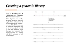
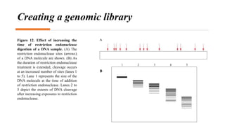
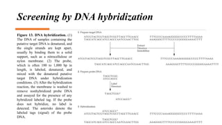
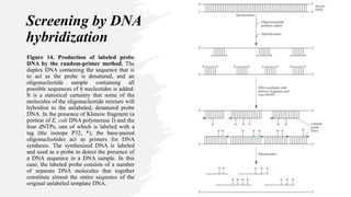
Recombinant DNA technology involves transferring genetic material from one organism to another. A common method is to extract DNA from a donor organism, cut it with restriction enzymes, and ligate it into a vector like a plasmid. This recombinant DNA is then introduced into host cells. Cells containing the recombinant DNA can be identified by screening, such as using hybridization probes to detect the target DNA sequence. Creating a genomic library involves cutting an organism's entire genome into fragments, inserting them into vectors, and transforming host cells to generate a collection containing all the organism's DNA.
This presentation deals with the introduction of Recombinant DNA Technology. The role of different enzymes. Specifically Restriction endonucleases and roles of various vectors.
Now a day's these technique is tremendously use for in lab by using foreign Dna to to producing insulin in bacteria , plant with high yielding capacity by using Gene from another species
The document discusses the process of synthesizing cDNA from mRNA. It involves isolating mRNA, using reverse transcriptase to copy the mRNA into single-stranded cDNA, then converting it to double-stranded cDNA using DNA polymerase. The double-stranded cDNA can then be inserted into a vector and used to create a cDNA library through cloning in bacteria or phage. The library can be screened by hybridization or assays to identify clones containing genes of interest.
Gene cloning involves isolating a particular gene or DNA fragment of interest from an organism's total DNA and producing many copies of just that fragment. There are several reasons for cloning DNA, such as determining a gene's nucleotide sequence, identifying control sequences, investigating protein/enzyme function, and engineering organisms for specific purposes like insulin production. Common tools used in cloning include restriction enzymes, ligase, vectors like plasmids and bacteriophages, and host cells. DNA is cut with restriction enzymes, ligated into a vector, and introduced into host cells to replicate the exogenous DNA fragment.
Vectors part 1 | molecular biology | biotechnologyatul azad
This document discusses various types of plasmid and bacteriophage vectors used for cloning DNA fragments. It describes the features and selection methods of commonly used vectors like pBR322, pUC18, pGEM3Z, lambda phage and M13 phage vectors. Plasmid vectors like pBR322 are advantageous for having small size, high copy number and antibiotic resistance markers. Lambda phage vectors can accommodate larger inserts compared to plasmids and allow easy screening of recombinant phages. M13 vectors are useful for obtaining single-stranded DNA copies for sequencing.
The document discusses various methods for screening and selecting recombinant cells. Direct selection methods include antibiotic resistance screening and blue-white color screening. Indirect selection methods include screening by nucleic acid hybridization, colony hybridization, immunological assays, and detecting protein/enzyme activity. These screening methods allow identification of recombinant cells that contain the gene of interest from a mixture of transformed cells.
Recombinant DNA technology involves transferring genes between organisms using artificial means. It works by combining DNA from different sources into a single molecule. The process involves generating DNA fragments, inserting the fragments into vectors, introducing the vectors into host cells, and selecting clones containing the recombinant DNA. Common tools used include restriction enzymes to cut DNA, vectors like plasmids to carry DNA, bacterial hosts like E. coli, and techniques like transformation and selection to introduce and identify recombinant DNA. Applications include analyzing gene structure, producing pharmaceuticals, genetically modified organisms, and gene therapy.
This document discusses rDNA technology and methods for gene cloning. It describes how recombinant DNA is formed by combining DNA sequences as desired. Gene cloning involves isolating a specific DNA fragment from an organism and introducing it into a plasmid vector that can replicate in a host cell, producing multiple copies of the fragment. Various methods are described for isolating DNA fragments, including mechanical shearing, restriction enzyme digestion, and reverse transcriptase. Different types of vectors like plasmids, bacteriophages, cosmids, BACs, and YACs that can be used are also summarized.
This document is a biology investigatory project submitted by Arajit Kumar Pati on the topic of genetic engineering. It provides an overview of genetic engineering and recombinant DNA technology. It describes various techniques used in genetic engineering like DNA cloning, restriction digestion, use of vectors, recombinant DNA formation, insertion of genes into hosts, and production of gene products. It discusses the wide applications of genetic engineering in fields like medicine, agriculture, and industry. It concludes that genetic engineering has revolutionized our understanding of inheritance and molecular biology.
1. Molecular cloning involves cutting DNA from one organism and inserting it into a vector that can replicate in a host organism, allowing the DNA fragment to be amplified. Recombinant DNA technology uses restriction enzymes to cut DNA into fragments that are then ligated into cloning vectors like plasmids or bacteriophages.
2. After transforming host bacteria with the recombinant vector, clones containing the inserted DNA fragment can be selected for and amplified. Colonies containing the insert are identified through antibiotic resistance or colorimetric markers present on the vector.
3. cDNA libraries provide a way to clone and study eukaryotic genes. mRNA is isolated and reverse transcribed into cDNA, which is then ligated into vectors and
The document discusses the principles and processes of biotechnology. It describes two core techniques that enabled modern biotechnology: genetic engineering and maintaining sterile environments for microbial growth. It then discusses various tools used in recombinant DNA technology, including restriction enzymes, vectors, competent hosts, PCR amplification, and downstream processing to obtain recombinant products.
Vectors are DNA molecules that can carry foreign DNA fragments into host cells. There are two major classes of vectors: plasmids and phages. Plasmid vectors like pBR322 were some of the earliest cloning vectors and have replication origins, antibiotic resistance genes, and multiple cloning sites. PUC plasmids are derived from pBR322 and use blue-white screening. Lambda phages can accommodate larger DNA fragments than plasmids. Cosmids and phagemids have characteristics of both plasmids and phages, allowing larger DNA fragments to be cloned and packaged. M13 phages produce single-stranded DNA clones. Different vector types are suited for various cloning and expression purposes.
A restriction map is a map of known restriction sites within a sequence of DNA. Restriction mapping requires the use of restriction enzymes. In molecular biology, restriction maps are used as a reference to engineer plasmids or other relatively short pieces of DNA, and sometimes for longer genomic DNA. There are other ways of mapping features on DNA for longer length DNA molecules, such as mapping by transduction (Bitner, Kuempel 1981).
Restriction mapping is a useful way to characterise a particular DNA molecule. It enables us to locate and isolate DNA fragments for further study and manipulation. The relative location of different restriction enzyme sites to each other are determined by enzymatic digest of the DNA with different restriction enzymes, alone and in various combinations.The digested DNA is separated by gel electrophoresis and the fragment sizes that have been generated are used to build the 'map' of sites of the fragment. The map lets us know 'where we are' in the linear DNA macromolecule.
The document discusses gene cloning, expression, and functional study. It describes different types of vectors used for cloning genes, including cloning vectors, expression vectors, and integration vectors. It provides details on various cloning vectors such as plasmid vectors, bacteriophage vectors, cosmids, BACs, and eukaryotic vectors. It also describes expression vectors and components required for gene expression. Finally, it discusses bacteriophage vectors, cosmids, YAC vectors, and BAC vectors which are used to clone large DNA fragments from eukaryotes.
Recombinant DNA technology involves manipulating DNA from different species to form new combinations. DNA is extracted from a donor organism and joined to a cloning vector using restriction enzymes and DNA ligase. This recombinant DNA construct is transferred into host cells for replication. The protein encoded by the cloned DNA can then be produced in the host cells. Plasmids are commonly used as cloning vectors because they contain origins of replication and antibiotic resistance genes for selection of transformed cells. The basic steps are gene isolation, restriction digestion, ligation, transformation of host cells, and selection/screening to identify cells containing the recombinant DNA.
DNA libraries allow for the storage and organization of genetic information, similar to how physical libraries store books. There are two main types of DNA libraries: genomic libraries, which are created from genomic DNA and contain entire genes with exons and introns, and cDNA libraries, which are created from mRNA and contain only exons. To create a genomic library, genomic DNA is isolated, fragmented, and inserted into cloning vectors within host bacteria. For cDNA libraries, mRNA is isolated, reverse transcribed into cDNA, which is then amplified and inserted into vectors. Both library types are screened to find clones containing desired DNA sequences.
Genomic and cDNA libraries allow for the representation of genomic sequences as multiple small fragments. A genomic library contains fragments from all DNA sources, including coding and non-coding regions. A cDNA library contains only expressed coding sequences, as it is synthesized from mRNA. The process involves isolating mRNA, synthesizing cDNA, incorporating it into a vector, and cloning the fragments. Libraries are important tools for studying genomes, genes, and gene expression.
Plasmids are extrachromosomal DNA molecules found in bacteria that can replicate independently of the bacterial chromosome. They contain an origin of replication and may have genes for antibiotic resistance, virulence factors, or degradation of molecules. Plasmids can be classified based on their ability to integrate into chromosomes, copy number, ability to conjugate, and compatibility with other plasmids. Common plasmids used for cloning include pBR322, pUC, and pGEM3Z. These vectors contain antibiotic resistance genes and can be used to select for recombinant plasmids using antibiotic selection or blue/white screening of beta-galactosidase activity. pGEM3Z also allows for in vitro transcription of cloned genes.
Genomic DNA libraries contain representative copies of all DNA fragments in an organism's genome, including both expressed and non-expressed sequences. They are constructed by isolating genomic DNA, fragmenting it, and cloning the fragments into suitable vectors like lambda phage or BACs. cDNA libraries contain only expressed sequences, as they are constructed by isolating mRNA from tissues, reverse transcribing it to cDNA, and cloning the cDNA fragments. Both library types are useful for gene discovery, sequencing, mapping genomes, and studying regulatory sequences.
Gene Cloning Vectors - Plasmids, Bacteriophages and Phagemids.Ambika Prajapati
A cloning vector is a small piece of DNA that can be stably maintained in an organism, and into which a foreign DNA fragment can be inserted for cloning purposes. The cloning vector may be DNA taken from a virus, the cell of a higher organism, or it may be the plasmid of a bacterium.
They allow the exogenous DNA to be inserted, stored, and manipulated mainly at DNA level.
Types -
1.Plasmid vectors.
2.Bacteriophage vectors .
3.Phagemids.
Biotechnology: Principles and Processes Class XII Chapter 11.pptxBhoomikaDhiman2
Highly descriptive and illustrative presentation based on Biotechnology chapter 11 of NCERT class XII.
This is an important topic especially from biological research point of view.
This is to help students thoroughly understand the topic for exams as well as for future practical applications.
Genome Editing Techniques by Kainat RamzanKainatRamzan3
Genome technology has revolutionized biological science through techniques of Gene Editing in order to edit any organism's genome.MegNs and zinc-finger nucleases are commonly understood to be used, as is the effector's transcriptional activator-like nucleases. In CRISPR/Cas9, genetic alterations, and gene functionality have become a well-known tool for understanding gene targeting.
The document discusses different types of vectors used in recombinant DNA technology, specifically focusing on expression vectors and plasmid vectors. It defines an expression vector as a plasmid used to introduce a gene into a target cell so that the encoded protein is produced. Plasmid vectors are commonly used expression vectors that contain regulatory sequences to efficiently transcribe the gene. Key components of expression vectors that allow for transcription and translation are also outlined. The document further discusses features of common plasmid vectors like pBR322, pUC, and Ti plasmid vectors used for plant transformation.
Restriction enzymes cut DNA fragments at specific sites, leaving sticky or blunt ends. DNA ligase then joins the ends of DNA fragments together. Recombinant DNA technology uses these enzymes to isolate a gene of interest, cut it and a vector DNA, and join them via ligation to produce recombinant DNA that can be introduced into a host for replication. This allows for genetic analysis and applications in medicine, agriculture, and industry.
The procedure involves placing a foreign gene into bacterial cells using restriction enzymes and vectors. The modified bacteria are then grown to produce clones. Key aspects of gene cloning include restriction endonucleases to cut DNA, vectors to allow DNA replication, and probes to identify specific clones. DNA ligase is used to join cut DNA fragments.
Recombinant DNA technology allows scientists to isolate, amplify, and manipulate genes. It involves inserting a fragment of DNA into a vector, such as a plasmid, and introducing it into a host cell. This causes the gene of interest to be replicated in large quantities. Vectors contain an origin of replication, antibiotic resistance genes, and sites for inserting DNA. Common vectors include plasmids, bacteriophages, cosmids, BACs, and YACs, which can accommodate varying sizes of DNA inserts. Recombinant DNA is used to study gene structure and function, produce therapeutic proteins, and correct genetic defects.
Candidate young stellar objects in the S-cluster: Kinematic analysis of a sub...Sérgio Sacani
Context. The observation of several L-band emission sources in the S cluster has led to a rich discussion of their nature. However, a definitive answer to the classification of the dusty objects requires an explanation for the detection of compact Doppler-shifted Brγ emission. The ionized hydrogen in combination with the observation of mid-infrared L-band continuum emission suggests that most of these sources are embedded in a dusty envelope. These embedded sources are part of the S-cluster, and their relationship to the S-stars is still under debate. To date, the question of the origin of these two populations has been vague, although all explanations favor migration processes for the individual cluster members. Aims. This work revisits the S-cluster and its dusty members orbiting the supermassive black hole SgrA* on bound Keplerian orbits from a kinematic perspective. The aim is to explore the Keplerian parameters for patterns that might imply a nonrandom distribution of the sample. Additionally, various analytical aspects are considered to address the nature of the dusty sources. Methods. Based on the photometric analysis, we estimated the individual H−K and K−L colors for the source sample and compared the results to known cluster members. The classification revealed a noticeable contrast between the S-stars and the dusty sources. To fit the flux-density distribution, we utilized the radiative transfer code HYPERION and implemented a young stellar object Class I model. We obtained the position angle from the Keplerian fit results; additionally, we analyzed the distribution of the inclinations and the longitudes of the ascending node. Results. The colors of the dusty sources suggest a stellar nature consistent with the spectral energy distribution in the near and midinfrared domains. Furthermore, the evaporation timescales of dusty and gaseous clumps in the vicinity of SgrA* are much shorter ( 2yr) than the epochs covered by the observations (≈15yr). In addition to the strong evidence for the stellar classification of the D-sources, we also find a clear disk-like pattern following the arrangements of S-stars proposed in the literature. Furthermore, we find a global intrinsic inclination for all dusty sources of 60 ± 20◦, implying a common formation process. Conclusions. The pattern of the dusty sources manifested in the distribution of the position angles, inclinations, and longitudes of the ascending node strongly suggests two different scenarios: the main-sequence stars and the dusty stellar S-cluster sources share a common formation history or migrated with a similar formation channel in the vicinity of SgrA*. Alternatively, the gravitational influence of SgrA* in combination with a massive perturber, such as a putative intermediate mass black hole in the IRS 13 cluster, forces the dusty objects and S-stars to follow a particular orbital arrangement. Key words. stars: black holes– stars: formation– Galaxy: center– galaxies: star formation
PPT on Sustainable Land Management presented at the three-day 'Training and Validation Workshop on Modules of Climate Smart Agriculture (CSA) Technologies in South Asia' workshop on April 22, 2024.
Discovery of An Apparent Red, High-Velocity Type Ia Supernova at 𝐳 = 2.9 wi...Sérgio Sacani
We present the JWST discovery of SN 2023adsy, a transient object located in a host galaxy JADES-GS
+
53.13485
−
27.82088
with a host spectroscopic redshift of
2.903
±
0.007
. The transient was identified in deep James Webb Space Telescope (JWST)/NIRCam imaging from the JWST Advanced Deep Extragalactic Survey (JADES) program. Photometric and spectroscopic followup with NIRCam and NIRSpec, respectively, confirm the redshift and yield UV-NIR light-curve, NIR color, and spectroscopic information all consistent with a Type Ia classification. Despite its classification as a likely SN Ia, SN 2023adsy is both fairly red (
�
(
�
−
�
)
∼
0.9
) despite a host galaxy with low-extinction and has a high Ca II velocity (
19
,
000
±
2
,
000
km/s) compared to the general population of SNe Ia. While these characteristics are consistent with some Ca-rich SNe Ia, particularly SN 2016hnk, SN 2023adsy is intrinsically brighter than the low-
�
Ca-rich population. Although such an object is too red for any low-
�
cosmological sample, we apply a fiducial standardization approach to SN 2023adsy and find that the SN 2023adsy luminosity distance measurement is in excellent agreement (
≲
1
�
) with
Λ
CDM. Therefore unlike low-
�
Ca-rich SNe Ia, SN 2023adsy is standardizable and gives no indication that SN Ia standardized luminosities change significantly with redshift. A larger sample of distant SNe Ia is required to determine if SN Ia population characteristics at high-
�
truly diverge from their low-
�
counterparts, and to confirm that standardized luminosities nevertheless remain constant with redshift.
PPT on Alternate Wetting and Drying presented at the three-day 'Training and Validation Workshop on Modules of Climate Smart Agriculture (CSA) Technologies in South Asia' workshop on April 22, 2024.
Evidence of Jet Activity from the Secondary Black Hole in the OJ 287 Binary S...Sérgio Sacani
Wereport the study of a huge optical intraday flare on 2021 November 12 at 2 a.m. UT in the blazar OJ287. In the binary black hole model, it is associated with an impact of the secondary black hole on the accretion disk of the primary. Our multifrequency observing campaign was set up to search for such a signature of the impact based on a prediction made 8 yr earlier. The first I-band results of the flare have already been reported by Kishore et al. (2024). Here we combine these data with our monitoring in the R-band. There is a big change in the R–I spectral index by 1.0 ±0.1 between the normal background and the flare, suggesting a new component of radiation. The polarization variation during the rise of the flare suggests the same. The limits on the source size place it most reasonably in the jet of the secondary BH. We then ask why we have not seen this phenomenon before. We show that OJ287 was never before observed with sufficient sensitivity on the night when the flare should have happened according to the binary model. We also study the probability that this flare is just an oversized example of intraday variability using the Krakow data set of intense monitoring between 2015 and 2023. We find that the occurrence of a flare of this size and rapidity is unlikely. In machine-readable Tables 1 and 2, we give the full orbit-linked historical light curve of OJ287 as well as the dense monitoring sample of Krakow.
Embracing Deep Variability For Reproducibility and Replicability
Abstract: Reproducibility (aka determinism in some cases) constitutes a fundamental aspect in various fields of computer science, such as floating-point computations in numerical analysis and simulation, concurrency models in parallelism, reproducible builds for third parties integration and packaging, and containerization for execution environments. These concepts, while pervasive across diverse concerns, often exhibit intricate inter-dependencies, making it challenging to achieve a comprehensive understanding. In this short and vision paper we delve into the application of software engineering techniques, specifically variability management, to systematically identify and explicit points of variability that may give rise to reproducibility issues (eg language, libraries, compiler, virtual machine, OS, environment variables, etc). The primary objectives are: i) gaining insights into the variability layers and their possible interactions, ii) capturing and documenting configurations for the sake of reproducibility, and iii) exploring diverse configurations to replicate, and hence validate and ensure the robustness of results. By adopting these methodologies, we aim to address the complexities associated with reproducibility and replicability in modern software systems and environments, facilitating a more comprehensive and nuanced perspective on these critical aspects.
https://hal.science/hal-04582287
SDSS1335+0728: The awakening of a ∼ 106M⊙ black hole⋆Sérgio Sacani
Context. The early-type galaxy SDSS J133519.91+072807.4 (hereafter SDSS1335+0728), which had exhibited no prior optical variations during the preceding two decades, began showing significant nuclear variability in the Zwicky Transient Facility (ZTF) alert stream from December 2019 (as ZTF19acnskyy). This variability behaviour, coupled with the host-galaxy properties, suggests that SDSS1335+0728 hosts a ∼ 106M⊙ black hole (BH) that is currently in the process of ‘turning on’. Aims. We present a multi-wavelength photometric analysis and spectroscopic follow-up performed with the aim of better understanding the origin of the nuclear variations detected in SDSS1335+0728. Methods. We used archival photometry (from WISE, 2MASS, SDSS, GALEX, eROSITA) and spectroscopic data (from SDSS and LAMOST) to study the state of SDSS1335+0728 prior to December 2019, and new observations from Swift, SOAR/Goodman, VLT/X-shooter, and Keck/LRIS taken after its turn-on to characterise its current state. We analysed the variability of SDSS1335+0728 in the X-ray/UV/optical/mid-infrared range, modelled its spectral energy distribution prior to and after December 2019, and studied the evolution of its UV/optical spectra. Results. From our multi-wavelength photometric analysis, we find that: (a) since 2021, the UV flux (from Swift/UVOT observations) is four times brighter than the flux reported by GALEX in 2004; (b) since June 2022, the mid-infrared flux has risen more than two times, and the W1−W2 WISE colour has become redder; and (c) since February 2024, the source has begun showing X-ray emission. From our spectroscopic follow-up, we see that (i) the narrow emission line ratios are now consistent with a more energetic ionising continuum; (ii) broad emission lines are not detected; and (iii) the [OIII] line increased its flux ∼ 3.6 years after the first ZTF alert, which implies a relatively compact narrow-line-emitting region. Conclusions. We conclude that the variations observed in SDSS1335+0728 could be either explained by a ∼ 106M⊙ AGN that is just turning on or by an exotic tidal disruption event (TDE). If the former is true, SDSS1335+0728 is one of the strongest cases of an AGNobserved in the process of activating. If the latter were found to be the case, it would correspond to the longest and faintest TDE ever observed (or another class of still unknown nuclear transient). Future observations of SDSS1335+0728 are crucial to further understand its behaviour. Key words. galaxies: active– accretion, accretion discs– galaxies: individual: SDSS J133519.91+072807.4
Mending Clothing to Support Sustainable Fashion_CIMaR 2024.pdfSelcen Ozturkcan
Ozturkcan, S., Berndt, A., & Angelakis, A. (2024). Mending clothing to support sustainable fashion. Presented at the 31st Annual Conference by the Consortium for International Marketing Research (CIMaR), 10-13 Jun 2024, University of Gävle, Sweden.
2. Introduction to recombinant DNA technology
Recombinant DNA technology (gene cloning or molecular cloning) is a general term
that encompasses a number of experimental protocols leading to the transfer of genetic
information (DNA) from one organism to another.
A recombinant DNA experiment often has the following format:
1) The DNA from a donor organism (cloned DNA, insert DNA, target DNA, or foreign
DNA) is extracted, enzymatically cleaved (cut, or digested) by restriction
endonucleases, and joined (ligated) to another DNA entity (plasmid cloning vector)
cut with the same restriction endonuclease to form a new, recombined DNA molecule
(cloning vector–insert DNA construct, or DNA construct) with T4 DNA ligase.
2) This cloning vector–insert DNA construct is transferred or introduced into and
maintained within a bacterial host cell (usually E. coli), which is called
transformation.
3) Those host cells that take up the DNA construct (transformed cells) are identified and
selected (separated, or isolated) from those that do not.
4) If required, a DNA construct can be created so that the protein product encoded by the
cloned DNA sequence is produced in the host cell.
3. Figure 1. Recombinant DNA-cloning
procedure. DNA from a source
organism is
cleaved with a restriction
endonuclease and inserted into a
cloning vector. The
cloning vector–insert (target) DNA
construct is introduced into a host
cell, and
those cells that carry the construct
are identified and grown. If required,
the cloned
gene can be expressed (transcribed
and translated) in the host cell, and
the protein
(recombinant protein) can be
harvested.
4. Restriction endonucleases
Restriction endonucleases (type II restriction endonucleases) are
enzymes that recognize specific double-stranded DNA palindromic
sequences (recognition site, or binding site) and cleave the DNA in
both strands at these sequences.
For molecular cloning, both the source DNA that contains the target
sequence and the cloning vector must be consistently cut into discrete
and reproducible fragments.
Restriction endonucleases have two modes of cleavage:
1) Symmetrical staggered cleavage: restriction endonucleases digest
(cleave) DNA, producing two single-stranded, complementary cut
ends with nucleotide extensions, known as sticky ends or
protruding ends (5′ phosphate extensions with recessed 3′ hydroxyl
ends or 3′ hydroxyl extensions with recessed 5′ phosphate ends).
2) Blunt-end cleavage: restriction endonucleases cut the backbones of
both strands to produce blunt-ended (flush-ended) DNA molecules.
5. Figure 2. Symmetrical,
staggered cleavage of a
short fragment of DNA
by the type
II restriction
endonuclease EcoRI.
The large arrows show
the sites of cleavage in
the
DNA backbone. S,
deoxyribose sugar; P,
phosphate group; OH,
hydroxyl group.
The EcoRI recognition
sequence is highlighted
by the dashed line.
6. Figure 3. Blunt-end
cleavage of a short
fragment of DNA by
the type II restriction
endonuclease HindII.
The large arrows
show the sites of
cleavage in the DNA
backbone. The HindII
recognition sequence
is highlighted.
7. Figure 4. Neoschizomers. Four restriction
endonucleases bind to the same recognition
site and cleave at different positions. The
restriction endonucleases and cleavage sites
(arrows) are color coded: KasI, red; NarI,
blue; SfoI, black; BbeI, green. A number of
other restriction endonucleases, such as NdaI,
Mly113I, MchI, BinSII, DinI, EgeI, and EheI,
that bind to and cleave this sequence are not
shown.
8. Mapping of restriction
endonuclease sites
(restriction mapping)
Restriction mapping is a technique used
to the location of specific sites within a
DNA molecule that is recognized and cut
by restriction endonucleases.
Restriction mapping can be used for:
1. Mapping the location of genes.
2. Identifying genetic mutations.
3. Molecular cloning.
9. Mapping of restriction
endonuclease sites (restriction
mapping)
Figure 5. Mapping of restriction endonuclease sites. (A) Restriction
endonuclease digestions and electrophoretic separation of fragments.
A purified, linear piece of DNA is cut with EcoRI and BamHI
separately (single digestions) and then with both enzymes together
(double digestion). The horizontal lines under the digestion
conditions represent schematically the locations of the DNA
fragments (bands) in the lanes of the gel after electrophoresis and
staining of the DNA with ethidium bromide. The numbers denote the
lengths of the digestion products (fragments) in base pairs. (B)
Restriction endonuclease map derived from the digestions and
electrophoretic separation shown in panel A.
10. Mapping of restriction endonuclease sites (restriction mapping)
How to carry out restriction mapping ? (applying on the previous example)
Step 1: assign letters to the fragments generated from the double digestion.
(A = 6.0 kb) (B = 4.0 kb) (C = 3.0 kb) (D = 2.5 kb) (E = 1.0 kb)
Step 2: Design a table containing the fragments from the single digestions, summing up the fragments from
the double digestion that construct each of those fragments.
Step 3: Arrange the fragments from step 2 in such a way that there is proper overlapping between them,
then properly locate the REs according to the corresponding single digestions.
A
A+D
D+C+B
C B+E
E
EcoRI BamHI
8.5 = A+D 9.5 = A+D+E (╳) or B+C+D (✓)
5.0 = B+E 6.0 = A
3.0 = C 1.0 = E
E B C D A
RE
RE
RE
RE
Notice that: each
restriction enzyme
has two restriction
sites (REs).
15. Screening bacterial colonies for mutant strains by
replica plating
Figure 10. Screening bacterial colonies for
mutant strains by replica plating. (A)
Replica-plating (colony transfer) device;
(B) replica-plating technique. Cells from
each separated colony on a master plate (1)
adhere to the velveteen of the replicaplating
device after it is gently pressed against the
agar surface (2). The adhering cells are
transferred (3), in succession, to a petri plate
with complete medium (4) and to one with
selective medium (5). The pattern of the
colonies is consistent among the replicated
plates because the orientation markers (red
squares) are aligned for each transfer. In this
example, minimal medium is the selective
medium used to identify colonies that require
a nutritional supplement for growth, i.e.,
auxotrophic mutants. The missing colony
(dashed circle) on the minimal medium (5)
denotes an auxotrophic mutation. The
equivalent location on the plate with
complete medium (4) has the colony with the
auxotrophic mutation that can be picked and
grown (6).
16. The plasmid pUC19
When cells carrying unmodified pUC19 are grown in the
presence of isopropyl-β-dthiogalactopyranoside (IPTG), which is
an inducer of the lac operon, the protein product of the lacI gene
can no longer bind to the promoter–operator region of the lacZ′
gene, so the lacZ′ gene in the plasmid is transcribed and
translated. The LacZ′ protein combines with the LacZα protein,
which is encoded by chromosomal DNA (of the host cell), to
form an active hybrid β-galactosidase.
In pUC19, the multiple cloning site is incorporated into the lacZ′
gene in the plasmid without interfering with the production of the
functional hybrid β-galactosidase.
If the substrate 5-bromo-4-chloro-3-indolyl-β-dgalactopyranoside
(X-Gal) is present in the medium, it is hydrolyzed by this hybrid
β-galactosidase to a blue product. Under these conditions,
colonies containing unmodified pUC19 appear blue.
17. pUC19 cloning experiment
DNA from a source organism is cut with one of the restriction endonucleases for which there is a recognition
site in the multiple cloning site.
This source DNA is mixed with pUC19 plasmid that has been treated with the same restriction
endonuclease and then with alkaline phosphatase.
After ligation with T4 DNA ligase, the reaction mixture is introduced into a host cell which can synthesize
that part of β-galactosidase (LacZα) that combines with the product of the lacZ′ gene to form a functional
enzyme (hybrid β-galactosidase).
The treated host cells are plated onto medium that contains ampicillin, IPTG, and X-Gal.
Nontransformed cells cannot grow in the presence of ampicillin, as they don’t have the ampicillin resistance
gene of the pUC19 plasmid.
Both cells with recircularized original plasmids and those with plasmid–cloned DNA constructs can grow in
the medium with ampicillin. However, cells with recircularized original plasmids can form functional β-
galactosidase and, so, produce blue colonies, while cells with plasmid–cloned DNA constructs produce
white colonies.
Cells with plasmid–cloned DNA constructs produce white colonies because the DNA inserted into a
restriction endonuclease site within the multiple cloning site disrupts the correct sequence of DNA codons
(reading frame) of the lacZ′ gene and prevents the production of a functional LacZ′ protein. Therefore, no
active hybrid β-galactosidase is produced, and the X-Gal is not converted into the blue compound.
The white (positive) colonies subsequently must be screened to identify those that carry a specific target
DNA sequence.
19. Creating a genomic library
One of the fundamental objectives of molecular biotechnology is the isolation of genes that encode proteins for
industrial, agricultural, and medical applications.
In prokaryotic organisms, structural genes form a continuous coding domain in the genomic DNA, whereas in
eukaryotes, the coding regions (exons) of structural genes are separated by noncoding regions (introns).
Consequently, different cloning strategies have to be used for cloning prokaryotic and eukaryotic genes.
In a prokaryote, the desired sequence (target DNA, or gene of interest) is typically a minuscule portion (about
0.02%) of the total chromosomal DNA. To clone and select the targeted DNA sequence, the complete DNA of an
organism, i.e., the genome, is cut with a restriction endonuclease, and each fragment is inserted into a vector. Then,
the specific clone that carries the target DNA sequence must be identified, isolated, and characterized.
The process of subdividing genomic DNA into clonable elements and inserting them into host cells is called
creating a library (clone bank, gene bank, or genomic library). A complete library, by definition, contains all of
the genomic DNA of the source organism.
Partial digestion is a way used to create a genomic library by treating the DNA from a source organism with a four-
cutter restriction endonuclease, e.g., Sau3AI, which theoretically cleaves the DNA approximately once in every
256 bp. The conditions of the digestion reaction are set to give a partial, not a complete, digestion, generating all
possible fragment sizes.
20. Creating a genomic library
Figure 11. Partial digestion of
a fragment of DNA with a type
II restriction endonuclease.
Partial digestions are usually
performed by varying either the
length of time or the amount of
enzyme used for the digestion.
In some of the DNA molecules,
the restriction endonuclease has
cut at all sites (each labeled
RE1). In other molecules, fewer
cleavages have occurred. The
desired outcome is a sample
with DNA molecules of all
possible lengths.
21. Creating a genomic library
Figure 12. Effect of increasing the
time of restriction endonuclease
digestion of a DNA sample. (A) The
restriction endonuclease sites (arrows)
of a DNA molecule are shown. (B) As
the duration of restriction endonuclease
treatment is extended, cleavage occurs
at an increased number of sites (lanes 1
to 5). Lane 1 represents the size of the
DNA molecule at the time of addition
of restriction endonuclease. Lanes 2 to
5 depict the extents of DNA cleavage
after increasing exposures to restriction
endonuclease.
B
22. Creating a genomic library
After a library is created, the clone(s) with the target sequence must be identified.
Four popular methods of identification are used:
1) DNA hybridization with a labeled DNA probe followed by radiographic screening for the probe label.
2) Immunological screening for the protein product.
3) Assaying for protein activity.
4) Functional (genetic) complementation.
23. Screening by DNA hybridization
Figure 13. DNA hybridization. (1)
The DNA of samples containing the
putative target DNA is denatured, and
the single strands are kept apart,
usually by binding them to a solid
support, such as a nitrocellulose or
nylon membrane. (2) The probe,
which is often 100 to 1,000 bp in
length, is labeled, denatured, and
mixed with the denatured putative
target DNA under hybridization
conditions. (3) After the hybridization
reaction, the membrane is washed to
remove nonhybridized probe DNA
and assayed for the presence of any
hybridized labeled tag. If the probe
does not hybridize, no label is
detected. The asterisks denote the
labeled tags (signal) of the probe
DNA.
24. Screening by DNA
hybridization
Figure 14. Production of labeled probe
DNA by the random-primer method. The
duplex DNA containing the sequence that is
to act as the probe is denatured, and an
oligonucleotide sample containing all
possible sequences of 6 nucleotides is added.
It is a statistical certainty that some of the
molecules of the oligonucleotide mixture will
hybridize to the unlabeled, denatured probe
DNA. In the presence of Klenow fragment (a
portion of E. coli DNA polymerase I) and the
four dNTPs, one of which is labeled with a
tag (the isotope P32, *), the base-paired
oligonucleotides act as primers for DNA
synthesis. The synthesized DNA is labeled
and used as a probe to detect the presence of
a DNA sequence in a DNA sample. In this
case, the labeled probe consists of a number
of separate DNA molecules that together
constitute almost the entire sequence of the
original unlabeled template DNA.
25. Screening by DNA
hybridization
Figure 15. Screening a library with a labeled
DNA probe (colony hybridization). Cells from
the transformation reaction are plated onto solid
agar medium under conditions that permit
transformed, but not nontransformed, cells to
grow. (1) From each discrete colony formed on
the master plate, a sample is transferred to a solid
matrix, such as a nitrocellulose or nylon
membrane. The pattern of the colonies on the
master plate is retained on the matrix. (2) The
cells on the matrix are lysed, and the released
DNA is denatured, deproteinized, and
irreversibly bound to the matrix. (3) A labeled
DNA probe is added to the matrix under
hybridization conditions. After the nonhybridized
probe molecules are washed away, the matrix is
processed by autoradiography to determine
which cells have bound labelled DNA. (4) A
colony on the master plate that corresponds to the
region of positive response on the X-ray film is
identified. Cells from the positive colony on the
master plate are subcultured because they may
carry the desired plasmid–cloned DNA construct.
26. Screening by
Immunological Assay
Figure 16. Immunological screening of
a gene library (colony immunoassay).
Cells from the transformation reaction are
plated onto solid agar medium under
conditions that permit transformed, but
not nontransformed, cells to grow.
Cells from the positive colony on the
master plate are subcultured because
they may carry the plasmid–insert
DNA construct that encodes the protein
that binds the primary antibody.
27. Screening by
Protein Activity
If the target gene produces an
enzyme that is not normally
made by the host cell, a direct
(in situ) plate assay can be
devised to identify members of
a library that carry the
particular gene encoding that
enzyme.
The genes for α-amylase,
endoglucanase, β-glucosidase,
and many other enzymes from
various organisms have been
isolated in this way.
Figure 17. Screening a genomic library for
enzyme activity. Cells of a genomic library
are plated onto solid medium containing the
substrate for the enzyme of interest. If a
functional enzyme is produced by a colony
that carries a cloned gene encoding the
enzyme, the substrate is converted to a
colored product that can be easily detected.
Note that other, noncolored colonies on the
medium also contain fragments of the
genomic library, but they do not carry the
gene for the enzyme of interest.
28. Screening by functional
(genetic) complementation
Figure 18. Gene cloning by functional
complementation. Host cells that are
defective in a certain function, e.g., A−, are
transformed with plasmids from a genomic
library derived from cells that are normal
with respect to function A, i.e., A+. Only
transformed cells that carry a cloned gene
that confers the A+ function will grow on
minimal medium. The cells that show
complementation are isolated, and the
insert of the vector is studied to
characterize the gene that corrects the
defect in the mutant host cells.
29. Cloning DNA sequences that encode
eukaryotic proteins
An eukaryotic structural gene will
not function in a prokaryotic
organism because there is no
mechanism for removing introns
from transcribed RNA.
The “intron problem” is
overcome by synthesizing double-
stranded DNA copies
(complementary DNA, cDNA) of
purified messenger RNA (mRNA)
molecules that lack introns and
cloning the cDNA molecules into a
vector to create a cDNA library.