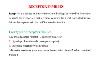
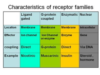
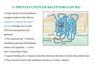
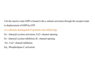
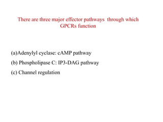
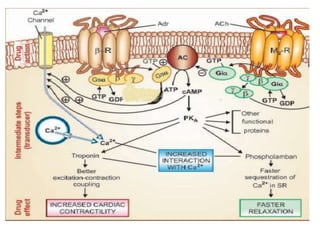
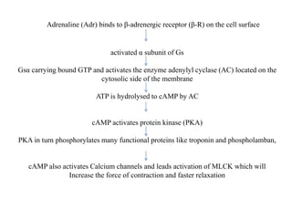
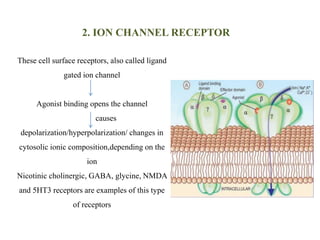
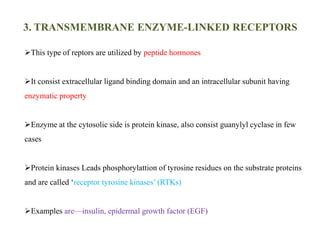
RECEPTORS
By: Raveena Chauhan
Pharmacology Department
Himalayan College of Pharmacy Kala Amb
RECEPTOR FAMILIES
Receptor: It is defined as a macromolecule or binding site located on the surface
or inside the effector cell that serves to recognize the signal molecule/drug and
initiate the response to it, but itself has no other function.
Four types of receptors families
1.G-protien coupled receptor (Metabotropic receptors)
2. Ligand-gated ion channels (inotropic receptors)
3. Enzymatic receptors (tyrosine kinase)
4.Receptor regulating gene expression (transcription factors/Nuclear receptors/
Steroid )
G-PROTEIN COUPLED RECEPTORS (GPCRS)
Large family of cell membrane
receptors linked to the effector
enzymes or channel or carrier
proteins through one or more
GTP activated proteins (G-
proteins)
The molecule has 7 α-helical
membrane spanning hydrophobic
amino acid segments – 3 extra
and 3 intracellular loops
Agonist binding site is located somewhere between the helices on the extracellular face
The G proteins float in the membrane and have α, β and γ subunits
In the inactive state GDP is bound to the α subunit activation through the receptor leads
to displacement of GDP by GTP
α subunits distinguish G proteins into following:
Gs : Adenylyl cyclase activation, Ca2+ channel opening
Gi : Adenylyl cyclase inhibition, K+ channel opening
Go : Ca2+ channel inhibition
Gq : Phospholipase C activation
There are three major effector pathways through which
GPCRs function
(a)Adenylyl cyclase: cAMP pathway
(b) Phospholipase C: IP3-DAG pathway
(c) Channel regulation
Adrenaline (Adr) binds to β-adrenergic receptor (β-R) on the cell surface
activated α subunit of Gs
Gsα carrying bound GTP and activates the enzyme adenylyl cyclase (AC) located on the
cytosolic side of the membrane
ATP is hydrolysed to cAMP by AC
cAMP activates protein kinase (PKA)
PKA in turn phosphorylates many functional proteins like troponin and phospholamban,
cAMP also activates Calcium channels and leads activation of MLCK which will
Increase the force of contraction and faster relaxation
Phospholipase C: IP3-DAG pathway
histamine binds to its H1 receptor (H1 R) and activates the G-protein Gq unit
α subunit binds GTP
activate membrane bound PLc
Leads to activation of inositol 1, 4, 5-trisphosphate (IP3) and diacylglycerol
(DAG) pathway
IP3 facilitate opning of Ca2+ channel , while DAG in conjunction with Ca2+
activates protein kinase C
Ca2+ combines with calmodulin (CAM) to activate myosin light chain kinase
(MLCK) and leads to Contraction
Channel Regulation
Activated G-proteins can open or close ion channels – Ca++, Na+ or K+ etc.
leads depolarization, hyperpolrization or Ca ++ changes etc.
2. ION CHANNEL RECEPTOR
These cell surface receptors, also called ligand
gated ion channel
Agonist binding opens the channel
causes
depolarization/hyperpolarization/ changes in
cytosolic ionic composition,depending on the
ion
Nicotinic cholinergic, GABA, glycine, NMDA
and 5HT3 receptors are examples of this type
of receptors
3. TRANSMEMBRANE ENZYME-LINKED RECEPTORS
This type of reptors are utilized by peptide hormones
It consist extracellular ligand binding domain and an intracellular subunit having
enzymatic property
Enzyme at the cytosolic side is protein kinase, also consist guanylyl cyclase in few
cases
Protein kinases Leads phosphorylattion of tyrosine residues on the substrate proteins
and are called ‘receptor tyrosine kinases’ (RTKs)
Examples are—insulin, epidermal growth factor (EGF)
Hormone binding induces dimerization of receptor molecules
which activate the kinase to autophosphorylate tyrosine residues on each other,
increasing their affinity for binding substrate proteins which have SH2 domain
substrate protein
phosphorylations leading to the response.
4. TRANSMEMBRANE JAK-STAT BINDING
RECEPTORS
These receptors differ from RTKs in not having any intrinsic catalytic domain
Cytokines/hormones binding to the extracellular domain
which activates the intracellular domain to bind free moving JAK
(Janus Kinase) molecules
activated JAK phosphorylate tyrosine residues on the receptor binds
another protein STAT (signal transducer and activator of transcription)
Tyrosine residues of STAT also get phosphorylated by JAK
phosphorylated STAT get dissociated from the receptor
move to the nucleus
regulate transcription of target genes.
5. RECEPTORS REGULATING GENE EXPRESSION
(TRANSCRIPTION FACTORS, NUCLEAR
RECEPTORS)
GR has a steroid binding domain near the carboxy terminus and a mid region DNA
binding domain joined by a ‘hinge region’
DNA binding domain has two ‘zinc fingers’
Binding of the steroid to GR dissociates the complexed protein (HSP90) removing
their inhibitory influence on it
steroid bound receptor diamer translocates to the nucleus, binds
coactivator/corepressor proteins and interacts with ‘glucocorticoid responsive
elements’ (GREs)
Leads transcription of DNA
Specific mRNA is produced and directed to the ribosome
protein synthesis
which inturn modifies cell function.
THANK YOU

Receptors

  • 1.
    RECEPTORS By: Raveena Chauhan PharmacologyDepartment Himalayan College of Pharmacy Kala Amb
  • 2.
    RECEPTOR FAMILIES Receptor: Itis defined as a macromolecule or binding site located on the surface or inside the effector cell that serves to recognize the signal molecule/drug and initiate the response to it, but itself has no other function. Four types of receptors families 1.G-protien coupled receptor (Metabotropic receptors) 2. Ligand-gated ion channels (inotropic receptors) 3. Enzymatic receptors (tyrosine kinase) 4.Receptor regulating gene expression (transcription factors/Nuclear receptors/ Steroid )
  • 4.
    G-PROTEIN COUPLED RECEPTORS(GPCRS) Large family of cell membrane receptors linked to the effector enzymes or channel or carrier proteins through one or more GTP activated proteins (G- proteins) The molecule has 7 α-helical membrane spanning hydrophobic amino acid segments – 3 extra and 3 intracellular loops Agonist binding site is located somewhere between the helices on the extracellular face The G proteins float in the membrane and have α, β and γ subunits
  • 5.
    In the inactivestate GDP is bound to the α subunit activation through the receptor leads to displacement of GDP by GTP α subunits distinguish G proteins into following: Gs : Adenylyl cyclase activation, Ca2+ channel opening Gi : Adenylyl cyclase inhibition, K+ channel opening Go : Ca2+ channel inhibition Gq : Phospholipase C activation
  • 6.
    There are threemajor effector pathways through which GPCRs function (a)Adenylyl cyclase: cAMP pathway (b) Phospholipase C: IP3-DAG pathway (c) Channel regulation
  • 8.
    Adrenaline (Adr) bindsto β-adrenergic receptor (β-R) on the cell surface activated α subunit of Gs Gsα carrying bound GTP and activates the enzyme adenylyl cyclase (AC) located on the cytosolic side of the membrane ATP is hydrolysed to cAMP by AC cAMP activates protein kinase (PKA) PKA in turn phosphorylates many functional proteins like troponin and phospholamban, cAMP also activates Calcium channels and leads activation of MLCK which will Increase the force of contraction and faster relaxation
  • 9.
  • 10.
    histamine binds toits H1 receptor (H1 R) and activates the G-protein Gq unit α subunit binds GTP activate membrane bound PLc Leads to activation of inositol 1, 4, 5-trisphosphate (IP3) and diacylglycerol (DAG) pathway IP3 facilitate opning of Ca2+ channel , while DAG in conjunction with Ca2+ activates protein kinase C Ca2+ combines with calmodulin (CAM) to activate myosin light chain kinase (MLCK) and leads to Contraction
  • 11.
    Channel Regulation Activated G-proteinscan open or close ion channels – Ca++, Na+ or K+ etc. leads depolarization, hyperpolrization or Ca ++ changes etc.
  • 12.
    2. ION CHANNELRECEPTOR These cell surface receptors, also called ligand gated ion channel Agonist binding opens the channel causes depolarization/hyperpolarization/ changes in cytosolic ionic composition,depending on the ion Nicotinic cholinergic, GABA, glycine, NMDA and 5HT3 receptors are examples of this type of receptors
  • 13.
    3. TRANSMEMBRANE ENZYME-LINKEDRECEPTORS This type of reptors are utilized by peptide hormones It consist extracellular ligand binding domain and an intracellular subunit having enzymatic property Enzyme at the cytosolic side is protein kinase, also consist guanylyl cyclase in few cases Protein kinases Leads phosphorylattion of tyrosine residues on the substrate proteins and are called ‘receptor tyrosine kinases’ (RTKs) Examples are—insulin, epidermal growth factor (EGF)
  • 14.
    Hormone binding inducesdimerization of receptor molecules which activate the kinase to autophosphorylate tyrosine residues on each other, increasing their affinity for binding substrate proteins which have SH2 domain substrate protein phosphorylations leading to the response.
  • 15.
    4. TRANSMEMBRANE JAK-STATBINDING RECEPTORS These receptors differ from RTKs in not having any intrinsic catalytic domain
  • 16.
    Cytokines/hormones binding tothe extracellular domain which activates the intracellular domain to bind free moving JAK (Janus Kinase) molecules activated JAK phosphorylate tyrosine residues on the receptor binds another protein STAT (signal transducer and activator of transcription) Tyrosine residues of STAT also get phosphorylated by JAK phosphorylated STAT get dissociated from the receptor move to the nucleus regulate transcription of target genes.
  • 17.
    5. RECEPTORS REGULATINGGENE EXPRESSION (TRANSCRIPTION FACTORS, NUCLEAR RECEPTORS)
  • 18.
    GR has asteroid binding domain near the carboxy terminus and a mid region DNA binding domain joined by a ‘hinge region’ DNA binding domain has two ‘zinc fingers’ Binding of the steroid to GR dissociates the complexed protein (HSP90) removing their inhibitory influence on it steroid bound receptor diamer translocates to the nucleus, binds coactivator/corepressor proteins and interacts with ‘glucocorticoid responsive elements’ (GREs) Leads transcription of DNA Specific mRNA is produced and directed to the ribosome protein synthesis which inturn modifies cell function.
  • 19.