This case involves a patient presenting with a 3 month history of progressive purpuric rash, dyspnea, and fatigue. Examination revealed tender bruising over the thighs and petechial lesions. Laboratory results including normal platelet count and coagulation profile. The patient had a poor diet consisting only of peanut butter sandwiches and no multivitamins. A CT scan showed soft tissue infiltration in the thigh. The diagnosis was determined to be scurvy due to vitamin C deficiency based on symptoms, examination, dietary history, and resolution of symptoms with vitamin C supplementation.
There are several causes for purpuras..... How to clinically approach a patient with purpuric rash???? List of investigations which are helpful in reaching upto the clinical diagnosis....
A presentation about DIC (Disseminated Intravascular Coagulopathy).
Done by 4th year medical students at the University of Science and Technology, Sana'a, Republic of Yemen, in October 2010.
DIC during Pregnancy is the most dreaded complication and matter to clear the concepts is required.
the slides clear and give a better idea about disseminated intravascular coagulation.
hope you find all your answers to queries in these slides.
DIC during Pregnancy is the most dreaded complication and matter to clear the concepts is required.
the slides clear and give a better idea about disseminated intravascular coagulation.
hope you find all your answers to queries in these slides.
Cutaneous involvement is very common in the different types of vasculitis. Skin lesions may be the only manifestation or may occur in the context of systemic disease
Update on Patterns of Study in ANCA Associated Vasculitis presented at regional Northern Ireland Nephrology Meeting with Dr David Jayne as guest speaker..
Portal Hypertension in pediatric populationPrabinPaudyal3
PORTAL HYPERTENSION
OUTLINE:
Definition
Causes
Pathogenesis
Clinical features
Investigations
Management
Complications
Prognosis
Approach
Definition:
Defined as:
Portal Pressure > 10-12 mm Hg, with diameter >10mm Or
Hepatic Venous Pressure Gradient > 4 mm Hg
increased portal resistance or increased portal venous blood flow
major cause of morbidity and mortality in chronic liver diseases
Portal Vein:
Causes of Portal HTN:
Extrahepatic/Pre-hepatic
Hepatic
Pre-Sinusoidal
Sinusoidal
Post-Sinusoidal
Post-hepatic
A. Extra-hepatic:
Portal Vein Thrombosis- Most common
Neonates: Omphalitis, Umbilical Vein Catheterization, Dehydration, Sepsis
Older Children: Intra-abdominal infections e.g., Appendicitis, IBD, PSC
Hypercoagulable states: Deficiencies of factor V Leiden, protein C, S
Blunt Abdominal Trauma
Portal vein agenesis, atresia, stenosis
Splenic vein thrombosis
Biliary tract disease
Extrahepatic biliary atresia
Choledochal cyst
B. Intra-hepatic:
C. Post-hepatic:
Budd-Chiari Syndrome
IVC Webs
Chronic Constrictive Pericarditis
Pathogenesis And Consequence of Portal HTN
Portosystemic collaterals:
Sites:
Lower part of esophagus
Lower part of rectum
Around Umbilicus
Clinical Features:
Bleeding:
Most common presentation
risk of first bleed in cirrhosis is 22%
rises to 38% in with known varices >5-yr period
Pattern of bleeding
Hematemesis/Malena: Most common
worsened by Stress / Intercurrent illness
Size of varices → Bleeding
Splenomegaly:
2nd Most common presentation
asymptomatic or associated with cytopenia
Ascites:
Seen in 7-21% patients
Less common but important manifestations
Portal Hypertensive Biliopathy
Growth Failure
Hepatopulmonary Syndrome
Porto-pulmonary HTN
Caput Medusae:
Abnormal, dilated venous network on anterior abdominal wall, radiating from the umbilicus
Not seen in extra-hepatic portal HTN
Seen in intra-hepatic portal HTN
Continuous murmur between umbilicus and lower sternum
Cruveilhier-Baumgarten Murmur
Investigations
USG with Doppler
portal vein diameter > 10 mm
hepatic diseases, masses, presence of varices and ascites
ascertain pattern of flow
Reversal of portal blood flow (Hepatofugal flow) - Associated with bleeding varices
Cavernous transformation of the portal vein in EHPVO
Increased thickness of lesser omentum
CECT and MRA: Needed in selective cases
Selective Arteriography: When surgical decompression is being planned
GIT Endoscopy: Most reliable to detect varices
Other investigations:
CBC
LFT
Barium swallow
Portal angiogram
Percutaneous intrasplenic measurement of portal pressure
Venography
A. Emergency Management of Bleeding Varices
1st Step (Initial resuscitation):
airway protection
Obtain I/V Access
Restoration of IV volume: fluid and BT
PRBC: Target Hb: 7-9 g/dL
Correction of coagulopathy: vitamin K, FFP/PC
NG
Smoking is the strongest environmental exposure, triggers citrullination of proteins in the lung. Citrullination: Amino acids catalyzed to citrulline which is attacked by the immune system
In Early-stage RA smoking may accelerates joint damage.
There is role of microbiome in RA development.
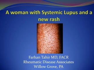
1. Farhan Tahir MD, FACR
Rheumatic Disease Associates
Willow Grove, PA
2. • Case review
• Purpura : causes and types
• Many faces of Purpuric rash
• Purpura from a Rheumatologist's eyes
• Discussion
• Conclusion
3. •Rash-Petechial purpura and ecchymosis (no prior history)
•Chronicity: over 3 months, progressive
•Preceding events: no infectious, traumatic or chemical
exposure, no herbal supplement exposure, no new drug
•Systemic features: dyspnea and fatigue, no
fever, diarrhea, hemoptysis
•Potential culprit medications: ASA, Prednisone
,Cellcept, Gabapentin
•Nutrition: Poor (peanut butter sandwich, no multivitamins)
•Co morbid conditions: SLE, chronic steroid use, osteoporosis
4. Med/Surg hx: Mitral valve prolapse, Hip replacement and
partial hysterectomy (no history of excessive bleeding)
Family: no bleeding diathesis
Social: lives alone, no drugs or alcohol abuse
Exam: Poor dental hygiene, tender bruising over
thighs, petechial lesions, 1+peripheral edema, normal
pulses in feet
5. •Purpura (from Latin: purpura, meaning
"purple")
• Bleeding under skin or into mucosal
membranes
• Pinpoint area, < 2mm: petechiae
• Larger confluent lesions: ecchymosis
(bruises)
•Causes of Purpura
• Disruption of vascular integrity
• Primary or secondary hemostatic abnormality
6. Destruction of vascular wall
• Trauma, inflammation or infection
Impaired platelet plug and fibrin clot
• Injury-> vasoconstriction & retraction->platelet
recognition vessel endothelial adhesion ->platelet granules-
>platelet plug
• Tissue factor-VII complex->activation of coagulation
cascade->Fibrin cross links->clot
Impaired collagen synthesis
• Disordered collagen and connective tissue synthesis and
structure, congenital versus acquired
10. • Palpable Purpura is usually inflammatory or
vasculitis
• Hypersensitivity vasculitis
• Henoch-Scolein purpura
• SLE, RA and small vessel vasculitis
• Infectious
• Drug induced
• Polyarteritis Nodosa
• Pseudovasculitis
11. • Non palpable purpura : Non vasculitic
Corticosteroid use
Idiopathic thrombocytopenic purpura
Thrombotic thrombocytopenic purpura
Disseminated intravascular coagulation
Vitamin K deficiency
Scurvy
24. Conditions that may present with a clinical syndrome mimicing
vasculitis:
(1) atrial myxoma
(2) septicemia
(3) chronic microthromboembolism
(4) infective endocarditis (both septic and microthrombemboli)
(5) rejection after organ transplantation
(6) drug or chemical related (ergotism, cocaine, etc)
32. • Normal platelet and wbc suggesting against thrombocytopenia
and bone marrow suppression : ITP, TTP
• Normal PT and PPT and bleeding time, goes against
coagulation factor deficiencies states DIC, clotting factor
inhibitors and vitamin K deficiency
• Normal inflammatory markers, absence of elevated dsDNA and
hypocomplementemia suggests against a lupus vasculitis
• Absence of pulmonary renal failure, negative serologies for
ANCA associated small vessels vasculitis
33. •Echocardiogram: normal ( excludes CHF, Pulm HTN)
•Doppler negative for DVT
•Colonoscopy- negative ( GI bleeding, HSP)
•V/Q scan: low probability for PE
•CT of the lower extremity showed a soft tissue
infiltration in the medial thigh (bleeding)
34. Risk of vitamin C deficiency
• Scurvy: dietary deficiency of vegetables and vitamins
• Deficiency can cause typical perifollicular hemorrhage, collagen
defect leads to superficial and deep tissue hemorrhage
38. •Syptoms including fatigue, purpuric rash, synovitis with effision,
anemia and markedly elevated ESR and CRP
•One patient presenetd with severe pulmonary hypertension
•Exam consistnt with hemarthrosis and classic skin findings
•Treatment with Vitamin C 500mg-1000mg daily adequately replensihes
•Body stoes
•1-3 weeks for resolution of skin findings
•1-3 moths for hematologic and hemarthrosis