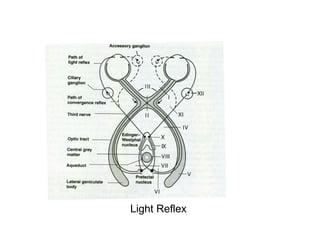
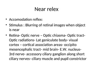
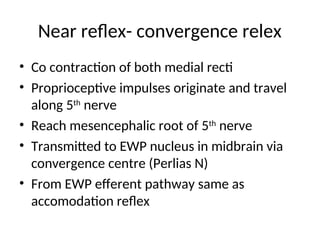
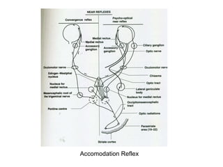
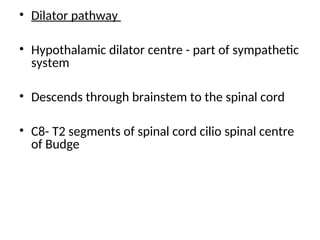
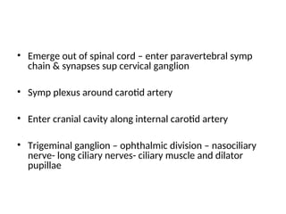
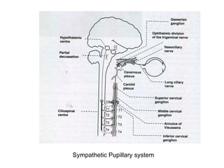
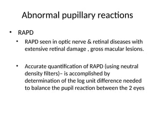
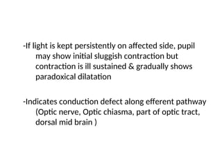
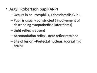
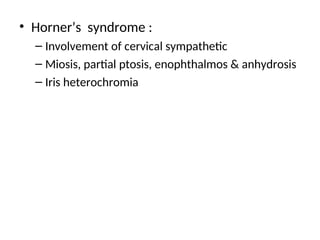
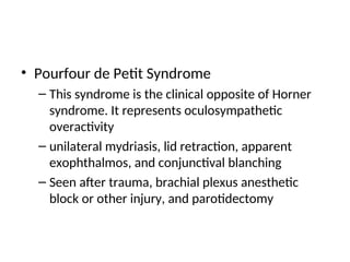
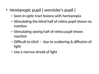
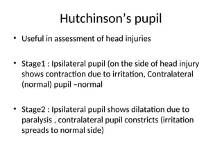
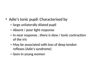
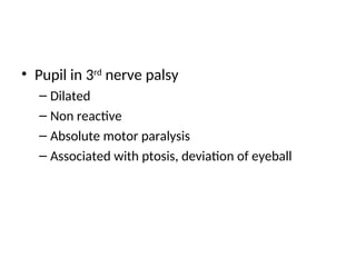
Pupillary pathways & reactions
Dr. C.R.Thirumalachar
• Pupillary constrictor/ spincter-innervated by
parasympathetic
• Pupillary dilator – innervated by sympathetic
• Evaluation of pupil- Diagnostic clue to ocular,
neurological, medical, surgical and paediatric
diseases
Light reflex: Direct & Consensual –
Afferent pathway
• Initiated by retinal photoreceptors
• Transmitted along optic nerve
• Undergo a hemidecussation at the optic chiasma
(nasal fibres cross over)
• Proceeds along optic tract
• Short of lateral geniculate body- enters midbrain
via sup. Brachium of sup. Colliculus
• Synapses at pre- tectal nucleus
• Ends in both Edinger westpal nucleui
• A second decussation occurs around aqueduct of sylvius
• Decussation at chiasma & midbrain level between pretectal
nucleus & Edinger Westpal nucleus accounts for consensual
light reflex
• E.W. nucleus (pupillo motor constrictor centre)
• Efferent fibres tract along 3rd
nerve-nerve to inf. Obl.
• Enter the ciliary ganglion through its short motor root
• Synapse & relay at ciliary ganglion
• Post ganglionic fibres reach ciliary muscle and iris spincter
through short ciliary nerves
Light Reflex
Near relex
• Accomodation reflex:
• Stimulus : Blurring of retinal images when object
is near
• Retina- Optic nerve – Optic chiasma- Optic tract-
Optic radiations- Lat geniculate body- visual
cortex – cortical association areas- occipito
mesencephalic tract- mid brain- E.W. nucleus-
3rd nerve- accessory ciliary ganglion along short
ciliary nerves- ciliary muscle and pupil constrictor
Near reflex- convergence relex
• Co contraction of both medial recti
• Proprioceptive impulses originate and travel
along 5th
nerve
• Reach mesencephalic root of 5th
nerve
• Transmitted to EWP nucleus in midbrain via
convergence centre (Perlias N)
• From EWP efferent pathway same as
accomodation reflex
Accomodation Reflex
• Dilator pathway
• Hypothalamic dilator centre - part of sympathetic
system
• Descends through brainstem to the spinal cord
• C8- T2 segments of spinal cord cilio spinal centre
of Budge
• Emerge out of spinal cord – enter paravertebral symp
chain & synapses sup cervical ganglion
• Symp plexus around carotid artery
• Enter cranial cavity along internal carotid artery
• Trigeminal ganglion – ophthalmic division – nasociliary
nerve- long ciliary nerves- ciliary muscle and dilator
pupillae
Sympathetic Pupillary system
Abnormal pupillary reactions
• RAPD
• RAPD seen in optic nerve & retinal diseases with
extensive retinal damage , gross macular lesions.
• Accurate quantification of RAPD (using neutral
density filters)– is accomplished by
determination of the log unit difference needed
to balance the pupil reaction between the 2 eyes
Marcus Gunn Pupil
-When the contralateral/normal eye is covered, pupil on the
affected side dilates
-When the affected eye is covered pupil of the normal eye
remains unaffected.
– Light is thrown on ipsilateral side(affected
side);Ipsilateral direct reflex & contralateral consensual
reflex- sluggish and ill sustained.
– Light thrown on contralateral side (normal side) direct &
consensual (affected side) is normal & well sustained
-If light is kept persistently on affected side, pupil
may show initial sluggish contraction but
contraction is ill sustained & gradually shows
paradoxical dilatation
-Indicates conduction defect along efferent pathway
(Optic nerve, Optic chiasma, part of optic tract,
dorsal mid brain )
• Argyll Robertson pupil(ARP)
– Occurs in neurosyphilis, Tabesdorsalis,G.P.I.
– Pupil is usually constricted ( involvement of
descending sympathetic dilator fibres)
– Light reflex is absent
– Accomodation reflex , near reflex retained
– Site of lesion –Pretectal nucleus. (dorsal mid
brain)
• Horner’s syndrome :
– Involvement of cervical sympathetic
– Miosis, partial ptosis, enophthalmos & anhydrosis
– Iris heterochromia
• Pourfour de Petit Syndrome
– This syndrome is the clinical opposite of Horner
syndrome. It represents oculosympathetic
overactivity
– unilateral mydriasis, lid retraction, apparent
exophthalmos, and conjunctival blanching
– Seen after trauma, brachial plexus anesthetic
block or other injury, and parotidectomy
• Hemianopic pupil ( wernicke’s pupil )
– Seen in optic tract lesions with hemianopia
– Stimulating the blind half of retina pupil shows no
reaction
– Stimulating seeing half of retina pupil shows
reaction
– Difficult to elicit – due to scattering & diffusion of
light
– Use a narrow streak of light
Hutchinson’s pupil
• Useful in assessment of head injuries
• Stage1 : Ipsilateral pupil (on the side of head injury
shows contraction due to irritation, Contralateral
(normal) pupil –normal
• Stage2 : Ipsilateral pupil shows dilatation due to
paralysis , contralateral pupil constricts (irritation
spreads to normal side)
• Stage3 : Both pupils dilate. Stage of bilateral
paralysis. To assess pupil repeatedly is
important, therefore mydriatics should be
avoided in case of head injuries
• Adie’s tonic pupil: Characterised by
– large unilaterally dilated pupil
– Absent / poor light response
– In near response , there is slow / tonic contraction
of the iris
– May be associated with loss of deep tendon
reflexes (Adie’s syndrome)
– Seen in young women
• Pupil in 3rd
nerve palsy
– Dilated
– Non reactive
– Absolute motor paralysis
– Associated with ptosis, deviation of eyeball
• Pupil in diabetes
– Constricted
– Sluggishly reactive due to
• Glycogen infiltration of spincter
• Autonomic denervation
• Arteriosclerosis of radial iris vessels
Thank You

The Pupillary Pathways ophthalmology.ppt

  • 1.
    Pupillary pathways &reactions Dr. C.R.Thirumalachar
  • 2.
    • Pupillary constrictor/spincter-innervated by parasympathetic • Pupillary dilator – innervated by sympathetic • Evaluation of pupil- Diagnostic clue to ocular, neurological, medical, surgical and paediatric diseases
  • 3.
    Light reflex: Direct& Consensual – Afferent pathway • Initiated by retinal photoreceptors • Transmitted along optic nerve • Undergo a hemidecussation at the optic chiasma (nasal fibres cross over) • Proceeds along optic tract • Short of lateral geniculate body- enters midbrain via sup. Brachium of sup. Colliculus • Synapses at pre- tectal nucleus • Ends in both Edinger westpal nucleui
  • 4.
    • A seconddecussation occurs around aqueduct of sylvius • Decussation at chiasma & midbrain level between pretectal nucleus & Edinger Westpal nucleus accounts for consensual light reflex • E.W. nucleus (pupillo motor constrictor centre) • Efferent fibres tract along 3rd nerve-nerve to inf. Obl. • Enter the ciliary ganglion through its short motor root • Synapse & relay at ciliary ganglion • Post ganglionic fibres reach ciliary muscle and iris spincter through short ciliary nerves
  • 5.
  • 6.
    Near relex • Accomodationreflex: • Stimulus : Blurring of retinal images when object is near • Retina- Optic nerve – Optic chiasma- Optic tract- Optic radiations- Lat geniculate body- visual cortex – cortical association areas- occipito mesencephalic tract- mid brain- E.W. nucleus- 3rd nerve- accessory ciliary ganglion along short ciliary nerves- ciliary muscle and pupil constrictor
  • 7.
    Near reflex- convergencerelex • Co contraction of both medial recti • Proprioceptive impulses originate and travel along 5th nerve • Reach mesencephalic root of 5th nerve • Transmitted to EWP nucleus in midbrain via convergence centre (Perlias N) • From EWP efferent pathway same as accomodation reflex
  • 8.
  • 9.
    • Dilator pathway •Hypothalamic dilator centre - part of sympathetic system • Descends through brainstem to the spinal cord • C8- T2 segments of spinal cord cilio spinal centre of Budge
  • 10.
    • Emerge outof spinal cord – enter paravertebral symp chain & synapses sup cervical ganglion • Symp plexus around carotid artery • Enter cranial cavity along internal carotid artery • Trigeminal ganglion – ophthalmic division – nasociliary nerve- long ciliary nerves- ciliary muscle and dilator pupillae
  • 11.
  • 12.
    Abnormal pupillary reactions •RAPD • RAPD seen in optic nerve & retinal diseases with extensive retinal damage , gross macular lesions. • Accurate quantification of RAPD (using neutral density filters)– is accomplished by determination of the log unit difference needed to balance the pupil reaction between the 2 eyes
  • 13.
    Marcus Gunn Pupil -Whenthe contralateral/normal eye is covered, pupil on the affected side dilates -When the affected eye is covered pupil of the normal eye remains unaffected. – Light is thrown on ipsilateral side(affected side);Ipsilateral direct reflex & contralateral consensual reflex- sluggish and ill sustained. – Light thrown on contralateral side (normal side) direct & consensual (affected side) is normal & well sustained
  • 14.
    -If light iskept persistently on affected side, pupil may show initial sluggish contraction but contraction is ill sustained & gradually shows paradoxical dilatation -Indicates conduction defect along efferent pathway (Optic nerve, Optic chiasma, part of optic tract, dorsal mid brain )
  • 15.
    • Argyll Robertsonpupil(ARP) – Occurs in neurosyphilis, Tabesdorsalis,G.P.I. – Pupil is usually constricted ( involvement of descending sympathetic dilator fibres) – Light reflex is absent – Accomodation reflex , near reflex retained – Site of lesion –Pretectal nucleus. (dorsal mid brain)
  • 16.
    • Horner’s syndrome: – Involvement of cervical sympathetic – Miosis, partial ptosis, enophthalmos & anhydrosis – Iris heterochromia
  • 17.
    • Pourfour dePetit Syndrome – This syndrome is the clinical opposite of Horner syndrome. It represents oculosympathetic overactivity – unilateral mydriasis, lid retraction, apparent exophthalmos, and conjunctival blanching – Seen after trauma, brachial plexus anesthetic block or other injury, and parotidectomy
  • 18.
    • Hemianopic pupil( wernicke’s pupil ) – Seen in optic tract lesions with hemianopia – Stimulating the blind half of retina pupil shows no reaction – Stimulating seeing half of retina pupil shows reaction – Difficult to elicit – due to scattering & diffusion of light – Use a narrow streak of light
  • 19.
    Hutchinson’s pupil • Usefulin assessment of head injuries • Stage1 : Ipsilateral pupil (on the side of head injury shows contraction due to irritation, Contralateral (normal) pupil –normal • Stage2 : Ipsilateral pupil shows dilatation due to paralysis , contralateral pupil constricts (irritation spreads to normal side)
  • 20.
    • Stage3 :Both pupils dilate. Stage of bilateral paralysis. To assess pupil repeatedly is important, therefore mydriatics should be avoided in case of head injuries
  • 21.
    • Adie’s tonicpupil: Characterised by – large unilaterally dilated pupil – Absent / poor light response – In near response , there is slow / tonic contraction of the iris – May be associated with loss of deep tendon reflexes (Adie’s syndrome) – Seen in young women
  • 22.
    • Pupil in3rd nerve palsy – Dilated – Non reactive – Absolute motor paralysis – Associated with ptosis, deviation of eyeball
  • 23.
    • Pupil indiabetes – Constricted – Sluggishly reactive due to • Glycogen infiltration of spincter • Autonomic denervation • Arteriosclerosis of radial iris vessels
  • 24.