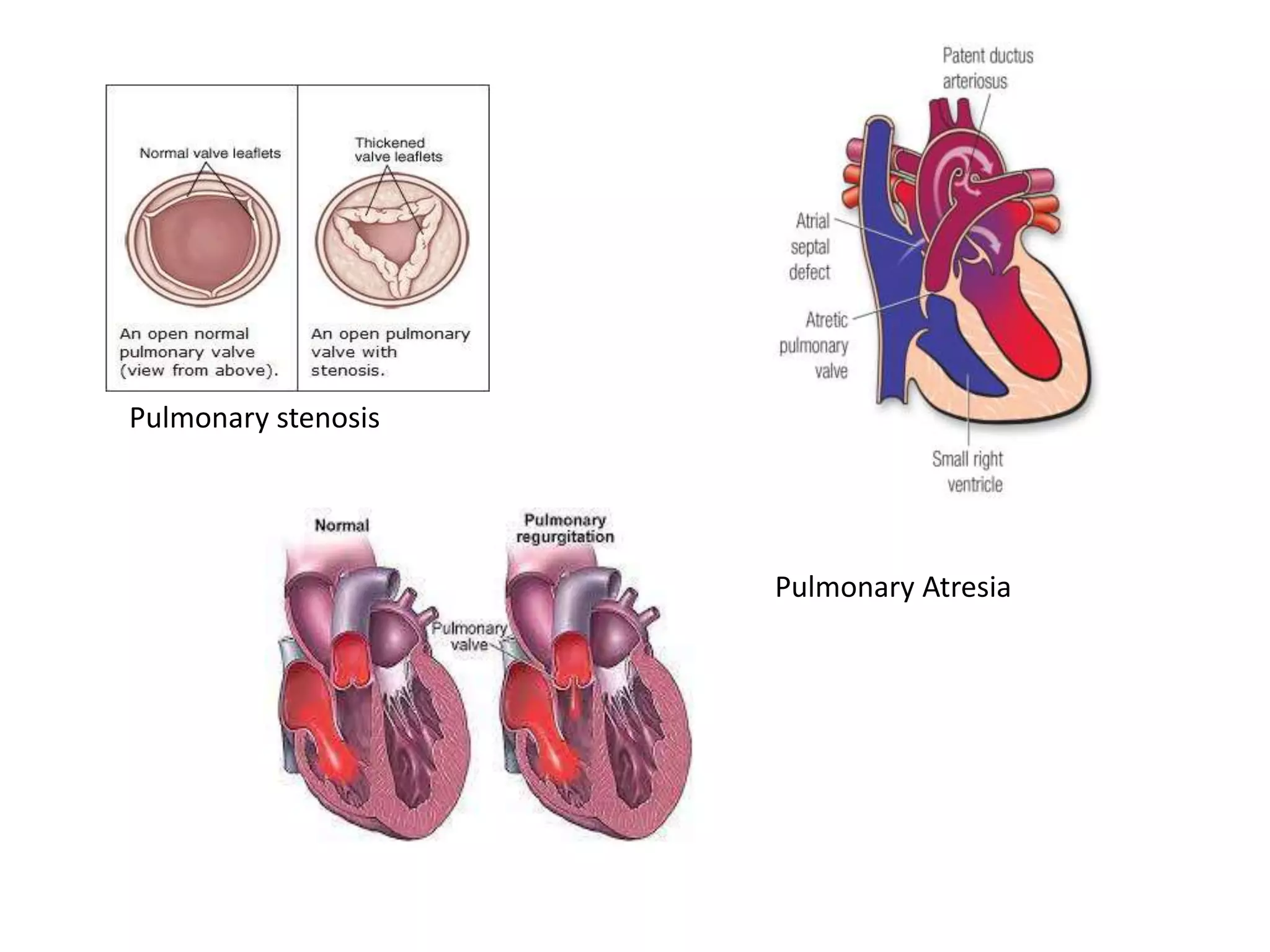
PULMONARY VALVE DISEASES
H. SYED ALI AFRIN
DEPT OF CCT
• Pulmonary valve disease is a condition in which the
lung (pulmonary valve ) located between your heart's
lower right chamber (right ventricle) and the artery
that delivers blood to the lungs (pulmonary artery)
doesn't work properly.
• The condition can interrupt blood flow from your
heart to your lungs.
• Many types of pulmonary valve disease are due to
heart conditions present at birth (congenital heart
defects). Treatment depends on the type and severity
of pulmonary valve disease.
Pulmonary stenosis
Pulmonary Atresia
PULMONARY VALVE STENOSIS
• Narrowing of the pulmonary valve reduces the blood flow
from the heart to the pulmonary artery and lungs.
TYPES
• MILD PS
• MODERATE PS
• SEVERE PS
Classification
• Valvular PS – most common
• Dome shaped ,narrow opening
• Thick leaflets- dysplastic valve
Causes
Occurs along with CHD such as
TOF
C-TGA
GENETIC SYNDROME – NOONAN’S SYNDROME
CLASSIFICATION
• Subvalvular stenosis
Muscle under the PV is thickened
• Supravalvular stenosis
• NARROWING OF THE PULMONARY ARTERY
ABOVE THE PULMONARY VALVE
• CAUSE
WILLIAM’S / ALAGILLE SYNDROME
PATHOPHYSIOLOGY
• NARROWING OF PULMONARY VALVE AREA
• OBSTRUCS FLOW TO PA
• INCREASES PRE LOAD
• RESULTS IN RVH
SIGNS AND SYMPTOMS
•A whooshing sound (murmur) that can be heard with a
stethoscope
•Fatigue
•Shortness of breath, especially during activity
•Chest pain
•Loss of consciousness (fainting)
•Babies with pulmonary valve stenosis and other congenital heart
defects may appear blue (cyanotic).
• Some patienis with mild pulmonary valve stenosis don't
notice any symptoms and may only require occasional doctor's
checkups.
• Moderate and severe pulmonary valve stenosis may
require a procedure to repair or replace the valve
Pulmonary valve regurgitation
•The flaps (leaflets) of the pulmonary valve don't
close tightly, causing blood to leak backward into the
right ventricle.
•The most common causes for a leaky pulmonary valve
is pulmonary hypertension.
•Less common causes are:
•Infective endocarditis
•Complications after surgery to repair TOF
•Carcinoid syndrome
•Rheumatic fever and complications after catheterization
Types
• Mild pulmonary regurgitation
• Moderate pulmonary regurgitation
• Severe pulmonary regurgitation
Clinical Manifestation
• Often asymptomatic
• Dyspnea on exertion
• Lethargy
• Peripheral edema
• Abdominal pain
• Thrill/murmur in pulmonary area
Investigation
• History and physical examination
• ECG: RVH
• CXR : ENLARGED PULMONARY ARTERIES AND
RIGHT VENTRICLE
• ECHOCARDIOGRAM: image may show rv
dilatation and hypertrophy
Abnormal septal motion if rv volume overload
Treatment (pv repair )
• Ring annuloplasty: To repair a leaking valve,
the surgeon sews a ring (made of metal, cloth
or tissue) around the valve to tighten it.
• Commissurotomy: The surgeon loosens a
valve with stenosis.
• Valve flap repair: The surgeon alters your
pulmonary valve flaps by trimming, patching,
shaping or rebuilding them so that they close
tightly.
Treatment
Pulmonary valve replacement
• Mechanical valve: A man-made valve can last as
long as 30 years. To prevent blood clots that may
accompany a mechanical valve, you doctor may
prescribe blood-thinning medications.
• Biological valve: The replacement valve is made
from cow, pig or human heart tissue. These valves
last 10 to 20 years.

PULMONARY VALVE DISEASES (2).pptx

  • 1.
    PULMONARY VALVE DISEASES H.SYED ALI AFRIN DEPT OF CCT
  • 2.
    • Pulmonary valvedisease is a condition in which the lung (pulmonary valve ) located between your heart's lower right chamber (right ventricle) and the artery that delivers blood to the lungs (pulmonary artery) doesn't work properly. • The condition can interrupt blood flow from your heart to your lungs. • Many types of pulmonary valve disease are due to heart conditions present at birth (congenital heart defects). Treatment depends on the type and severity of pulmonary valve disease.
  • 4.
  • 5.
    PULMONARY VALVE STENOSIS •Narrowing of the pulmonary valve reduces the blood flow from the heart to the pulmonary artery and lungs. TYPES • MILD PS • MODERATE PS • SEVERE PS
  • 6.
    Classification • Valvular PS– most common • Dome shaped ,narrow opening • Thick leaflets- dysplastic valve Causes Occurs along with CHD such as TOF C-TGA GENETIC SYNDROME – NOONAN’S SYNDROME
  • 7.
    CLASSIFICATION • Subvalvular stenosis Muscleunder the PV is thickened • Supravalvular stenosis • NARROWING OF THE PULMONARY ARTERY ABOVE THE PULMONARY VALVE • CAUSE WILLIAM’S / ALAGILLE SYNDROME
  • 8.
    PATHOPHYSIOLOGY • NARROWING OFPULMONARY VALVE AREA • OBSTRUCS FLOW TO PA • INCREASES PRE LOAD • RESULTS IN RVH
  • 10.
    SIGNS AND SYMPTOMS •Awhooshing sound (murmur) that can be heard with a stethoscope •Fatigue •Shortness of breath, especially during activity •Chest pain •Loss of consciousness (fainting) •Babies with pulmonary valve stenosis and other congenital heart defects may appear blue (cyanotic).
  • 11.
    • Some patieniswith mild pulmonary valve stenosis don't notice any symptoms and may only require occasional doctor's checkups. • Moderate and severe pulmonary valve stenosis may require a procedure to repair or replace the valve
  • 12.
    Pulmonary valve regurgitation •Theflaps (leaflets) of the pulmonary valve don't close tightly, causing blood to leak backward into the right ventricle. •The most common causes for a leaky pulmonary valve is pulmonary hypertension. •Less common causes are: •Infective endocarditis •Complications after surgery to repair TOF •Carcinoid syndrome •Rheumatic fever and complications after catheterization
  • 13.
    Types • Mild pulmonaryregurgitation • Moderate pulmonary regurgitation • Severe pulmonary regurgitation
  • 15.
    Clinical Manifestation • Oftenasymptomatic • Dyspnea on exertion • Lethargy • Peripheral edema • Abdominal pain • Thrill/murmur in pulmonary area
  • 16.
    Investigation • History andphysical examination • ECG: RVH • CXR : ENLARGED PULMONARY ARTERIES AND RIGHT VENTRICLE • ECHOCARDIOGRAM: image may show rv dilatation and hypertrophy Abnormal septal motion if rv volume overload
  • 17.
    Treatment (pv repair) • Ring annuloplasty: To repair a leaking valve, the surgeon sews a ring (made of metal, cloth or tissue) around the valve to tighten it. • Commissurotomy: The surgeon loosens a valve with stenosis. • Valve flap repair: The surgeon alters your pulmonary valve flaps by trimming, patching, shaping or rebuilding them so that they close tightly.
  • 18.
    Treatment Pulmonary valve replacement •Mechanical valve: A man-made valve can last as long as 30 years. To prevent blood clots that may accompany a mechanical valve, you doctor may prescribe blood-thinning medications. • Biological valve: The replacement valve is made from cow, pig or human heart tissue. These valves last 10 to 20 years.