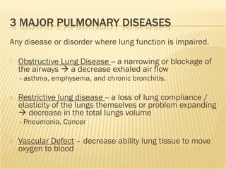
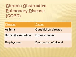
Pulmonary System and Exercise
Cough Wheeze Sputum Shortness of Breath History of exposure to smoking, pollution, etc Spirometry impairment
INSPIRATION AND EXPIRATION
 
 
Pulmonary Diffusion
Respiration INTERNAL Respiration  — Gas exchange between the blood and tissues Pulmonary diffusion  — exchange of oxygen and carbon dioxide between the lungs and the blood Pulmonary ventilation  — inflow and outflow of air between the lugs and atmosphere EXTERNAL Respiration
Any disease or disorder where lung function is impaired.  Obstructive Lung Disease  -- a narrowing or blockage of the airways    a decrease exhaled air flow - asthma, emphysema, and chronic bronchitis. Restrictive lung disease  -- a loss of lung compliance / elasticity of the lungs themselves or problem expanding    decrease in the total lungs volume - Pneumonia, Cancer  Vascular Defect  – decrease ability lung tissue to move oxygen to blood
C hronic  O bstructive  P ulmonary  D isease (COPD) Disease Cause Asthma Constriction airways Bronchitis secretion Excess mucus Emphysema Destruction of alveoli
Causes of COPD NOXIOUS AGENT (tobacco smoke, pollutants, occupational agent) Inflammation Airway inflammation Airway remodeling COPD Genetic factors Respiratory infection Other
Asthma Airway narrowing due to inflammation (bronchial hyperresponsiveness)     airflow obstruction  “ bronchoconstriction”  Less Constricted  More constricted
Bronchitis Inflammation or  thickened bronchial walls  within the lungs due to secretion of fluids acute: infection Chronic: +3 months to year
EMPHYSEMA Loss of elasticity  in the aveoli  Associated with exposure to toxic chemicals & long-term exposure to tobacco smoke.
RESTRICTIVE DISEASE Disease Cause Scoliosis Skeletal Origins Pulmonary Edema Plural Paralysis Neuromuscular Pneumonia  Alveolar blockage Cancer or Fibrotic  Lung Scarring or loss of tissue function  
Reversible Accumulation of inflammatory cells, mucus, in bronchi  Smooth muscle contraction in airways Increase functioning with dynamic hyperinflation during exercise  Irreversible Fibrosis and  narrowing  of the airways Loss of elastic recoil  due to  alveolar destruction Destruction of alveolar  support  that maintains patency of small airways
PULMONARY DISEASE Obstructive    Flow Restrictive    Volume
PULMONARY FUNCTIONS
PULMONARY FUNCTIONS
Diagnosis  Restrictive Disease (   Vol) Normal > 80% of predicted VC Mild 60 to 75% of predicted VC Moderate 50 to 60% of predicted VC Severe < 50% of predicted VC Diagnosis  Obstructive Disease (    Flow) Normal > 80% of predicted FEV1 Mild 60 to 70% of predicted FEV1 Moderate 40 to 59% of predicted FEV1 Severe < 40% of predicted FEV1
% of oxygen bound to hemoglobin Measures for Hypoxia (decreased O2) Hypoxic – not enough supply of 02 Anemic Hypoxia – not enough HGB Diagnosis  SaO2  Average 95-100% Normal 96 – 98 % Low <92% Dysfunction  < 88 %--need supplemental O2
Men  VC = 0.1626*Height(inches) - 0.031*Age(years) - 5.335  
 Women VC = 0.1321*Height(inches) - 0.018*Age(years) - 4.360 http://www.hopkinsmedicine.org/pftlab/predeqns.html
White males 15-79 years (Cherniack, 1972)  = (0.09107 * (height in inches)) - (0.0232 * (age in years)) - 1.50723 White females 15-79 years (Cherniack, 1972)  = (0.06029 * (height in inches)) - (0.01936 * (age in years)) - 0.18693 Black males 20-92 years years (Stinson, 1981)  = (0.096 * (height in inches)) - (0.021 * (age in years)) - 2.51  Black females 20-92 years (Stinson, 1981)  = (0.062 * (height in inches)) - (0.017 * (age in years)) - 0.951 http://www.medal.org/visitor/www%5CActive%5Cch8%5Cch8.01%5Cch8.01.01.aspx
Ventilation (L/min) VO 2  (ml/min kg) Max Max Ventilatory Adaptations to Graded Exercise Normal Pulmonary Impairment
THE VENTILATORY RESPONSE TO EXERCISE
COPD  complications include: Weight loss fat free mass (FFM) Osteoporosis Further reduces exercise capacity, quality of life and survival Loss excess energy  Increased energy cost of breathing Reduced tissue oxygen levels  Metabolic responses that enhance breakdown of body proteins
Identify and eliminate sources of bronchopulmonary inflammation cigarette smoking, inhaled irritants Inhale or oral bronchodilators and corticosteroids Establish individualized rehabilitation programs for stable patients
Rehabilitation programs generally similar to moderate physical and  breathing exercises Respiratory muscle training  may improve exercise performance Cardiovascular or selective respiratory muscle training  May improve oxygen delivery and endurance performance at submaximal exercise Chronic home oxygen therapy  for patients whose PaO 2  remains below 55 mmHg (the goal is to alleviate hypoxemia)
Smoking Exposure to pollution or other noxious agents Genetics Age History of childhood respiratory infections
Pay special attention to environmental conditions Follow GENERAL FIT recommended by ACSM Walking most similar to daily living activities Minimal goals for frequency is 3-5 d/wk— reduced function more frequent exercise training NO CONCENSUS AS TO THE OPTIMAL INTENSITY tolerated OR 50% of Max Start at few minutes…progress as able Pulmonary effect the lungs as well as muscles Follow older adults guidelines Shoulder girdle exercises
Monitor for signs and symptoms Use dyspnea scale for 2-3 on 4 point scale  May exhibit arterial desaturation w/ exercise May measure blood oxygenation Oximetry  May need O2 for patients with reduced PaO2
Complete extensive pulmonary function tests prior Only  stable patients  should exercise in a nonmedical setting RXEX Suggested that patients exercise at 50% VO 2 peak Emphasize progression over intensity To exercise Must be fully be symptom free from bronchitis Have a bronchodilating inhaler with them at all times Perform breathing exercises to help strengthen respiratory muscles
Avoid upper-body exercises initially because of the increased strain on the pulmonary system.   Some COPD individuals may require supplemental oxygen  during exercise. Generally, supplemental oxygen is recommended for patients with a PaO 2  < 55 mmHg or SaO 2  < 88%, while breathing room air COPD clients should not smoke The type and dose of medications should be reviewed with the clients physician, based on the clients response to exercise If a COPD clients exercise performance in a nonmedical supervised program worsens, they should be encouraged to participate in a pulmonary rehabilitation program, until signs and symptoms have improved

Pulmonary s11

  • 1.
  • 2.
    Cough Wheeze SputumShortness of Breath History of exposure to smoking, pollution, etc Spirometry impairment
  • 3.
  • 4.
  • 5.
  • 6.
  • 7.
    Respiration INTERNAL Respiration — Gas exchange between the blood and tissues Pulmonary diffusion — exchange of oxygen and carbon dioxide between the lungs and the blood Pulmonary ventilation — inflow and outflow of air between the lugs and atmosphere EXTERNAL Respiration
  • 8.
    Any disease ordisorder where lung function is impaired. Obstructive Lung Disease -- a narrowing or blockage of the airways  a decrease exhaled air flow - asthma, emphysema, and chronic bronchitis. Restrictive lung disease -- a loss of lung compliance / elasticity of the lungs themselves or problem expanding  decrease in the total lungs volume - Pneumonia, Cancer Vascular Defect – decrease ability lung tissue to move oxygen to blood
  • 9.
    C hronic O bstructive P ulmonary D isease (COPD) Disease Cause Asthma Constriction airways Bronchitis secretion Excess mucus Emphysema Destruction of alveoli
  • 10.
    Causes of COPDNOXIOUS AGENT (tobacco smoke, pollutants, occupational agent) Inflammation Airway inflammation Airway remodeling COPD Genetic factors Respiratory infection Other
  • 11.
    Asthma Airway narrowingdue to inflammation (bronchial hyperresponsiveness)  airflow obstruction “ bronchoconstriction” Less Constricted More constricted
  • 12.
    Bronchitis Inflammation or thickened bronchial walls within the lungs due to secretion of fluids acute: infection Chronic: +3 months to year
  • 13.
    EMPHYSEMA Loss ofelasticity in the aveoli Associated with exposure to toxic chemicals & long-term exposure to tobacco smoke.
  • 14.
    RESTRICTIVE DISEASE DiseaseCause Scoliosis Skeletal Origins Pulmonary Edema Plural Paralysis Neuromuscular Pneumonia Alveolar blockage Cancer or Fibrotic  Lung Scarring or loss of tissue function  
  • 15.
    Reversible Accumulation ofinflammatory cells, mucus, in bronchi Smooth muscle contraction in airways Increase functioning with dynamic hyperinflation during exercise Irreversible Fibrosis and narrowing of the airways Loss of elastic recoil due to alveolar destruction Destruction of alveolar support that maintains patency of small airways
  • 16.
    PULMONARY DISEASE Obstructive Flow Restrictive  Volume
  • 17.
  • 18.
  • 19.
    Diagnosis RestrictiveDisease (  Vol) Normal > 80% of predicted VC Mild 60 to 75% of predicted VC Moderate 50 to 60% of predicted VC Severe < 50% of predicted VC Diagnosis Obstructive Disease (  Flow) Normal > 80% of predicted FEV1 Mild 60 to 70% of predicted FEV1 Moderate 40 to 59% of predicted FEV1 Severe < 40% of predicted FEV1
  • 20.
    % of oxygenbound to hemoglobin Measures for Hypoxia (decreased O2) Hypoxic – not enough supply of 02 Anemic Hypoxia – not enough HGB Diagnosis SaO2 Average 95-100% Normal 96 – 98 % Low <92% Dysfunction < 88 %--need supplemental O2
  • 21.
    Men VC= 0.1626*Height(inches) - 0.031*Age(years) - 5.335 
 Women VC = 0.1321*Height(inches) - 0.018*Age(years) - 4.360 http://www.hopkinsmedicine.org/pftlab/predeqns.html
  • 22.
    White males 15-79years (Cherniack, 1972) = (0.09107 * (height in inches)) - (0.0232 * (age in years)) - 1.50723 White females 15-79 years (Cherniack, 1972) = (0.06029 * (height in inches)) - (0.01936 * (age in years)) - 0.18693 Black males 20-92 years years (Stinson, 1981) = (0.096 * (height in inches)) - (0.021 * (age in years)) - 2.51  Black females 20-92 years (Stinson, 1981) = (0.062 * (height in inches)) - (0.017 * (age in years)) - 0.951 http://www.medal.org/visitor/www%5CActive%5Cch8%5Cch8.01%5Cch8.01.01.aspx
  • 23.
    Ventilation (L/min) VO2 (ml/min kg) Max Max Ventilatory Adaptations to Graded Exercise Normal Pulmonary Impairment
  • 24.
  • 25.
    COPD complicationsinclude: Weight loss fat free mass (FFM) Osteoporosis Further reduces exercise capacity, quality of life and survival Loss excess energy Increased energy cost of breathing Reduced tissue oxygen levels Metabolic responses that enhance breakdown of body proteins
  • 26.
    Identify and eliminatesources of bronchopulmonary inflammation cigarette smoking, inhaled irritants Inhale or oral bronchodilators and corticosteroids Establish individualized rehabilitation programs for stable patients
  • 27.
    Rehabilitation programs generallysimilar to moderate physical and breathing exercises Respiratory muscle training may improve exercise performance Cardiovascular or selective respiratory muscle training May improve oxygen delivery and endurance performance at submaximal exercise Chronic home oxygen therapy for patients whose PaO 2 remains below 55 mmHg (the goal is to alleviate hypoxemia)
  • 28.
    Smoking Exposure topollution or other noxious agents Genetics Age History of childhood respiratory infections
  • 29.
    Pay special attentionto environmental conditions Follow GENERAL FIT recommended by ACSM Walking most similar to daily living activities Minimal goals for frequency is 3-5 d/wk— reduced function more frequent exercise training NO CONCENSUS AS TO THE OPTIMAL INTENSITY tolerated OR 50% of Max Start at few minutes…progress as able Pulmonary effect the lungs as well as muscles Follow older adults guidelines Shoulder girdle exercises
  • 30.
    Monitor for signsand symptoms Use dyspnea scale for 2-3 on 4 point scale May exhibit arterial desaturation w/ exercise May measure blood oxygenation Oximetry May need O2 for patients with reduced PaO2
  • 31.
    Complete extensive pulmonaryfunction tests prior Only stable patients should exercise in a nonmedical setting RXEX Suggested that patients exercise at 50% VO 2 peak Emphasize progression over intensity To exercise Must be fully be symptom free from bronchitis Have a bronchodilating inhaler with them at all times Perform breathing exercises to help strengthen respiratory muscles
  • 32.
    Avoid upper-body exercisesinitially because of the increased strain on the pulmonary system. Some COPD individuals may require supplemental oxygen during exercise. Generally, supplemental oxygen is recommended for patients with a PaO 2 < 55 mmHg or SaO 2 < 88%, while breathing room air COPD clients should not smoke The type and dose of medications should be reviewed with the clients physician, based on the clients response to exercise If a COPD clients exercise performance in a nonmedical supervised program worsens, they should be encouraged to participate in a pulmonary rehabilitation program, until signs and symptoms have improved

Editor's Notes

  • #8 Pulmonary ventilation is breathing